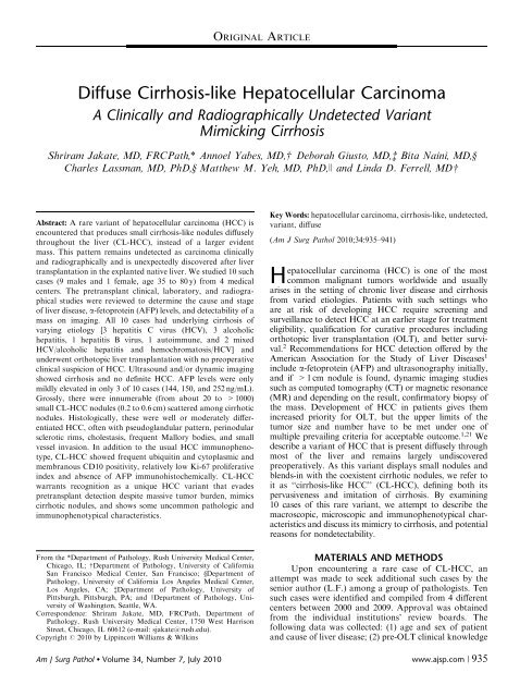
Diffuse Cirrhosis-like Hepatocellular Carcinoma - Rush University ...
Diffuse Cirrhosis-like Hepatocellular Carcinoma - Rush University ...
Diffuse Cirrhosis-like Hepatocellular Carcinoma - Rush University ...
Create successful ePaper yourself
Turn your PDF publications into a flip-book with our unique Google optimized e-Paper software.
<strong>Diffuse</strong> <strong>Cirrhosis</strong>-<strong>like</strong> <strong>Hepatocellular</strong> <strong>Carcinoma</strong><br />
A Clinically and Radiographically Undetected Variant<br />
Mimicking <strong>Cirrhosis</strong><br />
Shriram Jakate, MD, FRCPath,* Annoel Yabes, MD,w Deborah Giusto, MD,z Bita Naini, MD,y<br />
Charles Lassman, MD, PhD,y Matthew M. Yeh, MD, PhD,J and Linda D. Ferrell, MDw<br />
Abstract: A rare variant of hepatocellular carcinoma (HCC) is<br />
encountered that produces small cirrhosis-<strong>like</strong> nodules diffusely<br />
throughout the liver (CL-HCC), instead of a larger evident<br />
mass. This pattern remains undetected as carcinoma clinically<br />
and radiographically and is unexpectedly discovered after liver<br />
transplantation in the explanted native liver. We studied 10 such<br />
cases (9 males and 1 female, age 35 to 80 y) from 4 medical<br />
centers. The pretransplant clinical, laboratory, and radiographical<br />
studies were reviewed to determine the cause and stage<br />
of liver disease, a-fetoprotein (AFP) levels, and detectability of a<br />
mass on imaging. All 10 cases had underlying cirrhosis of<br />
varying etiology [3 hepatitis C virus (HCV), 3 alcoholic<br />
hepatitis, 1 hepatitis B virus, 1 autoimmune, and 2 mixed<br />
HCV/alcoholic hepatitis and hemochromatosis/HCV] and<br />
underwent orthotopic liver transplantation with no preoperative<br />
clinical suspicion of HCC. Ultrasound and/or dynamic imaging<br />
showed cirrhosis and no definite HCC. AFP levels were only<br />
mildly elevated in only 3 of 10 cases (144, 150, and 252 ng/mL).<br />
Grossly, there were innumerable (from about 20 to >1000)<br />
small CL-HCC nodules (0.2 to 0.6 cm) scattered among cirrhotic<br />
nodules. Histologically, these were well or moderately differentiated<br />
HCC, often with pseudoglandular pattern, perinodular<br />
sclerotic rims, cholestasis, frequent Mallory bodies, and small<br />
vessel invasion. In addition to the usual HCC immunophenotype,<br />
CL-HCC showed frequent ubiquitin and cytoplasmic and<br />
membranous CD10 positivity, relatively low Ki-67 proliferative<br />
index and absence of AFP immunohistochemically. CL-HCC<br />
warrants recognition as a unique HCC variant that evades<br />
pretransplant detection despite massive tumor burden, mimics<br />
cirrhotic nodules, and shows some uncommon pathologic and<br />
immunophenotypical characteristics.<br />
From the *Department of Pathology, <strong>Rush</strong> <strong>University</strong> Medical Center,<br />
Chicago, IL; wDepartment of Pathology, <strong>University</strong> of California<br />
San Francisco Medical Center, San Francisco; yDepartment of<br />
Pathology, <strong>University</strong> of California Los Angeles Medical Center,<br />
Los Angeles, CA; zDepartment of Pathology, <strong>University</strong> of<br />
Pittsburgh, Pittsburgh, PA; and JDepartment of Pathology, <strong>University</strong><br />
of Washington, Seattle, WA.<br />
Correspondence: Shriram Jakate, MD, FRCPath, Department of<br />
Pathology, <strong>Rush</strong> <strong>University</strong> Medical Center, 1750 West Harrison<br />
Street, Chicago, IL 60612 (e-mail: sjakate@rush.edu).<br />
Copyright r 2010 by Lippincott Williams & Wilkins<br />
ORIGINAL ARTICLE<br />
Key Words: hepatocellular carcinoma, cirrhosis-<strong>like</strong>, undetected,<br />
variant, diffuse<br />
(Am J Surg Pathol 2010;34:935–941)<br />
<strong>Hepatocellular</strong> carcinoma (HCC) is one of the most<br />
common malignant tumors worldwide and usually<br />
arises in the setting of chronic liver disease and cirrhosis<br />
from varied etiologies. Patients with such settings who<br />
are at risk of developing HCC require screening and<br />
surveillance to detect HCC at an earlier stage for treatment<br />
eligibility, qualification for curative procedures including<br />
orthotopic liver transplantation (OLT), and better survival.<br />
2 Recommendations for HCC detection offered by the<br />
American Association for the Study of Liver Diseases1 include a-fetoprotein (AFP) and ultrasonography initially,<br />
and if >1 cm nodule is found, dynamic imaging studies<br />
such as computed tomography (CT) or magnetic resonance<br />
(MR) and depending on the result, confirmatory biopsy of<br />
the mass. Development of HCC in patients gives them<br />
increased priority for OLT, but the upper limits of the<br />
tumor size and number have to be met under one of<br />
multiple prevailing criteria for acceptable outcome. 1,21 We<br />
describe a variant of HCC that is present diffusely through<br />
most of the liver and remains largely undiscovered<br />
preoperatively. As this variant displays small nodules and<br />
blends-in with the coexistent cirrhotic nodules, we refer to<br />
it as ‘‘cirrhosis-<strong>like</strong> HCC’’ (CL-HCC), defining both its<br />
pervasiveness and imitation of cirrhosis. By examining<br />
10 cases of this rare variant, we attempt to describe the<br />
macroscopic, microscopic and immunophenotypical characteristics<br />
and discuss its mimicry to cirrhosis, and potential<br />
reasons for nondetectability.<br />
MATERIALS AND METHODS<br />
Upon encountering a rare case of CL-HCC, an<br />
attempt was made to seek additional such cases by the<br />
senior author (L.F.) among a group of pathologists. Ten<br />
such cases were identified and compiled from 4 different<br />
centers between 2000 and 2009. Approval was obtained<br />
from the individual institutions’ review boards. The<br />
following data was collected: (1) age and sex of patient<br />
and cause of liver disease; (2) pre-OLT clinical knowledge<br />
Am J Surg Pathol Volume 34, Number 7, July 2010 www.ajsp.com | 935
Jakate et al Am J Surg Pathol Volume 34, Number 7, July 2010<br />
or detection of HCC; (3) serum AFP values; (4) pre-OLT<br />
imaging results; (5) gross photograph and/or gross<br />
description of the explanted liver, specifically the size<br />
ranges and distribution of cirrhotic-<strong>like</strong> abnormal pale<br />
or cholestatic nodules; (6) detailed review of the routine<br />
formalin-fixed, paraffin-embedded, and hematoxylin<br />
and eosin stained 4 mm thick microscopic sections; (7)<br />
immunohistochemical studies on formalin-fixed and<br />
paraffin-embedded representative sections of the tumor<br />
for the following markers: CD10, CD34, HepPar-1,<br />
Ki-67, cytokeratins 7 and 19, AFP, glypican-3, and<br />
ubiquitin; and (8) available follow-up including recurrence<br />
of HCC in the graft. The demographic, clinical,<br />
pathologic, and follow-up information was collected from<br />
individual sites. All microscopic sections were reviewed by<br />
4 participating pathologists as a panel with concurrence<br />
of findings.<br />
Macroscopically, the livers were examined and<br />
sampled for microscopic evaluation according to the<br />
individual medical center’s protocol. At all places, the<br />
livers were thinly sliced at approximately 0.5 cm intervals<br />
and random protocol sections from both lobes were<br />
taken, a minimum of 5 and a maximum of 14. In all cases,<br />
abnormal appearing cholestatic or pale nodules were<br />
documented for their size range and extent, and<br />
additional sections (5 to 10) from these abnormal nodules<br />
were taken. Formalin-fixed paraffin-embedded sections<br />
were processed, embedded, cut at 4 mm, and stained<br />
routinely with hematoxylin and eosin. The sections were<br />
examined microscopically with emphasis on presence of<br />
HCC in random sections of cirrhotic nodules, differentiation,<br />
and additional histologic tumor characteristics.<br />
Immunohistochemical studies were performed on one<br />
representative tumor bearing section for the following<br />
markers: CD10 (Leica Microsystems, Bannockburn, IL),<br />
HepPar-1 (Cell Marque, Rocklin, CA), CD34 (Cell<br />
Marque, Rocklin, CA), Ki-67 (Dako, Carpinteria, CA),<br />
cytokeratin 19 (Cell Marque, Rocklin, CA), cytokeratin 7<br />
(Cell Marque, Rocklin, CA), AFP (Cell Marque, Rocklin,<br />
CA), glypican-3 (Cell Marque, Rocklin, CA), and<br />
ubiquitin (Invitrogen, Carlsbad, CA). Cytokeratins 7 and<br />
19 were performed to determine if there was admixed<br />
cholangiocarcinoma component. In patients where follow<br />
up was available, it was studied for tumor recurrence,<br />
metastases and graft and patient survival.<br />
RESULTS<br />
Clinical Features<br />
The patient demographic data included 9 males and<br />
1 female in the age range of 35 to 80 years with the mean<br />
age of 61 years. All 10 patients had cirrhosis from varying<br />
etiologies [3 hepatitis C virus (HCV), 3 alcoholic hepatitis,<br />
1 hepatitis B virus (HBV), 1 autoimmune, and 2 mixed<br />
HCV/alcoholic hepatitis and hemochromatosis/HCV].<br />
All patients met the clinical and laboratory criteria for<br />
OLT qualification and underwent OLT without pretransplant<br />
diagnosis of HCC. In particular, HCC appeared<br />
clinically silent and without stigmata such as rapid weight<br />
936 | www.ajsp.com<br />
loss, tumor-related ascites or hepatomegaly, palpable<br />
mass, or evidence of metastases. In all 10 cases, the first<br />
suspicion of diffuse HCC ensued after the explanted liver<br />
was examined grossly and cut surfaces showed abnormal<br />
appearing pale and/or cholestatic cirrhotic-<strong>like</strong> nodules<br />
scattered among the cirrhotic nodules.<br />
Serology<br />
The AFP values were below 20 ng/mL in 7 patients<br />
and increased in only 3 patients at 144, 150, and 252<br />
ng/mL, respectively. In these cases, HCC was clinically<br />
suspected but as imaging studies did not identify a mass,<br />
these raised values were regarded as nonspecific and<br />
related to chronic liver disease.<br />
Imaging<br />
The ultrasonography performed on 6 of 10 cases<br />
showed normal hepatic echogenicity, diffuse coarseness<br />
compatible with cirrhosis, and no focal lesions. There was<br />
trace ascites or perihepatic fluid. The other 4 cases had<br />
dynamic imaging-CT of the abdomen with and without<br />
contrast and/or MR with and without gadolinium. In 2<br />
of these 4 cases, small lesion(s), 1000 in numbers)<br />
and diffusely scattered throughout the liver sometimes<br />
occupying up to 50% of the total volume (Fig. 1). Some<br />
cirrhotic nodules were partially pale suggestive of partial<br />
involvement of cirrhotic nodule by tumor (Fig. 2). In 2 of<br />
10 cases, the nodules were fewer, about 20, and more<br />
localized within the liver (Fig. 3). Aggregate tumor sizes<br />
were estimated between 5 and 23 cm. There was no<br />
necrosis and no tumor was grossly seen to invade the<br />
vessels or the bile ducts.<br />
Histologic Features<br />
Representative sections from all cases were reviewed<br />
and the tumor nodules in each case were classified and<br />
graded according to the World Health Organization<br />
criteria. 4 There was general uniformity of grade among<br />
multiple nodules in each individual patient. Almost all<br />
tumor nodules were either moderately or well-differentiated<br />
carcinoma of hepatocellular origin and often<br />
showed pseudoglandular pattern (Fig. 4A). Corresponding<br />
well with the macroscopically visible pallor and/or<br />
cholestasis, the tumor cells frequently showed ballooning<br />
r 2010 Lippincott Williams & Wilkins
Am J Surg Pathol Volume 34, Number 7, July 2010 <strong>Diffuse</strong> <strong>Cirrhosis</strong>-<strong>like</strong> <strong>Hepatocellular</strong> <strong>Carcinoma</strong><br />
FIGURE 1. Cut surface of liver showing pale and cholestatic<br />
cirrhosis-<strong>like</strong> hepatocellular carcinoma nodules (highlighted<br />
with the rectangles) no bigger than darker non-neoplastic<br />
cirrhotic nodules and occupying about 50% of the liver<br />
volume.<br />
and/or cholestasis and there were focally numerous<br />
Mallory bodies (Fig. 4B) in 8 of 10 cases. Many tumor<br />
nodules showed perinodular fibrous rims (Fig. 4C) greatly<br />
mimicking cirrhotic nodules and invasion in small<br />
intratumoral and peritumoral vessels (Fig. 4D). However,<br />
larger segmental and hilar vessels showed no invasion.<br />
Occasionally, cirrhotic nodules were partially and<br />
abruptly invaded by the tumor cells (Fig. 5A). Purported<br />
preneoplastic changes such as dysplasia (large cell and<br />
small cell change) or foci or nodules of altered hepatocytes<br />
were not noticeable in the intervening cirrhotic<br />
nodules. Although individually, none of these characteristics<br />
signify histologic uniqueness, the combined gross<br />
FIGURE 2. Close-up of the liver cut surface showing darker<br />
non-neoplastic cirrhotic nodules (highlighted by the red<br />
rectangle) and partial involvement of cirrhotic nodule by paler<br />
cirrhosis-<strong>like</strong> hepatocellular carcinoma (highlighted by the<br />
blue rectangle).<br />
FIGURE 3. Series of slices showing more localized and fewer<br />
pale nodules of cirrhosis-<strong>like</strong> hepatocellular carcinoma (highlighted<br />
with the rectangles).<br />
and microscopic pattern is quite consistent and distinctive<br />
of this CL-HCC variant.<br />
Immunohistochemical Features<br />
The immunophenotypical profile showed results<br />
anticipated for well and moderately differentiated HCC<br />
in most cases. All tumor cells were positive for HepPar-1<br />
and glypican-3 (Fig. 5B), negative for AFP and cytokeratins<br />
7 and 19 and showed strong CD34 expression<br />
by sinusoidal endothelial cells (Fig. 5C). All cases with<br />
Mallory bodies (8 of 10) showed expected ubiquitin<br />
positivity. Apart from the usual pericanalicular CD-10<br />
expression, 6 of 10 cases showed strong membranous and<br />
cytoplasmic positivity (Fig. 5D). Ki-67 proliferative index<br />
was low (1% to 25%, mean of 14%) conforming to the<br />
better differentiation of HCC.<br />
Follow Up<br />
At the time of transplant, not only was there no<br />
tumor suspected within the liver, but also no metastatic<br />
tumor was described clinically. Considering the extensiveness<br />
of the tumor, surprisingly no tumor was found in<br />
the major branches of hepatic or portal veins. Follow up<br />
was available in 5 out of 10 cases. Two of these 5 patients<br />
expired within 3 years–one from cardiac complications<br />
related to hemochromatosis but without tumor recurrence<br />
or metastasis and the second with tumor recurrence<br />
in the allograft and metastases to lungs. The remaining 3<br />
patients are surviving for more than 3 years without<br />
known recurrence or metastases. One of these 3 surviving<br />
patients had fewer, more localized tumor nodules while<br />
the other 2 patients had diffuse involvement. The clinical<br />
data is summarized in Table 1.<br />
DISCUSSION<br />
Multiplicity of primary neoplasm in the liver,<br />
especially with underlying chronic liver disease and cirrhosis<br />
is not uncommon. However, the CL-HCC variant seems to<br />
r 2010 Lippincott Williams & Wilkins www.ajsp.com | 937
Jakate et al Am J Surg Pathol Volume 34, Number 7, July 2010<br />
FIGURE 4. Well-differentiated hepatocellular carcinoma showing pseudoglandular pattern (A) and moderately differentiated<br />
hepatocellular carcinoma showing ballooning, cholestasis, and several Mallory bodies (B) (hematoxylin and eosin, magnification<br />
400). <strong>Cirrhosis</strong>-<strong>like</strong> hepatocellular carcinoma nodule showing perinodular sclerotic rim (C) and foci showing small vessel<br />
invasion (D) (hematoxylin and eosin, magnification 200).<br />
be a unique neoplastic process that necessitates understanding<br />
of phenomena such as macroscopic small<br />
nodular growth pattern extensively through the liver,<br />
microscopic and immunophenotypical preference for<br />
certain uncommon findings, multiclonal tumor growth<br />
versus intrahepatic metastases from a single primary<br />
neoplasm, ability to evade clinical detection, and implications<br />
of recurrence in the graft from such unforeseen<br />
pretransplant tumor mass. Our collection of only 10 cases<br />
over 9 years of assessment and from multiple medical<br />
centers are <strong>like</strong>ly reflective of the rarity of this variant.<br />
And although statistical determinations are not possible<br />
with this relatively minute population, some insight may<br />
be gained from their unifying characteristics.<br />
References have been made in the literature to<br />
macroscopic confluent multinodular growth pattern<br />
(sometimes called ‘‘cirrhotomimetic’’) of HCC 3,5,14,16<br />
but without emphasis on clinical undetectability, microscopic<br />
or immunophenotypical features, or incidental<br />
eligibility for OLT. Grossly, most HCCs may display 1<br />
938 | www.ajsp.com<br />
of 3 of the following described patterns in the decreasing<br />
order of frequency: expanding, generally solitary nodule<br />
(‘‘massive’’ type) with discrete margins and apparent<br />
encapsulation, multiple discrete nodules which may or<br />
may not be satellite nodules associated a solitary large<br />
nodule (‘‘extranodular’’ or ‘‘nodular’’ type) with margins<br />
and generally apparent encapsulation, and small ‘‘spreading’’<br />
nodules (‘‘infiltrative’’ or ‘‘diffuse’’ type) without<br />
apparent margins and a ‘‘replacing’’ rather than ‘‘expanding’’<br />
growth pattern. Previous studies with such gross<br />
descriptions and classifications have been based upon<br />
autopsy or surgical resections rather than native explanted<br />
livers at transplantation. CL-HCC matches closely with the<br />
diffuse or infiltrative growth pattern. However, most CL-<br />
HCC nodules do have a well-defined peritumoral fibrous<br />
border un<strong>like</strong> the original description of small nodular<br />
diffuse HCC. Having fibrous encapsulation actually<br />
enhances the chances of tumor visibility on dynamic<br />
imaging but the small size rather than fibrous encapsulation<br />
may be the primary reason for radiographic nondetection. 6<br />
r 2010 Lippincott Williams & Wilkins
Am J Surg Pathol Volume 34, Number 7, July 2010 <strong>Diffuse</strong> <strong>Cirrhosis</strong>-<strong>like</strong> <strong>Hepatocellular</strong> <strong>Carcinoma</strong><br />
FIGURE 5. Partial involvement of a cirrhotic nodule by CL-HCC (A) (hematoxylin and eosin, magnification 100).<br />
Immunohistochemically, CL-HCC cells showing glypican-3 positivity (B), CD34 reactivity in endothelial cells contrasting with<br />
the adjacent non-neoplastic cirrhotic nodule (C), and CD10 showing the pericanalicular reactivity seen in HCC but additional<br />
strong membranous and cytoplasmic positivity (D) (magnifications 200 for B and C, and 400 for D). CL-HCC indicates<br />
cirrhosis-<strong>like</strong> hepatocellular carcinoma.<br />
In addition, CL-HCC nodules on gross examination appear<br />
consistently pale and/or cholestatic corresponding with the<br />
microscopic findings of ballooning and/or cholestasis. In<br />
TABLE 1. Summary of Clinical Data<br />
No.<br />
Age (y)<br />
and Sex<br />
Cause of<br />
<strong>Cirrhosis</strong><br />
fact, this gross finding may be the principal way of<br />
discriminating potential tumor nodules from adjacent<br />
non-neoplastic cirrhotic nodules as their sizes are identical.<br />
AFP<br />
>20 ng/mL<br />
Pre-OLT >1 cm<br />
Mass on Imaging<br />
Follow-up of<br />
>3 y<br />
1 62, Male HCV No No Living, tumor free<br />
2 60, Male EtOH No No NA<br />
3 80, Male HCV 144 No NA<br />
4 73, Male EtOH 150 No NA<br />
5 65, Male HCV No No Living, tumor free<br />
6 59, Male EtOH No No Living, tumor free<br />
7 35, Male HBV No No NA<br />
8 60, Male HCV/HH No No Expired, cardiac HH<br />
complications<br />
9 61, Female AIH No No Expired, tumor recurrence in graft<br />
10 59, Male HCV/EtOH 252 No NA<br />
AFP indicates a-fetoprotein; AIH, autoimmune hepatitis; EtOH, alcoholic hepatitis; HBV, hepatitis B virus; HCV, hepatitis C virus; HH, hereditary hemochromatosis;<br />
NA, not available; OLT, orthotopic liver transplantation.<br />
r 2010 Lippincott Williams & Wilkins www.ajsp.com | 939
Jakate et al Am J Surg Pathol Volume 34, Number 7, July 2010<br />
As opposed to ambiguous neoplastic nodularity on<br />
gross examination, microscopically, CL-HCC cases demonstrate<br />
unequivocal hepatocytic malignancy with easy<br />
discrimination from intervening non-neoplastic cirrhotic<br />
nodules. Almost all cases show well to moderate differentiation<br />
(mostly moderate differentiation or grade 2) with<br />
distinctly higher nuclear/cytoplasmic ratios, stratified neoplastic<br />
cell layers, and mixed trabecular and pseudoglandular<br />
architecture with frequent cholestasis. Most tumor<br />
nodules show thin perinodular sclerotic rims. The tumor<br />
cells also show rather frequent ballooning and Mallory<br />
body formation and small vessel invasion. Although<br />
individually each microscopic finding is not uncommon in<br />
HCC, their collective frequency in CL-HCC seems noteworthy.<br />
An epiphenomenon of injury such as Mallory<br />
body, usually seen in a minority of HCC (about 16%<br />
cases), 18 appears quite common (80% cases) in CL-HCC.<br />
Immunophenotypically, CL-HCC matches conventional<br />
HCC for several markers such as positivity for<br />
HepPar-1 and glypican-3 and sinusoidal endothelial<br />
CD34 reactivity and negative staining for cytokeratins<br />
7 and 19. There is expected Mallory body-associated<br />
ubiquitin reactivity, general lack of AFP immunostaining,<br />
and low MIB-1 proliferative index common for better<br />
differentiated hepatocellular tumors. 10 CD10 shows distinctive<br />
cytoplasmic and cell membrane staining apart<br />
from usual pericanalicular pattern in majority of CL-<br />
HCC. This pattern has been previously correlated with<br />
poorly differentiated HCC and shorter survival compared<br />
with usual pericanalicular and noncytoplasmic CD10. 8<br />
However, in CL-HCC, this pattern is associated with<br />
better differentiation and survival yet to be determined,<br />
but probably not dismal.<br />
Several studies have addressed the technically<br />
challenging question of molecular relationships between<br />
multiple HCC nodules; whether these are intrahepatic<br />
metastases from a single original primary neoplasm or<br />
multiclonal separate tumor foci. This has been analyzed<br />
in the literature by varied methods such as comparing<br />
genetic information of loss of heterozygosity, microsatellite<br />
instability, hypermethylation of multiple genes,<br />
comparative genomic hybridization, and HBV integration<br />
patterns as indicators of clonality in two or more separate<br />
HCC tumor nodules. Conditions best suited for such<br />
clonality studies include oligonumeric nodules where each<br />
nodule can be individually studied, recurrent tumor in the<br />
graft which is presumed to belong to the original clone<br />
and HBV-related cirrhosis where integrated HBV DNA<br />
can be compared. As these opportune conditions are<br />
lacking in our cases, such clonality studies are not<br />
attempted. Synchronous multiclonal carcinogenesis appears<br />
overall more common (36% to 75%, mean of 60%)<br />
than intrahepatic metastases. 7,9,11,12,20 The majority of<br />
cases with intrahepatic metastases tend to be large<br />
discrete tumors whereas multiclonal tumors tend to be<br />
small diffuse nodules. Based upon these trends, the<br />
<strong>like</strong>lihood of CL-HCC being multiclonal is greater given<br />
the lack of a dominant massive nodule. And although not<br />
planned preoperatively, OLT is an optimal therapeutic<br />
940 | www.ajsp.com<br />
choice as it removes not only just the tumor but also the<br />
entire cirrhotic liver seemingly predisposed to extensive<br />
preneoplastic changes.<br />
The ability to evade clinical and radiographic<br />
detection seems to be the defining characteristic of this<br />
variant. Most HCC patients manifest some of the clinical<br />
signs and symptoms related to hepatic malignancy such as<br />
weight loss, abdominal distension, hepatomegaly, hepatic<br />
decompensation, ascites, or metastasis. CL-HCC patients<br />
do not seem to evoke such clinical suspicion. However,<br />
if the pretransplant needle biopsies show incidental HCC<br />
foci in liver needle cores when there is no radiologic<br />
tumor mass and the biopsies are random and not image<br />
guided, diagnosis of CL-HCC should be suspected<br />
(unpublished observation). Additionally, a CL growth<br />
pattern may also be associated with a more conventional<br />
HCC with a dominant large mass (unpublished observation).<br />
Most CL-HCC cases show either normal AFP or<br />
mild elevation that is not considered to signify high<br />
positive predictive value for HCC (below 200 ng/mL). 2<br />
Radiographically, on ultrasound as well as dynamic<br />
imaging (CT or MR), CL-HCC shows cirrhosis but fails<br />
to demonstrate a discrete mass or focally enhancing lesion<br />
in the arterial or venous phase. Although perinodular<br />
fibrous rim increases the probability of radiographic<br />
detection, the relatively small sizes of the nodules are<br />
below the radar for visibility on imaging. This nondetectability<br />
is in fact the very basis for eligibility for OLT as<br />
awareness of such massive tumor would disqualify<br />
patients under any of the multiple criteria. 1<br />
The demographics of the patient population of this<br />
variant does not suggest any age bias (mean age 61 y). And<br />
although 9 of 10 patients are males, the population is<br />
too small to hypothesize sex predisposition at this time<br />
particularly as there is overall male predominance in HCC<br />
of 2.4 to 3.7:1.0. 15 The causes of cirrhosis in CL-HCC are<br />
varied and there seems to be no single etiologic predilection.<br />
Given this unanticipated tumor burden in the<br />
explanted liver, there is a natural suspicion for poor<br />
outcome and potential recurrence of HCC in the graft.<br />
Overall, recurrence rates in HCC exceed 70% at 5 years<br />
and more <strong>like</strong>ly to appear during the first 3 years of<br />
follow-up. 1 Some studies have shown that preoperatively<br />
undetected foci of HCC are un<strong>like</strong>ly to affect prognosis<br />
17,19 and survival and tumor-free survival in HCC<br />
may be better with lower histologic grade regardless of<br />
the size of the tumor. 13,22,23 Three years of follow-up is<br />
available in only 5 of our 10 cases and 3 of these are living<br />
without tumor recurrence. Given the extensiveness of the<br />
original tumor this appears an encouraging sign.<br />
CL-HCC variant is a cryptic and extensive tumor that<br />
masquerades as cirrhotic nodules and evades pre-OLT<br />
detection. It appears that such patients inadvertently qualify<br />
and probably should qualify for OLT regardless of the<br />
tumormassastheyseemtohavesomefavorablefeatures<br />
such as better histologic differentiation, low proliferative<br />
tumor activity, and lack of metastases at the time of OLT.<br />
Further studies are required to understand carcinogenesis of<br />
this variant and its peculiar clinicopathologic manifestations.<br />
r 2010 Lippincott Williams & Wilkins
Am J Surg Pathol Volume 34, Number 7, July 2010 <strong>Diffuse</strong> <strong>Cirrhosis</strong>-<strong>like</strong> <strong>Hepatocellular</strong> <strong>Carcinoma</strong><br />
REFERENCES<br />
1. Bruix J, Sherman M. AASLD practice guideline. Management of<br />
hepatocellular carcinoma. Hepatology. 2005;42:1208–1236.<br />
2. Cabibbo G, Craxi A. <strong>Hepatocellular</strong> cancer: optimal strategies for<br />
screening and surveillance. Dig Dis. 2009;27:142–147.<br />
3. Edmondson HA, Steiner PE. Primary carcinoma of the liver. A study<br />
of 100 cases among 48,900 necropsies. Cancer. 1954;7:462–503.<br />
4. Hamilton SR, Aaltonen LA. World Health Organization Classification<br />
of Tumours, Tumours of the Digestive System. Lyon: IARC<br />
Press; 2000.<br />
5. Ishak KG, Goodman ZD, Stocker JT. <strong>Hepatocellular</strong> carcinoma.<br />
In: Rosai J, Sobin L, eds. Tumors of the Liver and Intrahepatic Bile<br />
Ducts. Washington DC: Armed Forces Institute of Pathology; 2001:<br />
203–205.<br />
6. Kitao A, Zen Y, Matsui O, et al. Hepatocarcinogenesis: multistep<br />
changes of drainage vessels at CT during arterial portography and<br />
hepatic arteriography—radiologic-pathologic correlation. Radiology.<br />
2009;252:605–614.<br />
7. Lin YW, Lee HS, Chen CH, et al. Clonality analysis of multiple<br />
hepatocellular carcinomas by loss of heterozygosity pattern determined<br />
by chromosomes 16q and 13q. J Gastroenterol Hepatol. 2005;<br />
20:536–546.<br />
8. Mondada D, Bosman FT, Fontolliet C, et al. Elevated hepatocyte<br />
paraffin 1 and neprilysin expression in hepatocellular carcinoma are<br />
correlated with longer survival. Virchows Arch. 2006;448:35–45.<br />
9. Morimoto O, Nagano H, Sakon M, et al. Diagnosis of intrahepatic<br />
metastasis and multicentric carcinogenesis by microsatellite loss of<br />
heterozygosity in patients with multiple and recurrent hepatocellular<br />
carcinomas. J Hepatol. 2003;39:215–221.<br />
10. Ng IO, Na J, Lai EC, et al. Ki-67 antigen expression in hepatocellular<br />
carcinoma using monoclonal antibody MIB1. Am J Clin Pathol.<br />
1995;104:313–318.<br />
11. Ng IO, Guan X, Poon RT, et al. Determination of the molecular<br />
relationship between multiple tumour nodules in hepatocellular<br />
carcinoma differentiates multicentric origin from intrahepatic<br />
metastasis. J Pathol. 2003;199:345–353.<br />
12. Nomoto S, Kinoshita T, Katao K, et al. Hypermethylation of<br />
multiple genes as clonal markers in multicentric hepatocellular<br />
carcinoma. Br J Cancer. 2007;97:1260–1265.<br />
13. Nzeako UC, Goodman ZD, Ishak KG. Comparison of tumor<br />
pathology the duration of survival of North American patients with<br />
hepatocellular carcinoma. Cancer. 1995;76:579–588.<br />
14. Okuda K, Peters RL, Simpson IW. Gross anatomic features of<br />
hepatocellular carcinoma from three disparate geographic areas.<br />
Proposal of new classification. Cancer. 1984;54:2165–2173.<br />
15. Parkin DM, Bray F, Ferlay J, et al. Global cancer statistics, 2002.<br />
CA Cancer J Clin. 2005;55:74–108.<br />
16. Shimada M, Rikimaru T, Hamatsu T, et al. The role of macroscopic<br />
classification in nodular-type hepatocellular carcinoma. Am J Surg.<br />
2001;182:177–182.<br />
17. Sotiropoulos GC, Malago M, Molmenti EP, et al. Liver transplantation<br />
and incidentally found hepatocellular carcinoma in liver explants:<br />
need for a new definition? Transplantation. 2006;81:531–535.<br />
18. Strnad P, Zatloukal K, Stumptner C, et al. Mallory-Denk bodies:<br />
lessons from keratin-containing hepatic inclusion bodies. Biochim<br />
Biophys Acta. 2008;1782:764–774.<br />
19. Urahashi T, Lynch SV, Kim YH, et al. Undetected hepatocellular<br />
carcinoma in patients undergoing liver transplantation is associated<br />
with favorable outcome. Hepatogastroenterology. 2007;54:1192–1195.<br />
20. Yamamoto T, Kajino K, Kudo M, et al. Determination of the clonal<br />
origin of multiple human hepatocellular carcinomas by cloning and<br />
polymerase chain reaction of the integrated hepatitis B virus DNA.<br />
Hepatology. 1999;29:1446–1452.<br />
21. Yao FY, Ferrell L, Bass NM, et al. Liver transplantation for<br />
hepatocellular carcinoma: extension of the tumor size limits does not<br />
adversely impact survival. Hepatology. 2001;33:1394–1403.<br />
22. Yao FY, Ferrell L, Bass NM, et al. Liver transplantation for<br />
hepatocellular carcinoma: comparison of the proposed UCSF<br />
criteria with the Milan criteria and the Pittsburgh modified TNM<br />
criteria. Liver Transpl. 2002;8:765–774.<br />
23. Zavaglia C, De Carlis L, Alberti AB, et al. Predictors of long-term<br />
survival after liver transplantation for hepatocellular carcinoma. Am<br />
J Gastroenterol. 2005;100:2708–2716.<br />
r 2010 Lippincott Williams & Wilkins www.ajsp.com | 941


