The Axial Skeleton Eighty bones segregated into three regions
The Axial Skeleton Eighty bones segregated into three regions
The Axial Skeleton Eighty bones segregated into three regions

You also want an ePaper? Increase the reach of your titles
YUMPU automatically turns print PDFs into web optimized ePapers that Google loves.
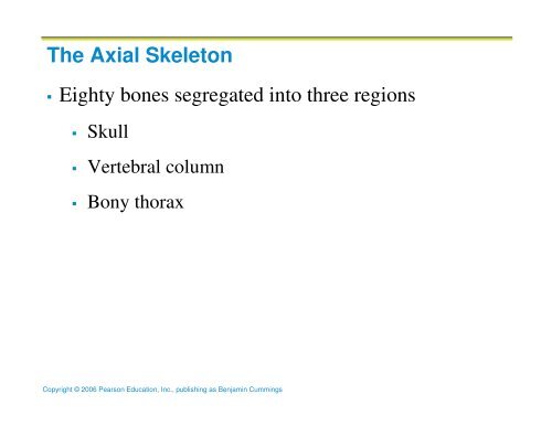
<strong>The</strong> <strong>Axial</strong> <strong>Skeleton</strong><br />
<strong>Eighty</strong> <strong>bones</strong> <strong>segregated</strong> <strong>into</strong> <strong>three</strong> <strong>regions</strong><br />
Skull<br />
Vertebral column<br />
Bony thorax<br />
Copyright © 2006 Pearson Education, Inc., publishing as Benjamin Cummings
Bones of the <strong>Axial</strong> <strong>Skeleton</strong><br />
Copyright © 2006 Pearson Education, Inc., publishing as Benjamin Cummings<br />
Figure 7.1
<strong>The</strong> Skull<br />
<strong>The</strong> skull, the body’s most complex bony structure,<br />
is formed by the cranium and facial <strong>bones</strong> (22 in<br />
all)<br />
Cranium<br />
Made up of cranial <strong>bones</strong><br />
protects the brain and is the site of attachment for<br />
head and neck muscles<br />
Copyright © 2006 Pearson Education, Inc., publishing as Benjamin Cummings
Facial <strong>bones</strong><br />
Facial <strong>bones</strong><br />
Form the framework of the face<br />
Contain cavities for special sense organs (smell,<br />
sight, taste, hearing)<br />
Provide openings for the passage of air and food<br />
Secure the teeth<br />
Anchor the facial muscles of expression<br />
Copyright © 2006 Pearson Education, Inc., publishing as Benjamin Cummings
Bones of the Skull<br />
Most of the bone of the skull are flat <strong>bones</strong><br />
All of the <strong>bones</strong> of the skull are tightly united by<br />
sutures (interlocking joints)…except the mandible<br />
Copyright © 2006 Pearson Education, Inc., publishing as Benjamin Cummings
Major Skull Sutures<br />
Sutures of the skull connect the cranial <strong>bones</strong><br />
<strong>The</strong> major sutures of the skull are:<br />
Coronal<br />
Sagittal<br />
Squamous<br />
Lambdoid<br />
w/ other sutures connecting the facial <strong>bones</strong><br />
Copyright © 2006 Pearson Education, Inc., publishing as Benjamin Cummings
Overview of Skull Geography<br />
<strong>The</strong> cranium can be divided <strong>into</strong> a vault and a base<br />
<strong>The</strong> cranial vault (calvarin) forms the superior,<br />
lateral, and posterior aspects of the skull (and<br />
forehead)<br />
<strong>The</strong> cranial base (floor) forms the skull’s inferior<br />
aspect<br />
Copyright © 2006 Pearson Education, Inc., publishing as Benjamin Cummings
Overview of Skull Geography<br />
<strong>The</strong> internal bony ridges divide the base <strong>into</strong> 3<br />
steps (or fossae):<br />
Anterior cranial fossae<br />
Middle cranial fossae<br />
Posterior cranial fossae<br />
<strong>The</strong> brain sits within these 3 cranial fossae<br />
Copyright © 2006 Pearson Education, Inc., publishing as Benjamin Cummings
Overview of Skull Geography<br />
Other cavities include:<br />
Middle & internal ear cavities<br />
Nasal cavity<br />
Orbits<br />
sinuses<br />
Copyright © 2006 Pearson Education, Inc., publishing as Benjamin Cummings
Anatomy of the Cranium<br />
Eight cranial <strong>bones</strong><br />
Parietal (2)<br />
Temporal (2)<br />
Frontal<br />
Occipital<br />
Sphenoid<br />
Ethmoid<br />
<strong>The</strong> curvatures of the cranial <strong>bones</strong> makes them strong and<br />
allows them to be thin (e.g. an eggshell)<br />
Copyright © 2006 Pearson Education, Inc., publishing as Benjamin Cummings
Frontal Bone<br />
Forms the anterior portion of the cranium<br />
Articulates posteriorly with the paired parietal <strong>bones</strong> via the coronal<br />
suture<br />
Most anterior part is the vertical frontal squama (otherwise known as the<br />
“forehead”)<br />
<strong>The</strong> frontal squama ends inferiorly at the supraorbital margins<br />
<strong>The</strong> supraorbital margin is pierced by the supraorbital foramen<br />
allowing passage of the supraorbital artery and nerve to the<br />
forehead<br />
Extending posteriorly, the frontal bone forms the superior wall of the<br />
orbits and the anterior cranial fossa<br />
<strong>The</strong> glabella is the smooth part of the bone between the orbits<br />
<strong>The</strong> frontonasal suture joins the glabella and the nasal <strong>bones</strong><br />
<strong>The</strong> frontal sinuses are found deep and lateral to the glabella<br />
Copyright © 2006 Pearson Education, Inc., publishing as Benjamin Cummings
Frontal Bone<br />
Copyright © 2006 Pearson Education, Inc., publishing as Benjamin Cummings<br />
Figure 7.2a
Parietal Bones and Major Associated Sutures<br />
<strong>The</strong> parietal <strong>bones</strong> form the superior and lateral aspects of the<br />
skull<br />
<strong>The</strong>y make up the bulk of the cranial vault<br />
<strong>The</strong> 4 largest sutures are associated with the parietal <strong>bones</strong>:<br />
Coronal suture – articulation between parietal <strong>bones</strong><br />
and frontal bone anteriorly<br />
Sagittal suture – where right and left parietal <strong>bones</strong><br />
meet superiorly<br />
Lambdoid suture – where parietal <strong>bones</strong> meet the<br />
occipital bone posteriorly<br />
Squamosal or squamous suture – where parietal and<br />
temporal <strong>bones</strong> meet<br />
Copyright © 2006 Pearson Education, Inc., publishing as Benjamin Cummings
Parietal Bones and Major Associated Sutures<br />
Form most of the superior and lateral aspects of the<br />
skull<br />
Copyright © 2006 Pearson Education, Inc., publishing as Benjamin Cummings<br />
Figure 7.3a
Occipital Bone and Its Major Markings<br />
<strong>The</strong> occipital bone forms most of the skulls posterior wall and base<br />
It articulates anteriorly w/ the paired parietal and temporal <strong>bones</strong> via the<br />
lambdoid and occipitomastoid sutures, respectively<br />
It articulates w/ the sphenoid bone via the basioccipital plate<br />
Internally, the occipital bone forms the walls of the posterior cranial fossa<br />
<strong>The</strong> foramen magnum found at the base of the occipital bone if where the<br />
inferior part of the brain connects with the spinal cord<br />
<strong>The</strong> occipital condyles flank the foramen magnum laterally and articulate w/<br />
the first vertebra permitting a “nodding” movement<br />
<strong>The</strong> external occipital proturbence (can you feel yours??)<br />
External occipital crest: secures the legamentum nuchae (an elastic ligament<br />
that connects the vertebrae to the skull)<br />
Superior & inferior nuchal lines: anchor many muscles of the neck & back<br />
Copyright © 2006 Pearson Education, Inc., publishing as Benjamin Cummings
Occipital Bone and Its Major Markings<br />
Forms most of skull’s<br />
posterior wall and base<br />
Major markings<br />
include the posterior<br />
cranial fossa, foramen<br />
magnum, occipital<br />
condyles, and the<br />
hypoglossal canal<br />
Copyright © 2006 Pearson Education, Inc., publishing as Benjamin Cummings<br />
Figure 7.2b
Temporal Bones<br />
Copyright © 2006 Pearson Education, Inc., publishing as Benjamin Cummings<br />
Figure 7.5
Temporal Bones<br />
Lie inferior to the parietal <strong>bones</strong> and join them at<br />
the squamous sutures<br />
Form the inferolateral aspects of the skull and parts<br />
of the cranial floor<br />
Divided <strong>into</strong> four major <strong>regions</strong><br />
Squamous<br />
Tympanic<br />
Mastoid<br />
petrous<br />
Copyright © 2006 Pearson Education, Inc., publishing as Benjamin Cummings
Temporal Bones: Squamous Region<br />
<strong>The</strong> squamous region has the bar-like zygomatic<br />
process that meets the zygomatic bone forming the<br />
zygomatic arch (the cheek bone)<br />
<strong>The</strong> mandibular fossa on the inferior surface of the<br />
zygomatic process receives the condyle of the<br />
mandible (lower jawbone) forming the<br />
temporomandibular joint<br />
Copyright © 2006 Pearson Education, Inc., publishing as Benjamin Cummings
Temporal Bones: Tympanic Region<br />
<strong>The</strong> Tympanic region surrounds the external<br />
acoustic meatus (external ear canal)<br />
Inferior to the exteranl acoustic meatus lies the<br />
styloid process, an attachment point for several<br />
tongue and neck muscles<br />
Copyright © 2006 Pearson Education, Inc., publishing as Benjamin Cummings
Temporal Bones: Mastoid Region<br />
<strong>The</strong> mastoid region contains the large mastoid<br />
process which anchors some neck muscles<br />
Feel it right behind your ear…go ahead, feel it!<br />
<strong>The</strong> stylomastoid foramen allows cranial nerve VII<br />
(the facial nerve) to leave the skull<br />
Copyright © 2006 Pearson Education, Inc., publishing as Benjamin Cummings
Temporal Bones: Petrous Region<br />
<strong>The</strong> petrous region contributes to the cranial base<br />
It lies between the occipital bone and the sphenoid<br />
bone<br />
Together w/ the sphenoid bone they form the<br />
middle cranial fossa which supports the temporal<br />
lobes of the brain<br />
This region also houses the middle & internal ear<br />
cavities<br />
Copyright © 2006 Pearson Education, Inc., publishing as Benjamin Cummings
Temporal Bones: Petrous Region<br />
Several foramen penetrate the petrous region:<br />
-the jugular foramen: internal jugular & 3 cranial nerves<br />
-the carotid canal: internal carotid artery<br />
-the foramen lacerum<br />
-the internal acoustic meatus: cranial nerves VII & VIII<br />
-the foramenia rotundum, ovale, and spinosum<br />
Copyright © 2006 Pearson Education, Inc., publishing as Benjamin Cummings
Sphenoid Bone<br />
Butterfly-shaped bone that spans the width of the<br />
middle cranial fossa<br />
<strong>The</strong> Keystone Bone: Forms the central wedge that<br />
articulates with all other cranial <strong>bones</strong><br />
Consists of a central body, greater wings, lesser<br />
wings, and pterygoid processes<br />
Contains the sphenoid sinuses<br />
Copyright © 2006 Pearson Education, Inc., publishing as Benjamin Cummings
Sphenoid Bone<br />
<strong>The</strong> superior surface bears the sella tucica which<br />
bears the hypophyseal fossa that forms the<br />
enclosure for the pituitary gland<br />
<strong>The</strong> hypophyseal fossa is abutted anteriorly by the<br />
tuberculum sellae and posteriorly by the dorsum<br />
sellae, which terminates in the posterior clinical<br />
processes<br />
Copyright © 2006 Pearson Education, Inc., publishing as Benjamin Cummings
Sphenoid Bone<br />
Copyright © 2006 Pearson Education, Inc., publishing as Benjamin Cummings<br />
Figure 7.6a
Sphenoid Bone<br />
Copyright © 2006 Pearson Education, Inc., publishing as Benjamin Cummings<br />
Figure 7.6b
Sphenoid Bone<br />
<strong>The</strong> greater wings project laterally from the body and form:<br />
Middle cranial fossa<br />
Dorsal walls of the orbits<br />
External wall of the skull (medial to the zygomatic arch)<br />
<strong>The</strong> lesser wings form:<br />
Part of the floor of the anterior cranial fossa<br />
Part of the medial walls of the orbits<br />
Terminate medially at the anterior clinoid process (an anchoring site<br />
for the brain)<br />
Copyright © 2006 Pearson Education, Inc., publishing as Benjamin Cummings
Sphenoid Bone<br />
<strong>The</strong> pterygoid processes project inferior from the body.<br />
Function to anchor the pterygoid muscles that we use when we<br />
chew<br />
Openings in the sphenoid bone:<br />
Optic canals: connected by the optic chiasmatic groove.<br />
Allow the optic nerves to pass<br />
Superior orbital fissure: Allow the passage of cranial nerves<br />
III, IV, and VI which control eye movement<br />
Copyright © 2006 Pearson Education, Inc., publishing as Benjamin Cummings
Sphenoid Bone<br />
More openings in the sphenoid bone:<br />
Foramen rotundum:<br />
Branch of cranial nerve V for the maxillary nerve<br />
Foramen ovale:<br />
Branch of cranial nerve V for the mandibullar nerve<br />
Foramen spinosum:<br />
Transmits the middle meningreal artery that serves internal<br />
faces of some cranial <strong>bones</strong><br />
Copyright © 2006 Pearson Education, Inc., publishing as Benjamin Cummings
Ethmoid Bone<br />
Copyright © 2006 Pearson Education, Inc., publishing as Benjamin Cummings<br />
Figure 7.7
Ethmoid Bone<br />
Deepest of the skull <strong>bones</strong>; lies between the sphenoid and nasal<br />
<strong>bones</strong><br />
Forms most of the bony area between the nasal cavity and the<br />
orbits<br />
<strong>The</strong> cribiform plates (seen from above) form the roof of the<br />
nasal cavities and the floor of the anterior cranial fossa<br />
<strong>The</strong> olfactory foramina: allow the olfactory nerves to pass<br />
Copyright © 2006 Pearson Education, Inc., publishing as Benjamin Cummings
Ethmoid Bone<br />
Crista galli: (triangular process)<br />
Attachment site for the dura mater (the<br />
outermost covering of the brain)<br />
Perpendicular plate<br />
Projects inferiorly in the median plane<br />
Forms the superior part of the nasal septum<br />
(which divides the nasal cavity right and left)<br />
Copyright © 2006 Pearson Education, Inc., publishing as Benjamin Cummings
Ethmoid Bone<br />
Right & left lateral masses:<br />
Riddled w/ ethmoid sinuses (acts as sieve)<br />
Superior/middle nasal conchae (turbinates)<br />
Protrude <strong>into</strong> the nasal cavity<br />
Orbital plate:<br />
Located on the lateral surfaces of the lateral masses<br />
Contribute to the medial walls of the orbits<br />
Copyright © 2006 Pearson Education, Inc., publishing as Benjamin Cummings
Wormian Bones<br />
Tiny irregularly shaped <strong>bones</strong> that appear within<br />
sutures<br />
Copyright © 2006 Pearson Education, Inc., publishing as Benjamin Cummings
Facial Bones<br />
14 <strong>bones</strong> (6 paired sets) of which only the mandible and<br />
vomer are unpaired<br />
<strong>The</strong> paired <strong>bones</strong> are:<br />
Maxillae<br />
Zygomatics<br />
Nasals<br />
Lacrimals<br />
Palatines<br />
inferior conchae<br />
Copyright © 2006 Pearson Education, Inc., publishing as Benjamin Cummings
Mandible and Its Markings<br />
<strong>The</strong> mandible (lower jawbone) is the largest,<br />
strongest bone of the face<br />
Its major markings include the coronoid process,<br />
mandibular condyle, the alveolar margin, and the<br />
mandibular and mental foramina<br />
Copyright © 2006 Pearson Education, Inc., publishing as Benjamin Cummings
Mandible and Its Markings<br />
Copyright © 2006 Pearson Education, Inc., publishing as Benjamin Cummings<br />
Figure 7.8a
Mandible and Its Markings<br />
<strong>The</strong> mandible (lower jaw) is the largest, strongest bone of the<br />
face<br />
<strong>The</strong> body forms the chin<br />
<strong>The</strong> rami meet the body posteriorly at a mandibular angle<br />
<strong>The</strong> mandibular notch separates the coronoid and condyle<br />
processes or the rami<br />
<strong>The</strong> coronoid process (anterior) is the insertion point for the<br />
large temporalis muscle that elevates the lower jaw during<br />
chewing<br />
Copyright © 2006 Pearson Education, Inc., publishing as Benjamin Cummings
Mandible and Its Markings<br />
<strong>The</strong> mandibular condyle (posterior) articulates<br />
with the mandibular fossa of the temporal bone<br />
forming the temporomandibular joint<br />
Mandibular body anchors the teeth<br />
Alveolar margin: the superior border of the body<br />
contains the sockets win which the teeth are<br />
embedded<br />
Copyright © 2006 Pearson Education, Inc., publishing as Benjamin Cummings
Mandible and Its Markings<br />
Mandibular symphysis:<br />
Site of fusion of the two mandibular <strong>bones</strong> ( in infancy)<br />
Mandibular foramina<br />
Found on the medial surface of each ramus<br />
Nerve passage for tooth sensation (injection site for<br />
Lidocaine)<br />
Mental foramina:<br />
Lateral aspects of body<br />
Allow blood vessels and nerves to pass to skin of the<br />
chin<br />
Copyright © 2006 Pearson Education, Inc., publishing as Benjamin Cummings
Maxillary Bone<br />
Copyright © 2006 Pearson Education, Inc., publishing as Benjamin Cummings<br />
Figure 7.8b
Maxillary Bones (maxillae)<br />
Medially fused <strong>bones</strong> that make up the upper jaw<br />
and the central portion of the facial skeleton<br />
Facial keystone <strong>bones</strong> that articulate with all other<br />
facial <strong>bones</strong> except the mandible<br />
Copyright © 2006 Pearson Education, Inc., publishing as Benjamin Cummings
Maxillary Bones: Major Markings<br />
Alveolar margins:<br />
Carry the upper teeth<br />
Anterior nasal spine:<br />
Site of fusion of maxillae (inferior to nose)<br />
Palatine processes:<br />
Project posteriorly from the alveolar margins<br />
Fuse medially forming the anterior 2/3 of the hard palate<br />
or bony roof of the mouth<br />
Copyright © 2006 Pearson Education, Inc., publishing as Benjamin Cummings
Maxillary Bones: Major Markings<br />
Incisive fossa (midline foramen)<br />
Posterior to teeth<br />
Passage for blood vessels/nerves<br />
Maxillary sinuses<br />
Largest of the paranasal sinuses<br />
Extend from the orbits to the roots of the upper teeth<br />
Copyright © 2006 Pearson Education, Inc., publishing as Benjamin Cummings
Maxillary Bones: Major Markings<br />
Zygomatic processes:<br />
Lateral articulation between the maxillae and zygomatic<br />
<strong>bones</strong><br />
Inferior orbital fissure:<br />
Located deep within the orbit at the junction of the<br />
maxilla and the greater wings of the sphenoid<br />
Permits zygomatic nerve, maxillary nerve (cranial nerve<br />
V branch) and blood vessels to pass to the face<br />
Copyright © 2006 Pearson Education, Inc., publishing as Benjamin Cummings
Maxillary Bones: Major Markings<br />
Infraorbital foramen:<br />
Allows the infraorbital nerve (a continuation of<br />
the maxillary nerve) and artery to reach the face<br />
Copyright © 2006 Pearson Education, Inc., publishing as Benjamin Cummings
Zygomatic Bones<br />
Irregularly shaped <strong>bones</strong> (cheek<strong>bones</strong>)<br />
Articulate with the zygomatic processes of the<br />
temporal <strong>bones</strong> posteriorly, the zygomatic process<br />
of the frontal bone superiorly, and the zygomatic<br />
process of the maxillae anteriorly<br />
For part of the inferolateral<br />
Margins of the orbits<br />
Copyright © 2006 Pearson Education, Inc., publishing as Benjamin Cummings
Other Facial Bones<br />
Nasal <strong>bones</strong><br />
Thin medially fused <strong>bones</strong> that form the bridge of the<br />
nose<br />
Articulate with the frontal bone superiorly, the maxillary<br />
<strong>bones</strong> laterally, and the perpendicular plate of the<br />
ethmoid bone posteriorly<br />
Cartilages form the “skeleton” of the external nose<br />
inferiorly<br />
Copyright © 2006 Pearson Education, Inc., publishing as Benjamin Cummings
Other Facial Bones<br />
Lacrimal <strong>bones</strong>:<br />
Contribute to the medial walls of the orbit<br />
Smallest and most fragile of the facial <strong>bones</strong><br />
Articulate with the frontal bone superiorly, ethmoid bone<br />
posteriorly, and the maxillae anteriorly<br />
Contains a deep groove (lacrimal fossa) that houses the lacrimal<br />
sac that is part of the passage that allows tears to drain from the<br />
eye to the nasal cavity<br />
Copyright © 2006 Pearson Education, Inc., publishing as Benjamin Cummings
Other Facial Bones<br />
Palatine <strong>bones</strong>:<br />
Fashioned from 2 bony plates<br />
Horizontal: complete posterior portion of the hard palate<br />
Perpendicular: forms the posterior walls of the nasal<br />
cavity and the small parts of the orbits<br />
3 articulating processes:<br />
Pyramidal<br />
Sphenoidal<br />
orbital<br />
Copyright © 2006 Pearson Education, Inc., publishing as Benjamin Cummings
Other Facial Bones<br />
Vomer – plow-shaped bone that forms part of the<br />
nasal septum<br />
Inferior nasal conchae – paired, curved <strong>bones</strong> in<br />
the nasal cavity that form part of the lateral walls<br />
of the nasal cavity<br />
Copyright © 2006 Pearson Education, Inc., publishing as Benjamin Cummings
KU Game Day!!! HOME GAMES THIS WEEK!<br />
Thurs. 7 pm<br />
Sat. 7 pm<br />
Mon. 7:30 pm<br />
Copyright © 2006 Pearson Education, Inc., publishing as Benjamin Cummings
Anterior Aspects of the Skull<br />
Parietal bone<br />
Frontal squama<br />
of frontal bone<br />
Nasal bone<br />
Sphenoid bone<br />
(greater wing)<br />
Temporal bone<br />
Ethmoid bone<br />
Lacrimal bone<br />
Zygomatic bone<br />
Infraorbital foramen<br />
Maxilla<br />
Mandible<br />
Mental<br />
foramen<br />
(a)<br />
Copyright © 2006 Pearson Education, Inc., publishing as Benjamin Cummings<br />
Mandibular symphysis<br />
Frontal bone<br />
Glabella<br />
Frontonasal suture<br />
Supraorbital foramen<br />
(notch)<br />
Supraorbital margin<br />
Superior orbital<br />
fissure<br />
Optic canal<br />
Inferior orbital<br />
fissure<br />
Middle nasal concha<br />
Perpendicular plate<br />
Inferior nasal concha<br />
Vomer bone<br />
Ethmoid<br />
bone<br />
Figure 7.2a
Posterior Aspects of the Skull<br />
Lambdoid<br />
suture<br />
Sutural<br />
bone<br />
Occipital bone<br />
Superior nuchal line<br />
External<br />
occipital<br />
protuberance<br />
Occipitomastoid<br />
suture<br />
(b)<br />
External<br />
occipital<br />
crest<br />
Copyright © 2006 Pearson Education, Inc., publishing as Benjamin Cummings<br />
Occipital<br />
condyle<br />
Sagittal suture<br />
Parietal bone<br />
Mastoid<br />
process<br />
Inferior<br />
nuchal<br />
line<br />
Figure 7.2b
External Lateral Aspects of the Skull<br />
Coronal suture Frontal bone<br />
Parietal bone<br />
Temporal bone<br />
Lambdoid<br />
suture<br />
Squamous suture<br />
Occipital bone<br />
Zygomatic process<br />
Occipitomastoid suture<br />
External acoustic meatus<br />
(a)<br />
Mastoid process<br />
Styloid process<br />
Mandibular condyle<br />
Mandibular notch<br />
Mandibular ramus<br />
Copyright © 2006 Pearson Education, Inc., publishing as Benjamin Cummings<br />
Sphenoid bone<br />
(greater wing)<br />
Ethmoid bone<br />
Lacrimal bone<br />
Lacrimal fossa<br />
Nasal bone<br />
Zygomatic bone<br />
Maxilla<br />
Alveolar margins<br />
Mandible<br />
Mental foramen<br />
Mandibular angle Coronoid process<br />
Figure 7.3a
Midsagittal Lateral Aspects of the Skull<br />
Parietal bone<br />
Squamous<br />
suture<br />
Temporal<br />
bone<br />
Lambdoid suture<br />
Occipital<br />
bone<br />
Occipitomastoid<br />
suture<br />
External occipital<br />
protuberance<br />
(b)<br />
Internal acoustic<br />
meatus<br />
Sella turcica<br />
of sphenoid<br />
bone<br />
Pterygoid<br />
process of<br />
sphenoid<br />
Mandibular<br />
bone<br />
foramen<br />
Copyright © 2006 Pearson Education, Inc., publishing as Benjamin Cummings<br />
Palatine<br />
bone<br />
Palatine<br />
process of<br />
maxilla<br />
Coronal suture<br />
Frontal bone<br />
Sphenoid bone<br />
(greater wing)<br />
Frontal sinus<br />
Crista galli<br />
Nasal bone<br />
Sphenoid sinus<br />
Ethmoid bone<br />
(perpendicular plate)<br />
Vomer bone<br />
Incisive fossa<br />
Maxilla<br />
Alveolar margins<br />
Mandible<br />
Figure 7.3b
Inferior Portion of the Skull<br />
Hard<br />
palate<br />
(a)<br />
Maxilla<br />
(palatine process)<br />
Zygomatic bone<br />
Temporal bone<br />
(zygomatic process)<br />
Vomer<br />
Mandibular<br />
fossa<br />
Styloid process<br />
Mastoid process<br />
Palatine bone<br />
(horizontal plate)<br />
Temporal bone<br />
(petrous part)<br />
Pharyngeal<br />
tubercle of<br />
basioccipital<br />
Parietal bone<br />
External occipital crest<br />
External occipital<br />
protuberance<br />
Copyright © 2006 Pearson Education, Inc., publishing as Benjamin Cummings<br />
Foramen magnum<br />
Incisive fossa<br />
Medial palatine suture<br />
Infraorbital foramen<br />
Maxilla<br />
Sphenoid bone<br />
(greater wing)<br />
Foramen ovale<br />
Foramen<br />
lacerum<br />
Carotid canal<br />
External acoustic meatus<br />
Stylomastoid<br />
foramen<br />
Jugular foramen<br />
Occipital condyle<br />
Inferior nuchal line<br />
Superior nuchal line<br />
Figure 7.4a
Inferior Portion of the Skull<br />
Olfactory foramina<br />
Anterior cranial fossa<br />
Sphenoid<br />
(b)<br />
Lesser wing<br />
Greater wing<br />
Tuberculum sellae<br />
Hypophyseal fossa<br />
Sella Dorsum sellae<br />
turcica Posterior clinoid process<br />
Middle cranial<br />
fossa<br />
Temporal bone<br />
(petrous part)<br />
Internal<br />
acoustic meatus<br />
Posterior<br />
cranial fossa<br />
Parietal bone<br />
Occipital bone<br />
Foramen magnum<br />
Copyright © 2006 Pearson Education, Inc., publishing as Benjamin Cummings<br />
(c)<br />
Frontal bone<br />
Cribriform plate<br />
Ethmoid<br />
Crista galli<br />
bone<br />
Optic canal<br />
Anterior clinoid process<br />
Foramen rotundum<br />
Foramen ovale<br />
Foramen spinosum<br />
Foramen lacerum<br />
Jugular foramen<br />
Hypoglossal canal<br />
Anterior<br />
cranial<br />
fossa<br />
Middle<br />
cranial<br />
fossa<br />
Posterior<br />
cranial<br />
fossa<br />
Figure 7.4b