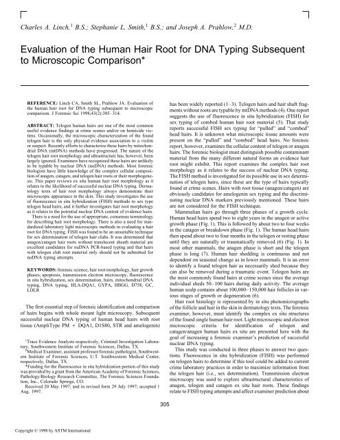
Evaluation of the human hair root for DNA typing ... - Library
Evaluation of the human hair root for DNA typing ... - Library
Evaluation of the human hair root for DNA typing ... - Library

You also want an ePaper? Increase the reach of your titles
YUMPU automatically turns print PDFs into web optimized ePapers that Google loves.
Charles A. Linch, 1 B.S.; Stephanie L. Smith, 1 B.S.; and Joseph A. Prahlow, 2 M.D.<br />
<strong>Evaluation</strong> <strong>of</strong> <strong>the</strong> Human Hair Root <strong>for</strong> <strong>DNA</strong> Typing Subsequent<br />
to Microscopic Comparison*<br />
REFERENCE: Linch CA, Smith SL, Prahlow JA. <strong>Evaluation</strong> <strong>of</strong><br />
<strong>the</strong> <strong>human</strong> <strong>hair</strong> <strong>root</strong> <strong>for</strong> <strong>DNA</strong> <strong>typing</strong> subsequent to microscopic<br />
comparison. J Forensic Sci 1998;43(2):305–314.<br />
has been widely reported (1–3). Telogen <strong>hair</strong>s and <strong>hair</strong> shaft frag-<br />
ments without <strong>root</strong>s are typable by mt<strong>DNA</strong> methods (4). One report<br />
suggests <strong>the</strong> use <strong>of</strong> fluorescence in situ hybridization (FISH) <strong>for</strong><br />
ABSTRACT: Telogen <strong>human</strong> <strong>hair</strong>s are one <strong>of</strong> <strong>the</strong> most common<br />
useful evidence findings at crime scenes and/or on homicide vic-<br />
tims. Occasionally, <strong>the</strong> microscopic characterization <strong>of</strong> <strong>the</strong> found<br />
telogen <strong>hair</strong> is <strong>the</strong> only physical evidence association to a victim<br />
sex <strong>typing</strong> <strong>of</strong> combed <strong>human</strong> <strong>hair</strong> <strong>root</strong> material (5). That study<br />
reports successful FISH sex <strong>typing</strong> <strong>for</strong> “pulled” and “combed”<br />
head <strong>hair</strong>s. It is unknown what microscopic tissue amounts were<br />
present on <strong>the</strong> “pulled” and “combed” head <strong>hair</strong>s. No <strong>for</strong>ensic<br />
or suspect. Recently ef<strong>for</strong>ts to characterize <strong>the</strong>se <strong>hair</strong>s by mitochondrial<br />
<strong>DNA</strong> (mt<strong>DNA</strong>) methods have progressed. The nature <strong>of</strong> <strong>the</strong><br />
telogen <strong>hair</strong> <strong>root</strong> morphology and ultrastructure has, however, been<br />
largely ignored. Examiners have recognized <strong>the</strong>se <strong>hair</strong>s are unlikely<br />
to be typable by nuclear <strong>DNA</strong> (nu<strong>DNA</strong>) methods. Most <strong>for</strong>ensic<br />
biologists have little knowledge <strong>of</strong> <strong>the</strong> complex cellular composi-<br />
report, however, examines <strong>the</strong> cellular content <strong>of</strong> telogen or anagen<br />
<strong>hair</strong>s. The <strong>for</strong>ensic biologist must distinguish possible contaminant<br />
material from <strong>the</strong> many different natural <strong>for</strong>ms an evidence <strong>hair</strong><br />
<strong>root</strong> might exhibit. This report examines <strong>the</strong> complex <strong>hair</strong> <strong>root</strong><br />
morphology as it relates to <strong>the</strong> success <strong>of</strong> nuclear <strong>DNA</strong> <strong>typing</strong>.<br />
tion <strong>of</strong> anagen, catagen, and telogen <strong>hair</strong> <strong>root</strong>s or <strong>the</strong>ir morphogene- The FISH method is investigated <strong>for</strong> its possible use in sex determisis.<br />
This paper reviews ex situ <strong>human</strong> <strong>hair</strong> <strong>root</strong> morphology as it<br />
relates to <strong>the</strong> likelihood <strong>of</strong> successful nuclear <strong>DNA</strong> <strong>typing</strong>. Derma-<br />
tology texts <strong>of</strong> <strong>hair</strong> <strong>root</strong> morphology always demonstrate <strong>the</strong>ir<br />
microscopic appearance in <strong>the</strong> skin. This study investigates <strong>the</strong> use<br />
<strong>of</strong> fluorescence in situ hybridization (FISH) methods to sex type<br />
telogen head <strong>hair</strong>s, and it fur<strong>the</strong>r investigates <strong>hair</strong> <strong>root</strong> morphology<br />
nation <strong>of</strong> telogen <strong>hair</strong>s, since <strong>the</strong>se are <strong>the</strong> type <strong>of</strong> <strong>hair</strong>s typically<br />
found at crime scenes. Hairs with <strong>root</strong> tissue (anagen/catagen) are<br />
obviously candidates <strong>for</strong> amelogenin sex <strong>typing</strong> and <strong>the</strong> discrimi-<br />
nating nuclear <strong>DNA</strong> markers previously mentioned. These <strong>hair</strong>s<br />
are not considered <strong>for</strong> <strong>the</strong> FISH technique.<br />
as it relates to <strong>the</strong> potential nuclear <strong>DNA</strong> content <strong>of</strong> evidence <strong>hair</strong>s.<br />
There is a need <strong>for</strong> <strong>the</strong> use <strong>of</strong> appropriate, consensus terminology<br />
<strong>for</strong> describing <strong>hair</strong> <strong>root</strong> morphology. There is also a need <strong>for</strong> stan-<br />
dardized laboratory light microscopic methods in evaluating a <strong>hair</strong><br />
<strong>root</strong> <strong>for</strong> <strong>DNA</strong> <strong>typing</strong>. FISH was found to be an unsuitable technique<br />
<strong>for</strong> sex determination <strong>of</strong> telogen <strong>hair</strong> clubs. It was determined that<br />
anagen/catagen <strong>hair</strong> <strong>root</strong>s without translucent sheath material are<br />
Mammalian <strong>hair</strong>s go through three phases <strong>of</strong> a growth cycle.<br />
Human head <strong>hair</strong>s spend two to eight years in <strong>the</strong> anagen or active<br />
growth phase (Fig. 1). This is followed by about two to four weeks<br />
in <strong>the</strong> catagen or breakdown phase (Fig. 1). The <strong>human</strong> head <strong>hair</strong>s<br />
<strong>the</strong>n spend about two to four months in <strong>the</strong> telogen or resting phase<br />
until <strong>the</strong>y are naturally or traumatically removed (6) (Fig. 1). In<br />
excellent candidates <strong>for</strong> nu<strong>DNA</strong> PCR-based <strong>typing</strong> and that <strong>hair</strong>s<br />
with telogen club <strong>root</strong> material only should not be submitted <strong>for</strong><br />
nu<strong>DNA</strong> <strong>typing</strong> attempts.<br />
most o<strong>the</strong>r mammals, <strong>the</strong> anagen phase is short and <strong>the</strong> telogen<br />
phase is long (7). Human <strong>hair</strong> shedding is continuous and not<br />
dependent on seasonal change as in lower mammals. It is an error<br />
KEYWORDS: <strong>for</strong>ensic science, <strong>hair</strong> <strong>root</strong> morphology, <strong>hair</strong> growth<br />
phases, apoptosis, transmission electron microscopy, fluorescence<br />
in situ hybridization, sex determination, <strong>hair</strong>s, mitochondrial <strong>DNA</strong><br />
<strong>typing</strong>, <strong>DNA</strong> <strong>typing</strong>, HLA-DQA1, GYPA, HBGG, D758, GC,<br />
LDLR<br />
to identify a found telogen <strong>hair</strong> as necessarily shed because <strong>the</strong>y<br />
can also be removed during a traumatic event. Telogen <strong>hair</strong>s are<br />
<strong>the</strong> most commonly found <strong>hair</strong>s at crime scenes since <strong>the</strong> average<br />
individual sheds 50–100 <strong>hair</strong>s during daily activity. The average<br />
<strong>human</strong> scalp contains about 100,000–150,000 <strong>hair</strong> follicles in various<br />
stages <strong>of</strong> growth or degeneration (6).<br />
Hair <strong>root</strong> histology is represented by in situ photomicrographs<br />
The first essential step <strong>of</strong> <strong>for</strong>ensic identification and comparison <strong>of</strong> <strong>the</strong> follicle and <strong>hair</strong> in <strong>the</strong> skin in dermatology texts. The <strong>for</strong>ensic<br />
<strong>of</strong> <strong>hair</strong>s begins with whole mount light microscopy. Subsequent examiner, however, must identify <strong>the</strong> complex ex situ structures<br />
successful nuclear <strong>DNA</strong> <strong>typing</strong> <strong>of</strong> <strong>human</strong> head <strong>hair</strong>s with <strong>root</strong> <strong>of</strong> <strong>the</strong> found single <strong>human</strong> <strong>hair</strong> <strong>root</strong>. Light microscopic and electron<br />
tissue (AmpliType PM DQA1, D1S80, STR and amelogenin) microscopic criteria <strong>for</strong> identification <strong>of</strong> telogen and<br />
1Trace Evidence Analysts respectively, Criminal Investigation Laboratory,<br />
Southwestern Institute <strong>of</strong> Forensic Sciences, Dallas, TX.<br />
2Medical Examiner, assistant pr<strong>of</strong>essor/<strong>for</strong>ensic pathologist, Southwest-<br />
ern Institute <strong>of</strong> Forensic Sciences, U.T. Southwestern Medical Center,<br />
respectively, Dallas, TX.<br />
catagen/anagen <strong>human</strong> <strong>hair</strong>s ex situ are presented here with <strong>the</strong><br />
goal <strong>of</strong> increasing a <strong>for</strong>ensic examiner’s prediction <strong>of</strong> successful<br />
nuclear <strong>DNA</strong> <strong>typing</strong>.<br />
This study was conducted in three phases to answer two questions.<br />
Fluorescence in situ hybridization (FISH) was per<strong>for</strong>med<br />
on telogen <strong>hair</strong>s to determine if this tool could be added to current<br />
*Funding <strong>for</strong> <strong>the</strong> fluorescence in situ hybridization portion <strong>of</strong> this study<br />
was provided by a grant from <strong>the</strong> American Academy <strong>of</strong> Forensic Sciences,<br />
Pathology/Biology Research Committee, The Forensic Sciences Founda-<br />
tion, Inc., Colorado Springs, CO.<br />
Received 20 May 1997; and in revised <strong>for</strong>m 29 July 1997; accepted 1<br />
Aug. 1997.<br />
crime laboratory practices in order to maximize in<strong>for</strong>mation from<br />
<strong>the</strong> telogen <strong>hair</strong> (i.e., sex determination). Transmission electron<br />
microscopy was used to explore ultrastructural characteristics <strong>of</strong><br />
anagen, telogen and catagen ex situ <strong>hair</strong> <strong>root</strong>s. These findings<br />
relate to FISH <strong>typing</strong> attempts and affect examiner prediction about<br />
Copyright © 1998 by ASTM International<br />
305
306 JOURNAL OF FORENSIC SCIENCES<br />
FIG. 1—Hair <strong>root</strong> growth and degeneration phases. (A) anagen (active) growth bulb with translucent <strong>root</strong> sheath (epi<strong>the</strong>lial) tissue. Note flameshaped,<br />
translucent dermal papilla at base <strong>of</strong> bulb. Sometimes a candidate <strong>for</strong> RFLP <strong>DNA</strong> <strong>typing</strong> if sheath found in excess. (B) Anagen/catagen bulbs<br />
(2) with no sheath tissue. Bulb, stem, and cortical shaft. Excellent candidates <strong>for</strong> PCR <strong>DNA</strong> <strong>typing</strong>. (C) Catagen (breakdown) phase <strong>root</strong> with bulb.<br />
Good candidate <strong>for</strong> PCR <strong>DNA</strong> <strong>typing</strong>. Note area <strong>of</strong> subsequent telogen club <strong>for</strong>mation. (Arrow). (D) late catagen, early telogen phase. (E) Telogen<br />
(resting) club with germinal nipple. No usable nu<strong>DNA</strong>. (F) Telogen club. No usable nu<strong>DNA</strong>. The process from (A) to (F) results in <strong>the</strong> total <strong>root</strong> length<br />
being reduced by about two-thirds due to cellular loss (apoptosis), fibrosis, and cornification. Whole mount (Permount) light microscopy. (400)<br />
which type <strong>hair</strong> <strong>root</strong> ends would yield nuclear <strong>DNA</strong> PCR product. locus DXZ1) and <strong>the</strong> CEP Y (satellite III) SpectrumGreen <strong>DNA</strong><br />
Finally, PCR amplifications were per<strong>for</strong>med on certain <strong>root</strong> end probe hybridizes to <strong>the</strong> satellite III sequence <strong>of</strong> <strong>human</strong> chromomorphologies<br />
to fur<strong>the</strong>r investigate nuclear <strong>DNA</strong> <strong>typing</strong> success some Y (band Yq12, locus DYX1). Both hybridized probes propredictability,<br />
using a modified <strong>hair</strong> extraction protocol which duce fluorescent signals in interphase nuclei and on metaphase<br />
involved <strong>hair</strong> shaft grinding. These PCR findings were compared chromosomes. Normal male and female control cells are recomto<br />
a <strong>for</strong>ensic casework retrospective study examining probative mended to be placed on <strong>the</strong> same slide as <strong>the</strong> specimen when using<br />
evidence <strong>hair</strong>s that were PCR typed by conventional extraction<br />
methods.<br />
<strong>the</strong> CEP X/Y (sat III) assay.<br />
Materials and Methods<br />
Samples<br />
Fluorescence In Situ Hybridization Sex Determination <strong>of</strong><br />
Telogen Hairs<br />
Telogen head <strong>hair</strong>s were gently removed from <strong>the</strong> scalps <strong>of</strong> a<br />
total <strong>of</strong> fifteen male and female individuals and mounted to glass<br />
The CEP X ( satellite) SpectrumOrange <strong>DNA</strong> probe hybridizes microscope slides with Permount resin <strong>for</strong> up to six weeks (whole<br />
to <strong>the</strong> centromere <strong>of</strong> <strong>human</strong> chromosome X (bands p11.1-q11, mount preparations). These <strong>hair</strong>s were microscopically screened
LINCH ET AL. • HUMAN HAIR ROOT FOR <strong>DNA</strong> TYPING 307<br />
<strong>for</strong> those with telogen <strong>root</strong> clubs and no adherent translucent germ with germ tissue, and one anagen/catagen head <strong>hair</strong>. These employcell<br />
tissue <strong>for</strong> testing. Hairs possessing visible tissue are candidates ees had known AmpliType PMDQA1 <strong>typing</strong>s from blood sam-<br />
<strong>for</strong> amelogenin <strong>DNA</strong> PCR sex <strong>typing</strong> and were not considered <strong>for</strong> ples on file at <strong>the</strong> Institute. These 24 <strong>hair</strong>s were mounted on glass<br />
this phase <strong>of</strong> <strong>the</strong> study. The <strong>hair</strong>s were removed by scoring and microscope slides in Permount <strong>for</strong> up to six weeks. After micro-<br />
breaking <strong>the</strong> coverslip, dissolving with xylene and washing with scopic screening (100) and photomicroscopy, a 2-cm section <strong>of</strong><br />
dH2O. Material was teased from <strong>the</strong> <strong>root</strong> club with a sterile scalpel <strong>the</strong> <strong>hair</strong> including <strong>the</strong> <strong>root</strong>, was removed by scoring and breaking<br />
blade in a drop <strong>of</strong> 60% acetic acid. The <strong>root</strong> club material was <strong>the</strong> coverslip. This area <strong>of</strong> breakage was dissolved with xylene to<br />
heat fixed to <strong>the</strong> microscope slide.<br />
Hybridization<br />
facilitate removal <strong>of</strong> <strong>the</strong> <strong>hair</strong>. A sterile scalpel blade was used to<br />
remove <strong>the</strong> proximal portion <strong>of</strong> <strong>the</strong> <strong>hair</strong> including <strong>the</strong> <strong>root</strong>.<br />
The fixed telogen <strong>root</strong> club material was prepared <strong>for</strong> FISH Modified Hair <strong>DNA</strong> Extraction<br />
using <strong>the</strong> Vysis Spectrum CEP X ( satellite), Y (satellite III)<br />
cocktail probes according to <strong>the</strong> manufacturers instructions.<br />
Briefly, <strong>the</strong> microscope slides with <strong>the</strong> telogen <strong>root</strong> club material<br />
were denatured at 75C <strong>for</strong> 5 min in 70% <strong>for</strong>mamide/2X SSC. The<br />
slides were dehydrated <strong>for</strong> 1 min each in 70%, 85%and 100%<br />
Each 2-cm piece <strong>of</strong> <strong>hair</strong> with attached <strong>root</strong> was placed in a 1.5-<br />
mL microcentrifuge tube with <strong>the</strong> addition <strong>of</strong> 500 L <strong>of</strong>2%SDS.<br />
Hairs were sonicated <strong>for</strong> 10 min and <strong>the</strong>n washed with dH2O. Each<br />
<strong>hair</strong> section was placed in a 0.2 mL Kontes glass grinder (Fisher<br />
K885470-0000) which contained 150 L TE4 ethanol and were maintained at a temperature <strong>of</strong> 45–50C. The<br />
Vysis CEP probe mixture <strong>of</strong> 7 L hybridization buffer, 1 L X,Y<br />
cocktail probe and 2 L<strong>of</strong>dH2Owas maintained at 45–50C after<br />
denaturation at 75C <strong>for</strong> 5 min. The dry telogen <strong>root</strong> club material<br />
was treated with 5 L <strong>of</strong> probe mix, <strong>the</strong>n coverslipped and incubated<br />
<strong>for</strong> 30–60 min at 42C in a non-humidified chamber. The<br />
coverslip was removed and <strong>the</strong> slide was placed in a coplin jar<br />
containing 0.4X SSC <strong>for</strong> 2 min at 75C <strong>for</strong> post hybridization wash.<br />
The glass slide was <strong>the</strong>n washed in 2X SSC/0.1% NP-40 at room<br />
temperature <strong>for</strong> 5–60 s. The microscope slide was dried in darkness<br />
and coverslipped after <strong>the</strong> application <strong>of</strong> 10 L <strong>of</strong> DAPI II counter-<br />
stain to <strong>the</strong> telogen club material.<br />
. Grinding was<br />
per<strong>for</strong>med so that no <strong>hair</strong> material was visible by stereomicroscopy.<br />
The homogenate, approximately 150 L, was added to 50 L<br />
<strong>of</strong> 20% (w/v) Chelex®100 (Perkin-Elmer P/N N808-0087). This<br />
material was vortexed <strong>for</strong> 5–10 s and micr<strong>of</strong>uged <strong>for</strong> about 10 s.<br />
The tubes were placed in a dry bath incubator at 100C <strong>for</strong> 8 min.<br />
The extracted material was vortexed <strong>for</strong> 5–10 s and micr<strong>of</strong>uged<br />
<strong>for</strong> 2–3 min at high speed.<br />
The careful cleaning <strong>of</strong> glass grinders has been described previously<br />
(4). The glass grinders were cleaned between uses to prevent<br />
cross-contamination. Each grinder was used <strong>for</strong> about 4–5 <strong>hair</strong>s<br />
from different individuals and <strong>the</strong>n discarded.<br />
Fluorescence Microscopy<br />
Amplification and Typing <strong>of</strong> <strong>the</strong> Extracted <strong>DNA</strong> Using <strong>the</strong><br />
The slides were examined <strong>for</strong> non-overlapping cells (an impor-<br />
AmpliType PMDQA1 Kit<br />
tant ancillary step to avoid false results) on a Zeiss fluorescence The manufacturer’s protocol <strong>for</strong> <strong>the</strong> AmpliType PMDQA1<br />
microscope with a triple bandpass filter system <strong>for</strong> <strong>the</strong> presence Kit was followed <strong>for</strong> amplification using 20 L <strong>of</strong> extracted <strong>hair</strong><br />
<strong>of</strong> X (orange) and Y (green) chromosome signals. The microscopist <strong>DNA</strong>. The 102-L total volume mixture (40-L PM reaction mix,<br />
did not know <strong>the</strong> identity <strong>of</strong> <strong>the</strong> <strong>hair</strong> donor(s). Photomicrographs 40-L primer mixture, 2-L BSA (8 mg/mL) and 20 L <strong>DNA</strong>)<br />
were taken when possible due to <strong>the</strong> low emission intensity <strong>of</strong> was amplified in a Perkin-Elmer GeneAmp PCR System 2400<br />
some fluorescent signals. Thermal Cycler. Five microliters <strong>of</strong> 200 mM disodium EDTA was<br />
added to each tube prior to strip hybridization. Twenty microliters<br />
Transmission Electron Microscopy (TEM) <strong>of</strong> denatured amplified <strong>DNA</strong> product was used <strong>for</strong> <strong>the</strong> reverse<br />
Several telogen head <strong>hair</strong>s (without germ cell tissue at <strong>the</strong> <strong>root</strong>)<br />
and several anagen head <strong>hair</strong>s (without shaft sheath tissue) were<br />
immediately placed in glutaraldehyde after removal from <strong>the</strong> scalp.<br />
Routine TEM fixation, embedding, ultramicrotomy, and staining<br />
techniques were used. This included osmium tetroxide post-fixadot-blot,<br />
allele-specific oligonucleotide (ASO) strip hybridization.<br />
Again, <strong>the</strong> manufacturer’s protocol was used <strong>for</strong> hybridization and<br />
strip development. Strip development was accomplished in a Bel-<br />
lco Glass, Inc., dual pan hot shaker (Cat 7746-32110).<br />
tion, ethanol dehydrations, propylene oxide infiltration, and Results and Discussion<br />
embedding with epoxy resins (Araldite, Epon). After ultramicrotomy,<br />
<strong>the</strong> sections were stained with uranyl acetate and lead citrate<br />
on <strong>for</strong>mvar-coated grids.<br />
Fluorescence In Situ Hybridization Sex Determination <strong>of</strong><br />
Telogen Hairs<br />
The approximate 80–90 nm thick sections were examined on a<br />
Jeol Jem 1200EX II transmission electron microscope operated at<br />
Results<br />
120 kV. Electron micrographs <strong>of</strong> one telogen head <strong>hair</strong> club, one<br />
anagen head <strong>hair</strong> bulb with adjacent stem and cortex, and one late<br />
anagen/early catagen head <strong>hair</strong> bulb with adjacent stem and cortex<br />
were obtained.<br />
Out <strong>of</strong> 15 telogen head <strong>hair</strong>s from 15 individuals, no telogen<br />
head <strong>hair</strong>s were correctly typed <strong>for</strong> sex identification by <strong>the</strong> FISH<br />
method. Most cells and cell remnants exhibited no signals, while<br />
a few cells exhibited more than two. Some male <strong>hair</strong> <strong>root</strong> materials<br />
exhibited female signals (two or more orange X chromosome sig-<br />
AmpliType PMDQA1 PCR Amplification and Typing nals fluorescing), and some female <strong>hair</strong> <strong>root</strong> materials exhibited<br />
Samples<br />
male signals (one orange X chromosome signal and one green Y<br />
chromosome signal fluorescing). One male and one female fresh<br />
Twelve Institute <strong>of</strong> Forensic Sciences employees contributed telogen <strong>hair</strong> <strong>root</strong> club, not exposed to Permount, were also tested<br />
one telogen head <strong>hair</strong> with club <strong>root</strong>, or one telogen head <strong>hair</strong> and yielded <strong>the</strong> same false hybridization results.
308 JOURNAL OF FORENSIC SCIENCES<br />
Fluorescence In Situ Hybridization Sex Determination <strong>of</strong> (10). Loss <strong>of</strong> target <strong>DNA</strong>, poor penetration <strong>of</strong> probe, and incom-<br />
Telogen Hairs<br />
plete or non-specific hybridization are problems associated with<br />
apoptotic, necrotic, and keratinizing cells (10). FISH requires<br />
Discussion examination <strong>of</strong> a large number <strong>of</strong> cells, <strong>the</strong> use <strong>of</strong> control cells on<br />
<strong>the</strong> same microscope slide as <strong>the</strong> evidence slide (due to critical<br />
These findings strongly indicate nuclear <strong>DNA</strong> degradation. The<br />
temperature requirements) and sophisticated statistical analysis<br />
telogen <strong>hair</strong> <strong>root</strong> club results from <strong>the</strong> shrinkage, detachment, and<br />
(10).<br />
descent <strong>of</strong> <strong>the</strong> viable cellular bulb material beneath it (dermal<br />
papilla, matrix cells, stem and fibrous outer <strong>root</strong> sheath) (7). The<br />
TEM <strong>of</strong> Telogen, Catagen, and Anagen Head Hairs<br />
late telogen club is encased in trichilemmal keratin and is completely<br />
surrounded by <strong>the</strong> shrunken outer <strong>root</strong> sheath while in <strong>the</strong> Results<br />
skin (8). The programmed cell death (apoptosis) <strong>of</strong> elements below<br />
No intact nuclei, nuclear remnants, mitochondria, melanosomes,<br />
and around <strong>the</strong> telogen club explains <strong>the</strong> non-specific probe hybridor<br />
keratin filaments were seen in <strong>the</strong> featureless trichilemmal s<strong>of</strong>t<br />
ization described above. Apoptosis results in double strand cleavkeratin<br />
material <strong>of</strong> <strong>the</strong> telogen <strong>root</strong> club (Fig. 3B). Numerous intact<br />
age <strong>of</strong> nuclear <strong>DNA</strong> at linker regions; while in necrosis <strong>the</strong>re is<br />
nuclei were seen in <strong>the</strong> anagen and early catagen <strong>root</strong> bulbs (Fig.<br />
random <strong>DNA</strong> degradation with digestion <strong>of</strong> histones (9). Cell<br />
4B). The hard keratin filament density increases as one goes from<br />
shrinkage and nuclear condensation is one <strong>of</strong> <strong>the</strong> electron microproximal<br />
to distal regions in <strong>the</strong> anagen stem and cortical shaft<br />
scopic characteristics <strong>of</strong> apoptosis (10). Degraded nuclear <strong>DNA</strong> is<br />
(Fig. 5). Nuclei were observed to become increasingly elongated<br />
not a suitable target <strong>for</strong> FISH probes since false, absent, or multiple<br />
as one goes from proximal to distal regions in <strong>the</strong> anagen/catagen<br />
signals may occur (10). Telogen <strong>hair</strong>s with visible translucent germ<br />
stem and cortical shaft. (The term “stem” is used here to designate<br />
(epi<strong>the</strong>lial) tissue surrounding <strong>the</strong> club were not tested because<br />
<strong>the</strong> ex situ slender, pigmented, usually longest section <strong>of</strong> <strong>the</strong> termi<strong>the</strong>se<br />
<strong>hair</strong>s would be better candidates <strong>for</strong> amelogenin PCR amplifinal<br />
<strong>hair</strong> <strong>root</strong> that stretches from <strong>the</strong> summit <strong>of</strong> a bulb to <strong>the</strong> base<br />
cation <strong>for</strong> sex determination and, o<strong>the</strong>r nuclear <strong>DNA</strong> markers. The<br />
<strong>of</strong> <strong>the</strong> <strong>hair</strong> cortex.) Areas <strong>of</strong> autophagic vacuole <strong>for</strong>mation were<br />
telogen <strong>root</strong> club has keratinized <strong>root</strong>lets which anchor into survisible<br />
in <strong>the</strong> upper regions <strong>of</strong> <strong>the</strong> late catagen bulb and stem.<br />
rounding germ cells and <strong>the</strong>se translucent cells may accompany<br />
<strong>the</strong> telogen club <strong>root</strong> upon pulling during early telogen/late catagen<br />
TEM <strong>of</strong> Telogen, Catagen, and Anagen Head Hairs<br />
(11). This translucent cornifying structure, seen in ex situ <strong>hair</strong>s, is<br />
also referred to as <strong>the</strong> telogen club nipple (8) (Fig. 1E). Discussion<br />
Intact interphase nuclei or metaphase chromosome preparations<br />
Empty vacuoles seen in <strong>the</strong> telogen club begin to <strong>for</strong>m during<br />
are a requirement <strong>for</strong> successful FISH results (10). Intact interphase<br />
<strong>the</strong> late catagen phase as <strong>the</strong> surrounding germ cells <strong>for</strong>m a capsule<br />
nuclei do not exist in <strong>the</strong> telogen <strong>root</strong> club because it consists<br />
sac around <strong>the</strong> club. These vacuoles are <strong>for</strong>med during cornifica<strong>of</strong><br />
trichilemmal keratin (see Transmission Electron Microscopy<br />
tion, a dehydrating process. A fully cornified <strong>hair</strong> contains less<br />
Results). This is a s<strong>of</strong>t keratin material as opposed to <strong>the</strong> downthan<br />
10% water (7). These empty vacuoles appear black with whole<br />
wardly pointed hard keratin <strong>of</strong> <strong>the</strong> <strong>hair</strong> shaft cortex above it. It is<br />
mount light microscopy (as do air-filled medullary structures and<br />
worthy to note that anagen <strong>hair</strong> <strong>root</strong>s digested with Proteinase-K<br />
cortical fusi) and should not be incorrectly identified as large pig-<br />
(20 mg/mL, 4 h at 56C) and mounted in Permount exhibit <strong>the</strong><br />
ment accumulations within <strong>the</strong> translucent club material.<br />
same light microscopic characteristics as <strong>the</strong> “dead man’s” <strong>root</strong><br />
Figure 1A, left, illustrates <strong>the</strong> viable anagen bulb which contains<br />
end (downwardly pointed shaft remnant with dark banding) (Fig.<br />
numerous matrix cells and melanocytes. The egg-shaped translu-<br />
2). This suggests that <strong>the</strong> “dead man’s” <strong>root</strong> end morphology is<br />
cent dermal papilla protrudes into <strong>the</strong> base <strong>of</strong> <strong>the</strong> bulb. The <strong>hair</strong><br />
possibly caused by <strong>the</strong> digestion <strong>of</strong> <strong>the</strong> s<strong>of</strong>t bulb by autolytic probulb<br />
is a structure rich in nuclear and mitochondrial <strong>DNA</strong>. The<br />
teinases released by <strong>the</strong> surrounding necrotic cells in <strong>the</strong> decomrate<br />
<strong>of</strong> turnover <strong>of</strong> <strong>the</strong> matrix cells is greater than that <strong>of</strong> any normal<br />
posing skin. Some <strong>hair</strong> examiners use <strong>the</strong> terms “putrid <strong>root</strong>” and<br />
tissue, with <strong>the</strong> possible exception <strong>of</strong> bone marrow (7). Scalp <strong>hair</strong>s<br />
“postmortem <strong>root</strong> banding” to describe this <strong>hair</strong> morphology.<br />
grow approximately 0.35 mm per day (7). Figure 5 demonstrates<br />
While a previous report suggests that FISH might be useful <strong>for</strong><br />
<strong>the</strong> <strong>for</strong>mation <strong>of</strong> hard keratin filaments as one moves from proximal<br />
gender determination <strong>of</strong> <strong>hair</strong>s (5), this study shows that FISH is<br />
to distal regions in <strong>the</strong> anagen <strong>hair</strong> shaft. Compare to <strong>the</strong> featureless<br />
unsuitable <strong>for</strong> sex <strong>typing</strong> <strong>of</strong> found telogen <strong>hair</strong> clubs. FISH probes<br />
s<strong>of</strong>t keratin material <strong>of</strong> Fig. 3B.<br />
have inherent problems even when used with fresh viable cells<br />
The onset <strong>of</strong> <strong>the</strong> catagen phase is not visible with light microscopy<br />
ex situ since it is diagnosed by <strong>the</strong> cessation <strong>of</strong> mitosis in <strong>the</strong><br />
bulb matrix cells, <strong>the</strong> collapse <strong>of</strong> <strong>the</strong> outer <strong>root</strong> sheath epi<strong>the</strong>lium<br />
(apoptosis) and <strong>the</strong> presence <strong>of</strong> pigment granules in <strong>the</strong> dermal<br />
papilla. (The outer <strong>root</strong> sheath actually represents a downward<br />
extension <strong>of</strong> <strong>the</strong> epidermis (8).) It is possible to see catagen <strong>hair</strong>s<br />
in skin that have fully developed club structures (telogen) but<br />
exhibit evidence <strong>of</strong> ongoing catagen activity (12). The whole mount<br />
light microscopic distinction between late anagen and early catagen,<br />
and late catagen and early telogen, is not always clear. It is<br />
<strong>of</strong> interest to note <strong>the</strong> future site <strong>of</strong> telogen club <strong>for</strong>mation in <strong>the</strong><br />
catagen <strong>hair</strong> <strong>root</strong> above <strong>the</strong> bulb and stem (elongation zone) (Fig.<br />
FIG. 2—Anagen <strong>root</strong> bulb (Fig. 1-B) after digestion with Proteinase 1C). During <strong>the</strong> growth cycle from anagen to telogen, <strong>the</strong> <strong>hair</strong><br />
K. Only hard keratin <strong>of</strong> cortex remains. Bulb and stem tissue has been<br />
digested. Black banding is an area <strong>of</strong> air vacuole <strong>for</strong>mation. Same micro-<br />
follicle shrinks to about one-third <strong>of</strong> its <strong>for</strong>mer length and <strong>the</strong><br />
scopic appearance as <strong>hair</strong>s from a decomposing <strong>human</strong> scalp. Light telogen club is <strong>for</strong>med at <strong>the</strong> level <strong>of</strong> attachment <strong>of</strong> <strong>the</strong> arrector<br />
microscopy, Permount embedding. (400) pili muscle (8). These authors assign anagen, catagen, or telogen
LINCH ET AL. • HUMAN HAIR ROOT FOR <strong>DNA</strong> TYPING 309<br />
FIG. 3—(A) Light microscopy <strong>of</strong> telogen club. Note numerous dark air vacuoles. (B) Transmission electron microscopy <strong>of</strong> telogen club. The club is<br />
composed <strong>of</strong> trichilemmal keratin. Note numerous air vacuoles and absence <strong>of</strong> cellular elements. The entire club consists <strong>of</strong> <strong>the</strong> homogenous amorphous<br />
material. (1200)<br />
FIG. 4—(A) Light microscopy <strong>of</strong> anagen bulb. (B) Transmission electron microscopy <strong>of</strong> anagen bulb matrix surrounding <strong>the</strong> dermal papilla. These<br />
cells give rise to <strong>the</strong> cortical cells. Note tightly packed, intact nuclei. The melanosomes (dark inclusions) are produced by melanocytes and are transferred<br />
to young cortical cells. (18000)<br />
growth status to ex situ whole mount <strong>hair</strong>s with regard to <strong>the</strong> AmpliType PMDQA1 Typing <strong>of</strong> Head Hair Roots<br />
dominant <strong>root</strong> histologic structures present (see Fig. 6).<br />
It should be remembered that <strong>the</strong> catagen (breakdown) phase <strong>of</strong><br />
Results<br />
<strong>the</strong> <strong>hair</strong> cycle lasts only about two to four weeks and that in <strong>the</strong> The AmpliType® Polymarker (LDLR, GYPA, HBGG, D7S8,<br />
non-balding scalp, approximately 80–90% <strong>of</strong> <strong>the</strong> <strong>hair</strong>s are in <strong>the</strong> GC) and HLA (DQA1) amplification <strong>typing</strong> kit detected no PCR<br />
anagen (active growth) phase and about 10% <strong>of</strong> <strong>the</strong> <strong>hair</strong>s are in product <strong>for</strong> head <strong>hair</strong>s with telogen clubs and head <strong>hair</strong>s with telo-<br />
<strong>the</strong> resting or telogen phase (6). Naturally shed telogen <strong>hair</strong>s accugen club and attached germinal nipples (Fig.(s) 1E, F). Telogen<br />
mulate over time in <strong>the</strong> victim or suspect’s environment and anagen head <strong>hair</strong>s with ample translucent germ/epi<strong>the</strong>lial tissue surround-<br />
<strong>hair</strong>s are more likely to be removed by trauma. As previously ing <strong>the</strong> keratin club produced detectable PCR product in 2 <strong>of</strong> <strong>the</strong><br />
noted, telogen <strong>hair</strong>s can also be lost due to trauma along with 3 head <strong>hair</strong>s tested.<br />
anagen/catagen <strong>hair</strong>s.<br />
Anagen/catagen head <strong>hair</strong>s with only a bulb produced detectable
310 JOURNAL OF FORENSIC SCIENCES<br />
FIG. 5—(A) Matrix cells <strong>of</strong> anagen bulb. (B) Higher level <strong>of</strong> cortical cells trans<strong>for</strong>med from matrix cells. Ton<strong>of</strong>ilaments are being produced.<br />
Prekeratinous zone (X6000). (C) Ton<strong>of</strong>ilaments becoming more dense at higher level <strong>of</strong> shaft than (B) keratogenous zone. (D) Dense keratin filaments<br />
<strong>of</strong> <strong>hair</strong> cortex with one elongated nucleus visible. (E) Higher magnification <strong>of</strong> (C). Mitochondrion, mt, keratin filaments, kf, elipsoid melanosome, me,<br />
and nucleus, nu.
LINCH ET AL. • HUMAN HAIR ROOT FOR <strong>DNA</strong> TYPING 311<br />
FIG. 6—Identification <strong>of</strong> pubic <strong>hair</strong> <strong>root</strong> structures. (A,B) Anagen/catagen bulb without sheath tissue. Excellent candidate <strong>for</strong> PCR <strong>DNA</strong> <strong>typing</strong>.<br />
(C,D) Telogen club with ample translucent germinal tissue (follicular tag). Good candidate <strong>for</strong> <strong>DNA</strong> PCR <strong>typing</strong>. (E) Telogen club with germinal nipple.<br />
Poor candidate <strong>for</strong> <strong>DNA</strong> PCR <strong>typing</strong>. The majority <strong>of</strong> <strong>hair</strong>s found in sexual assault cases are like (C,D) because pubic <strong>hair</strong>s, unlike head <strong>hair</strong>s, spend<br />
more time in telogen phase than anagen phase. Light microscopy, Permount embedding. (X400)<br />
PCR product in 6 <strong>of</strong> 7 head <strong>hair</strong>s tested (Fig. 1B). All head <strong>hair</strong>s<br />
with <strong>root</strong> sheath tissue and bulb produced detectable PCR product<br />
(Fig. 1A). Table 1 summarizes <strong>the</strong> results from this study. Table<br />
TABLE 2—Hair PCR <strong>DNA</strong> results from Dallas County Institute <strong>of</strong><br />
2 summarizes a retrospective study <strong>of</strong> <strong>hair</strong> nuclear <strong>DNA</strong> PCR<br />
Forensic Sciences casework retrospective study. Extractions were done<br />
results which involved probative evidence <strong>hair</strong>s in Dallas County by three different laboratories over an eight year period; but, none<br />
Institute <strong>of</strong> Forensic Sciences cases. The retrospective study <strong>hair</strong>s<br />
utilized <strong>the</strong> grinding method.<br />
were amplified over an eight year period, by several different<br />
laboratories, using different extraction methods. No labs in <strong>the</strong><br />
TABLE 1—Hair PCR <strong>DNA</strong> results from this study in which <strong>the</strong><br />
grinding method <strong>of</strong> extraction (Chelex 100) was used.<br />
Number with Detectable<br />
AmpliType<br />
Type <strong>of</strong> Hair Root Number TestedPMDQA1 Product<br />
Telogen with club only<br />
Telogen club with translucent<br />
6 0<br />
germinal nipple<br />
Telogen club with ample translucent<br />
6 0<br />
germinal tissue (follicular tag)<br />
Anagen/Catagen bulb only<br />
3 2<br />
(no translucent tissue present)<br />
Anagen/Catagen with bulb and trans-<br />
6 5<br />
lucent sheath tissue 3 3<br />
Total 24 10<br />
Number with Detectable<br />
PCR Product (Various<br />
Type <strong>of</strong> Hair Root Number Tested Genetic Loci)<br />
Telogen with club only<br />
Telogen club with translucent<br />
6 0<br />
germinal nipple<br />
Telogen club with ample translucent<br />
germinal tissue<br />
3 0<br />
(follicular tag)<br />
Anagen/Catagen bulb only<br />
4 3<br />
(no translucent tissue present)<br />
Anagen/Catagen with bulb and<br />
4 3<br />
translucent sheath tissue 0 0<br />
Total 17 6
312 JOURNAL OF FORENSIC SCIENCES<br />
retrospective study used <strong>the</strong> grinding method <strong>of</strong> extraction dis- obtaining mitochondrial <strong>DNA</strong> (4). No glass grinder cross contami-<br />
cussed herein.<br />
nation or o<strong>the</strong>r sources <strong>of</strong> PCR contamination were detected in<br />
this study which was limited to nu<strong>DNA</strong> <strong>typing</strong>. The same grinder<br />
AmpliType PMDQA1 Typing <strong>of</strong> Head Hair Roots pestal and tube was used <strong>for</strong> 4–5 <strong>hair</strong>s and discarded. The ground<br />
Discussion<br />
Half <strong>of</strong> <strong>the</strong> telogen club <strong>hair</strong>s used in this study had <strong>the</strong> “cotton<br />
glass inner surface <strong>of</strong> <strong>the</strong> tube is exhausted after about 4–5 <strong>hair</strong>s<br />
have been individually homogenized.<br />
swab” <strong>root</strong> end appearance and no surrounding translucent tissue<br />
present. It is possible to obtain mt<strong>DNA</strong> product from this type <strong>of</strong><br />
Conclusions<br />
late telogen <strong>hair</strong> by using <strong>the</strong> grinding extraction method <strong>for</strong> PCR Findings from this study were generally consistent with a 1981<br />
amplification (4). No appreciable difference has been seen however study by Hukkelhoven, et al., which detailed <strong>DNA</strong> content found<br />
in <strong>the</strong> amount <strong>of</strong> mt<strong>DNA</strong> obtained from <strong>hair</strong> shafts with attached in <strong>the</strong> <strong>hair</strong> sheath, elongation zone, and bulb using a fluorimetric<br />
telogen <strong>root</strong> clubs versus 2-cm <strong>hair</strong> shaft pieces with no <strong>root</strong> (per- microassay technique (14). The most <strong>DNA</strong> was found in <strong>the</strong> transsonal<br />
communication, J.A. DiZinno, FBI, April, 1997). lucent epi<strong>the</strong>lial sheath tissue adhering to <strong>the</strong> anagen head <strong>hair</strong><br />
The <strong>hair</strong> examiner should recognize that <strong>the</strong> telogen <strong>root</strong> club shaft above <strong>the</strong> elongation zone (stem) and bulb. The pigmented<br />
is fairly clear in whole mount light microscopy. The dark micro- anagen/catagen elongation zone (stem) contained <strong>the</strong> next best typ-<br />
scopic air vacuoles in <strong>the</strong> club should not be interpreted as pigment able <strong>DNA</strong>, followed by <strong>the</strong> successful <strong>typing</strong> <strong>of</strong> <strong>the</strong> anagen/catagen<br />
clumps (Fig.(s) 3A, 3B). Pigmentation does accompany <strong>the</strong> <strong>DNA</strong> <strong>hair</strong> bulb alone. The 1981 study only examined “freshly plucked<br />
rich anagen <strong>hair</strong> bulb (Fig.(s) 4A, 4B).<br />
head <strong>hair</strong>s with visible bulb and sheath material.” The 1988 R.<br />
Hairs with evenly pigmented bulbs (catagen/anagen) present Higuchi, et al., report used <strong>the</strong> terms “freshly plucked” <strong>hair</strong>s and<br />
showed a high degree <strong>of</strong> typability due to <strong>the</strong> presence <strong>of</strong> <strong>the</strong> <strong>DNA</strong>- “shed” <strong>hair</strong>s to report a maximum recovery <strong>of</strong> 200 ng <strong>of</strong> <strong>DNA</strong><br />
rich dermal papilla and surrounding intact matrix bulb cells. This from <strong>the</strong> <strong>for</strong>mer and a maximum recovery <strong>of</strong> 10 ng <strong>of</strong> <strong>DNA</strong> from<br />
structure can be incorrectly identified and <strong>the</strong>re<strong>for</strong>e underestimated <strong>the</strong> latter (1). The microscopic appearance <strong>of</strong> <strong>the</strong> <strong>root</strong> <strong>of</strong> <strong>the</strong> “shed”<br />
<strong>for</strong> <strong>DNA</strong> content by experienced microscopists as a telogen club <strong>hair</strong>s was not reported. It is doubtful that <strong>the</strong> “shed” <strong>hair</strong>s had<br />
(no usable nuclear <strong>DNA</strong>), since no translucent surrounding tissue telogen clubs or telogen clubs with germ nipples. It is unlikely<br />
may be present. This study and review <strong>of</strong> prior casework <strong>DNA</strong> that <strong>the</strong> “combed” <strong>hair</strong>s reported in <strong>the</strong> FISH study had telogen<br />
<strong>hair</strong>s showed that telogen <strong>hair</strong>s with ample translucent germ clubs only (5). In fact, one <strong>of</strong> <strong>the</strong> authors <strong>of</strong> this manuscript (JAP)<br />
cell/epi<strong>the</strong>lial tissue (follicular tag) were easily typable by PCR reports that <strong>hair</strong> type (telogen vs. anagen/catagen) was not recorded<br />
methods (Fig.(s) 6C, 6D).<br />
in that study. Uni<strong>for</strong>mity in nomenclature is strongly urged. With<br />
Hairs with anagen bulbs and inner/outer <strong>root</strong> sheath shaft tissue, practice, one can microscopically identify a single <strong>hair</strong>’s particular<br />
in varying states <strong>of</strong> degeneration, will usually produce a large growth stage in light microscopic whole mounts. Forensic <strong>hair</strong><br />
amount <strong>of</strong> nuclear <strong>DNA</strong> PCR amplified product <strong>for</strong> obvious rea- examiners prefer to report <strong>the</strong> terms anagen and telogen, ra<strong>the</strong>r<br />
sons. Found <strong>hair</strong>s with unusually large amounts <strong>of</strong> tissue present than “plucked and shed” or “pulled and combed.” The catagen<br />
within <strong>the</strong> bulb and on <strong>the</strong> shaft are candidates <strong>for</strong> restriction frag- growth phase is rarely reported because <strong>of</strong> its short duration (2–4<br />
ment length polymorphism (RFLP) analysis. Cases microscopi- weeks) and ambiguous whole mount microscopic appearance (6).<br />
cally identified <strong>for</strong> such analysis should not be extracted with All <strong>hair</strong>s with <strong>root</strong>s submitted to <strong>the</strong> laboratory could be classified<br />
Chelex 100 because that procedure results in a denatured <strong>DNA</strong> as telogen with club, telogen with germ nipple, telogen with ample<br />
sample unsuitable <strong>for</strong> RFLP (13). It is worthy to note that no proba- germ tissue, anagen/catagen bulb with a <strong>root</strong> sheath, and<br />
tive evidence <strong>hair</strong> with anagen/catagen bulb and translucent <strong>root</strong> anagen/catagen bulb without <strong>root</strong> sheath tissue (Fig. 1).<br />
sheath material has been submitted <strong>for</strong> <strong>DNA</strong> analysis in <strong>the</strong> last The anagen <strong>hair</strong> bulb, which is evenly pigmented, has numerous<br />
eight years by <strong>the</strong> Dallas County Institute <strong>of</strong> Forensic Sciences. viable cells (matrix and melanocytic) with intact nuclei, and con-<br />
Hairs with anagen bulbs or early catagen bulbs and no translucent tains <strong>the</strong> <strong>DNA</strong> rich dermal papilla, should not be confused with<br />
peripheral tissue, should be considered very good candidates <strong>for</strong> <strong>the</strong> telogen club, which is not pigmented, has no identifiable cells,<br />
PCR <strong>DNA</strong> amplification. It is possible that <strong>the</strong>se types <strong>of</strong> <strong>hair</strong>s and has a macroscopic cotton swab appearance. During catagen<br />
were referred to as “shed” or “combed” in earlier reports (1,5). <strong>the</strong> bulb shrinks, setting <strong>the</strong> dermal papilla free. The lower follicle<br />
Hairs microscopically identified as in <strong>the</strong> late telogen growth phase consists <strong>of</strong> a thin cord <strong>of</strong> epi<strong>the</strong>lial cells surrounded by <strong>the</strong> fibrous<br />
without abundant translucent germ/epi<strong>the</strong>lial cell tissue produced <strong>root</strong> sheath. The thin cord <strong>of</strong> epi<strong>the</strong>lial cells retract upward <strong>for</strong>ming<br />
no PCR amplified nu<strong>DNA</strong> product in this study. It is noted that a small nipple attached to <strong>the</strong> newly <strong>for</strong>med club. This is referred<br />
<strong>DNA</strong> concentration and <strong>DNA</strong> quantification was not attempted to as <strong>the</strong> secondary <strong>hair</strong> germ nipple tissue. This is <strong>the</strong> onset <strong>of</strong><br />
after extraction. telogen (8). Many examiners use <strong>the</strong> term “follicular tag” to<br />
A resinous mounting medium with a refractive index (R.I.) similar describe <strong>the</strong> abundant secondary translucent germ tissue attached<br />
to that <strong>of</strong> <strong>the</strong> cuticle <strong>of</strong> <strong>hair</strong> is used to reduce <strong>the</strong> diffraction <strong>of</strong> light to a found telogen <strong>hair</strong> club (15).<br />
at <strong>the</strong> <strong>hair</strong> edges in whole mount preparations (Permount has an R.I. The telogen club is <strong>for</strong>med after <strong>the</strong> trans<strong>for</strong>mation <strong>of</strong> <strong>the</strong> outer<br />
<strong>of</strong> 1.525 at 25C). Optical clarity is necessary <strong>for</strong> <strong>the</strong> microscopic <strong>root</strong> sheath into germ cells which encapsulate <strong>the</strong> club with a sac.<br />
comparisons and <strong>root</strong> end photographic documentation. These The telogen club rises toward <strong>the</strong> skin surface while <strong>the</strong>re is<br />
authors do not agree that <strong>hair</strong>s can be microscopically compared shrinkage and descent into <strong>the</strong> skin by <strong>the</strong> bulb and its adherent<br />
without suitable mounting medium prior to <strong>DNA</strong> analysis as collapsed fibrous structures. The telogen club is simply a product<br />
reported by Hochmeister, et al. (3). The <strong>for</strong>ensic examiner usually <strong>of</strong> apoptosis (cell deletion) and keratinization (8). The previous<br />
has to microscopically examine numerous <strong>hair</strong>s from a crime scene anagen/catagen <strong>root</strong> structures are not discernable at this point and<br />
be<strong>for</strong>e a possible suspect <strong>hair</strong> suitable <strong>for</strong> tedious comparison is <strong>the</strong> overall <strong>hair</strong> <strong>root</strong> length is decreased by two-thirds through cell<br />
found.<br />
deletion (apoptosis) (8). This is <strong>the</strong> type <strong>of</strong> <strong>hair</strong> resting in <strong>the</strong> scalp<br />
Grinding <strong>of</strong> <strong>hair</strong> <strong>root</strong>s and <strong>hair</strong> shafts is <strong>the</strong> optimal method <strong>for</strong> waiting to be naturally shed or <strong>for</strong>cibly plucked. In <strong>the</strong> absence
LINCH ET AL. • HUMAN HAIR ROOT FOR <strong>DNA</strong> TYPING 313<br />
<strong>of</strong> translucent <strong>root</strong> sheath material, an experienced <strong>hair</strong> examiner financial waste to <strong>the</strong> equally critical waste <strong>of</strong> analytical time.<br />
may confuse telogen club and anagen bulb structures and <strong>the</strong>re<strong>for</strong>e For those circumstances in which <strong>the</strong> victim and <strong>the</strong> <strong>of</strong>fender paths<br />
lack judgment about a found <strong>hair</strong>’s potential <strong>DNA</strong> content. The did not previously cross, <strong>the</strong> microscopic examination <strong>of</strong> <strong>hair</strong><br />
translucent germ nipple tissue <strong>of</strong>ten accompanies <strong>the</strong> early telogen remains strong associative evidence.<br />
head <strong>hair</strong> upon removal (Fig. 1E). The usable nuclear <strong>DNA</strong> content These authors urge con<strong>for</strong>mity among researchers and exam-<br />
<strong>of</strong> this nipple structure is minimal. There are <strong>for</strong>ensic literature iners when describing a particular <strong>hair</strong> <strong>root</strong> type. Pulled, plucked,<br />
reports which confuse <strong>the</strong> use <strong>of</strong> <strong>the</strong> terms “club” and “bulb.” A combed and shed descriptions <strong>of</strong> <strong>hair</strong> <strong>root</strong>s are misleading. Terms<br />
bulb is a viable structure at anagen/catagen <strong>root</strong> ends and a club such as anagen/catagen bulb with sheath, anagen/catagen bulb with<br />
is <strong>the</strong> nonviable structure at <strong>the</strong> late telogen <strong>hair</strong> <strong>root</strong> end. no sheath tissue, telogen club with ample germinal tissue (follicular<br />
Few mitochondria were identified in <strong>the</strong> <strong>hair</strong> bulb matrix or tag), telogen club with germ tissue nipple and telogen club are<br />
cortical cells <strong>of</strong> this study by transmission electron microscopy more appropriate terms (Fig.(s) 1 and 6). Some <strong>for</strong>ensic examiners<br />
(Fig. 5E). Mitochondria are difficult to demonstrate in <strong>hair</strong> <strong>root</strong> use <strong>the</strong> terms club and bulb interchangeably. These structures are<br />
cells. Poor penetration <strong>of</strong> TEM fixative through ton<strong>of</strong>ilament/ very different, macroscopically, ultrastructurally, and functionally.<br />
keratin filament material and <strong>the</strong> masking <strong>of</strong> mitochondria by tri- The following is a summary <strong>of</strong> <strong>the</strong> conclusions presented herein:<br />
chohyalin granules and melanosomes are a few reasons <strong>for</strong> this (1) FISH does not appear to be a useful <strong>for</strong>ensic test <strong>for</strong> routine incor-<br />
(16). When small pieces <strong>of</strong> skin with <strong>hair</strong>s are fixed and sectioned, poration into <strong>hair</strong> examination. (2) Microscopic <strong>hair</strong> evaluation and<br />
a few mitochondria can be seen near <strong>the</strong> nucleus <strong>of</strong> matrix cells comparison using a suitable mounting media should always precede<br />
in <strong>the</strong> anagen <strong>hair</strong> bulb. Mitochondria are more numerous in <strong>the</strong> <strong>DNA</strong> testing. (3) Hair examiners should use appropriate (and consis<strong>root</strong><br />
upper bulb where cells are larger. Mitochondria are seen more<br />
frequently in <strong>the</strong> epidermal telogen club epi<strong>the</strong>lial sac (follicular<br />
tag) than in <strong>the</strong> telogen <strong>hair</strong> germ nipple tissues (16).<br />
This laboratory does not attempt to microscopically associate<br />
tent) dermatologic terminology when describing <strong>hair</strong> <strong>root</strong> morphol-<br />
ogy. (4) Telogen <strong>hair</strong> clubs should not be submitted <strong>for</strong> nu<strong>DNA</strong> PCR<br />
<strong>typing</strong>. (5) Anagen/catagen <strong>hair</strong> bulbs absent translucent sheath tis-<br />
sue are excellent candidates <strong>for</strong> nu<strong>DNA</strong> PCR <strong>typing</strong>.<br />
found limb <strong>hair</strong>s to individuals. These <strong>hair</strong>s are indistinctly pigmented<br />
and do not <strong>of</strong>fer <strong>the</strong> examiner a “pigment pattern” from<br />
Acknowledgments<br />
which meaningful associative judgments can be made. Limb <strong>hair</strong>s Funding <strong>for</strong> <strong>the</strong> fluorescence in situ hybridization portion <strong>of</strong> this<br />
are simply reported <strong>of</strong> possible arm or leg origin. Although <strong>the</strong> ana- study was provided by a grant from <strong>the</strong> American Academy <strong>of</strong><br />
gen phase <strong>of</strong> limb <strong>hair</strong> growth is only several months long, <strong>the</strong> same Forensic Sciences, AAFS Pathology/Biology Research Commit<strong>root</strong><br />
end morphologic descriptions are applicable when considering tee, The Forensic Sciences Foundation, Inc., P.O. Box 669, Colo-<br />
<strong>DNA</strong> PCR amplification <strong>of</strong> that material (6). This laboratory does rado Springs, CO 80901-0669.<br />
include found pubic <strong>hair</strong>s, along with head <strong>hair</strong>s, as candidates <strong>for</strong> The authors wish to thank Dallas County, Jeffrey J. Barnard<br />
microscopic comparison to known pubic and head <strong>hair</strong> standards M.D., Director and Chief Medical Examiner, Institute <strong>of</strong> Forensic<br />
respectively. Pubic <strong>hair</strong> <strong>root</strong>s can be evaluated <strong>for</strong> <strong>the</strong> same morpho- Sciences, Dallas, TX, and Irving C. Stone, Ph.D., Chief, Physical<br />
logic structures described here <strong>for</strong> determining PCR <strong>DNA</strong> potential, Evidence Section, Criminal Investigation Laboratory, Southwest-<br />
although pubic <strong>hair</strong> periods <strong>of</strong> rest (telogen) are longer than <strong>the</strong>ir ern Institute <strong>of</strong> Forensic Sciences <strong>for</strong> <strong>the</strong>ir support <strong>of</strong> this endeavor.<br />
periods <strong>of</strong> growth (anagen) (16). Anagen pubic <strong>hair</strong>s are found less Dennis Belloto, B.S., Dept. Of Pathology, U.T. Southwestern Med-<br />
frequently than telogen pubic <strong>hair</strong>s with ample translucent germ cell ical Center, Dallas, TX., provided expert transmission electron<br />
tissue on physical evidence items (Fig. 6).<br />
microscopy technical services. Dr. David A. Whiting kindly pro-<br />
Because evidence <strong>hair</strong>s can become contaminated with <strong>for</strong>eign vided technical review <strong>of</strong> <strong>the</strong> manuscript.<br />
body fluids and skin cells, <strong>the</strong> <strong>hair</strong> microscopist must be able to<br />
correctly distinguish all <strong>root</strong> structures from <strong>for</strong>eign material in<br />
References<br />
order to defend contamination issues in <strong>the</strong> courts when <strong>DNA</strong> 1. Higuchi RG, Beroldingen CH von, Sensabaugh GF, Ehrlich HA.<br />
PCR <strong>typing</strong> has been done. Exact determination <strong>of</strong> anagen versus<br />
catagen growth stage is not always possible in resinous whole<br />
mount preparations. Potential <strong>DNA</strong> containing <strong>root</strong> structures can<br />
<strong>DNA</strong> <strong>typing</strong> from single <strong>hair</strong>s. Nature 1988;332:543–6.<br />
2. Budowle B, Koons BW, Errera JD. Multiplex amplification and<br />
<strong>typing</strong> procedure <strong>for</strong> <strong>the</strong> loci D1S80 and amelogenin. J Forensic<br />
Sci 1995;41(4):660–3.<br />
always be correctly identified. The amount <strong>of</strong> innate nuclear <strong>DNA</strong> 3. Hochmeister MN, Budowle B, Eisenberg A, Borer UV, Dirnh<strong>of</strong>er<br />
obtained from a <strong>hair</strong> <strong>root</strong> is an indicator <strong>of</strong> that <strong>hair</strong>’s particular<br />
growth phase when it left <strong>the</strong> scalp.<br />
Observations reported in this paper regarding <strong>hair</strong> <strong>root</strong>s and <strong>the</strong>ir<br />
R. Using multiplex PCR amplification and <strong>typing</strong> kits <strong>for</strong> <strong>the</strong> analy-<br />
sis <strong>of</strong> <strong>DNA</strong> evidence in a serial killer case. J Forensic Sci 1996;<br />
41(1):155–62.<br />
4. Wilson MR, DiZinno JA, Polenskey D, Replogie J, Budowle B.<br />
nu<strong>DNA</strong> PCR typability are <strong>the</strong> result <strong>of</strong> this study and a review Validation <strong>of</strong> mitochondrial <strong>DNA</strong> sequencing <strong>for</strong> <strong>for</strong>ensic casework<br />
<strong>of</strong> eight years <strong>of</strong> casework. Hair nu<strong>DNA</strong> PCR <strong>typing</strong> results that<br />
were completed by several different laboratories <strong>for</strong> Institute <strong>of</strong><br />
Forensic Sciences cases were reviewed. Findings from this study<br />
and <strong>the</strong> retrospective study indicate that evidence <strong>hair</strong>s do not<br />
analysis. Int J Legal Med 1995;108:68–74.<br />
5. Prahlow JA, Lantz PE, Cox-Jones K, Rao PN, Pettenati MJ. Gender<br />
identification <strong>of</strong> <strong>human</strong> <strong>hair</strong> using fluorescence in situ hybridization.<br />
J Forensic Sci 1996;41(6):1035–37.<br />
6. Olsen EA, editor. Disorders <strong>of</strong> <strong>hair</strong> growth, diagnosis and treatment.<br />
appear to be compromised by mounting in Permount prior to<br />
nuclear <strong>DNA</strong> amplification. (Telogen club <strong>hair</strong>s and telogen club<br />
<strong>hair</strong>s with germ nipples not placed in Permount also produced no<br />
nu<strong>DNA</strong> product upon PCR amplifications.)<br />
New York: McGraw-Hill, Inc., 1994.<br />
7. Ackerman AB, DeViragh PA, Chongchitnant N. Neoplasms with<br />
follicular differentiation. Ch. 4, anatomic, histologic, and biologic<br />
aspects <strong>of</strong> <strong>hair</strong> follicles and <strong>hair</strong>s. Philadelphia: Lea and Febiger,<br />
1993.<br />
The focus <strong>of</strong> <strong>for</strong>ensic biology on <strong>DNA</strong> <strong>typing</strong> methods has led<br />
to a gradual decline in <strong>hair</strong> microscopy practice and training. The<br />
cost <strong>of</strong> indiscriminate <strong>DNA</strong> analyses without benefit <strong>of</strong> a meaningful<br />
microscopic assessment <strong>of</strong> <strong>the</strong> <strong>hair</strong> goes beyond <strong>the</strong> obvious<br />
8. Lever WF, Schaumberg-Lever G. Histopathology <strong>of</strong> <strong>the</strong> skin. Philadelphia:<br />
J.B. Lippincott Co., 1983.<br />
9. Lavin M. Programmed cell death: <strong>the</strong> cellular and molecular<br />
biology <strong>of</strong> apoptosis. Philadelphia: Harwood Academic Publishers,1993.
314 JOURNAL OF FORENSIC SCIENCES<br />
10. Studzinski GP, Cell growth and apoptosis. Ox<strong>for</strong>d: Ox<strong>for</strong>d University<br />
Press. 1995.<br />
11. Orfanos CE, Happle R, Editor. Hair and <strong>hair</strong> diseases. Berlin:<br />
Springer-Verlag, 1990.<br />
12. Kobori T, Montagna W, Editors. Biology and disease <strong>of</strong> <strong>the</strong> <strong>hair</strong>.<br />
Baltimore: University Park Press, 1975.<br />
15. Petraco N, Fraas C, Callery FX, DeForest PR. The morphology and<br />
evidential significance <strong>of</strong> <strong>human</strong> <strong>hair</strong> <strong>root</strong>s. J Forensic Sci 1988;<br />
33(1):68–76.<br />
16. Montagna W. The structure and function <strong>of</strong> skin. New York: Academic<br />
Press Inc., 1956.<br />
13. Walsh PS, Metzger DA, Higuchi R. Chelex® 100 as a medium <strong>for</strong><br />
simple extraction <strong>of</strong> <strong>DNA</strong> <strong>for</strong> PCR based <strong>typing</strong> from <strong>for</strong>ensic material.<br />
Biotechniques 1991;10(4):506–13.<br />
14. Hukkelhoven MWAC, Vromans E, Markslag AMG, Vermorken<br />
Additional in<strong>for</strong>mation and reprint requests:<br />
Charles A. Linch<br />
Southwestern Institute <strong>of</strong> Forensic Sciences<br />
AJM. A simple fluorimetric microassay <strong>of</strong> <strong>DNA</strong> in <strong>hair</strong> follicles 5230 Medical Center Drive<br />
or fractions <strong>of</strong> <strong>hair</strong> follicles. Anticancer Research 1981;1:341–4. Dallas, TX 75235