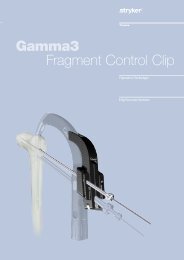
Hoffmann II MRI External Fixation Systems - Stryker
Hoffmann II MRI External Fixation Systems - Stryker
Hoffmann II MRI External Fixation Systems - Stryker

You also want an ePaper? Increase the reach of your titles
YUMPU automatically turns print PDFs into web optimized ePapers that Google loves.
Introduction<br />
In the ever changing world of<br />
medicine, new technology is<br />
advancing the treatment of the<br />
patient. The Magnetic Resonance<br />
Imaging (<strong>MRI</strong>) scanner uses<br />
electromagnetic fields to make exact<br />
images of the human body. More and<br />
more hospitals are investing in <strong>MRI</strong><br />
suites and using the <strong>MRI</strong> scanners<br />
on a routine basis to more accurately<br />
assess their patients.<br />
For this reason, <strong>Stryker</strong> offers a<br />
line extension of <strong>External</strong> <strong>Fixation</strong><br />
which may be used safely in <strong>MRI</strong><br />
scanners up to 3.0 Tesla 1 . This means<br />
that the patient can be placed in the<br />
<strong>MRI</strong> scanner with the fixator in situ<br />
without additional risk for the patient<br />
or the <strong>MRI</strong> scanner itself.<br />
With non <strong>MRI</strong> safe fixators, the<br />
patient can be injured in two<br />
ways. The static magnetic field<br />
generated by <strong>MRI</strong> systems will<br />
attract ferromagnetic objects with<br />
considerable force.<br />
This can lead to displacement of<br />
the components which can cause<br />
serious injuries to the patient and<br />
damage to the scanner. In addition<br />
to this, conductive devices may<br />
experience heating by the induction of<br />
electromotive forces when subjected to<br />
gradient magnetic fields. This can lead<br />
to temperature increases of the fixator<br />
pins which can be significant and<br />
reach critical levels. These increases<br />
may cause patient discomfort and may<br />
even lead to bone necrosis.<br />
The engineers at <strong>Stryker</strong> have created<br />
<strong>MRI</strong> systems which are made of nonferromagnetic<br />
and non-conductive<br />
materials. This allows the surgeon to<br />
build a construct with limited risk for<br />
the patient in an <strong>MRI</strong> environment up<br />
to 3.0 Tesla and is therefore rated MR-<br />
Conditional.<br />
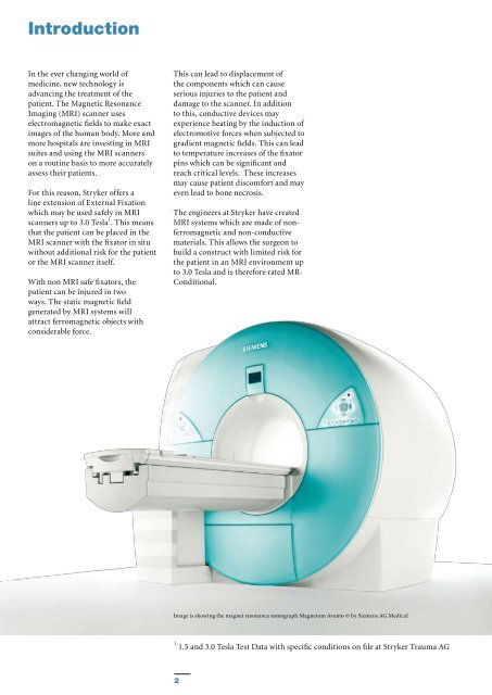
Image is showing the magnet resonance tomograph Magnetom Avanto © by Siemens AG Medical<br />
1 1.5 and 3.0 Tesla Test Data with specific conditions on file at <strong>Stryker</strong> Trauma AG<br />
2