Cutaneous Polyarteritis Nodosa – A Rare, Benign Vasculitis - medIND
Cutaneous Polyarteritis Nodosa – A Rare, Benign Vasculitis - medIND
Cutaneous Polyarteritis Nodosa – A Rare, Benign Vasculitis - medIND

You also want an ePaper? Increase the reach of your titles
YUMPU automatically turns print PDFs into web optimized ePapers that Google loves.
Abstract<br />
CASE REPORT<br />
<strong>Cutaneous</strong> <strong>Polyarteritis</strong> <strong>Nodosa</strong> <strong>–</strong><br />
A <strong>Rare</strong>, <strong>Benign</strong> <strong>Vasculitis</strong><br />
Smita Gupta*, Lalit Duggal**, Neeraj Jain**<br />
JIACM 2006; 7(2): 159-60<br />
A 26-year-old lady presented with discolouration, subcutaneous nodules over limbs, and early gangrene of toe. She had incidental<br />
urinary tract infection. Biopsy of the nodule revealed a picture of cutaneous polyarteritis nodosa. UTI was treated and she was<br />
managed with steroids and methotrexate to which she responded in four months time.<br />
Introduction<br />
<strong>Cutaneous</strong> polyarteritis nodosa (PAN) is a rare form of<br />
vasculitis which involves small and medium sized arteries<br />
of dermis and subcutaneous tissue 1 . It is characterised by<br />
tender subcutaneous nodules and livedo reticularis that<br />
may ulcerate 2 . The pathogenesis of cutaneous polyarteritis<br />
nodosa is not clearly understood. Systemic involvement is<br />
uncommon except for peripheral neuropathy, and<br />
progression to classical polyarteritis nodosa is an exception.<br />
It is essentially a benign disorder and should be<br />
distinguished from systemic PAN, as the clinical course and<br />
management of the two conditions is different.<br />
Case report<br />
A 26-years-old lady presented with a history of reddish<br />
discolouration over feet, lower limbs, and both forearms<br />
alongwith painful nodules over lower limbs since last 6<br />
months. She also had pain and bluish discolouration of<br />
right second toe since 7 days. There was no associated<br />
history of fever, joint pains, oral ulcerations, dysuria or<br />
bloody/high coloured urine, abdominal pain, headache,<br />
or paraesthesias of limbs. She was not a known<br />
hypertensive. General physical and systemic examination<br />
were normal, and her blood pressure was 120/80 mm Hg.<br />
On local examination, she had hyperpigmented<br />
maculopapular rashes, and tender subcutaneous nodules<br />
over feet and lower legs alongwith bluish discolouration<br />
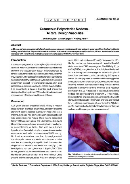
of right second toe which was tender and cold (Fig. 1). On<br />
investigations, her haemoglobin was 11.8 gm%, TLC-7,300/<br />
cu mm, platelet count-3,08,000 and ESR-34 mm/1st hr.<br />
Her kidney and liver function tests were normal. Urine<br />
(routine examination) revealed WBC-60 - 80/hpf with no<br />
casts. Urine culture showed E. coli (colony count > 10 5 ).<br />
Her 24 hr urinary protein was normal. Hepatitis B and C<br />
viral markers and CRP were negative. The ANA (by IF), p-<br />
ANCA (ELISA), cryoglobulins, anticardiolipin antibody, and<br />
lupus anticoagulants were negative. Arterial doppler of<br />
lower limb, and nerve conduction velocity (NCV) were<br />
normal. Skin biopsy taken from skin nodule was suggestive<br />
of nodular arteritis with a polymorphonuclear infiltrate<br />
involving medium sized arteries in deep reticular dermis<br />
alongwith extensive fibrinoid necrosis and vascular<br />
destruction (Fig. 2). A diagnosis of cutaneous polyarteritis<br />
nodosa with early gangrene of toe with UTI was made.<br />
She was started on prednisolone 0.5 mg/kg of body weight<br />
alongwith methotrexate 10 mg/week and was also treated<br />
for UTI. Steroids were tapered-off over 3 months. At followup<br />
of 4 months she had residual erythema over feet, no<br />
nodules, and the gangrenous toe was normal.<br />
Fig. 1: Reddish discolouration of both lower limbs and feet, and bluish<br />
discolouration of right second toe.<br />
* Senior Resident, ** Consultant, Department of Medicine, Sir Ganga Ram Hospital, Rajinder Nagar, New Delhi - 110 060.
Fig. 2: Polymorphonuclear infiltrates in deep reticularis dermis<br />
involving medium sized arteries with extensive fibrinoid necrosis.<br />
Discussion<br />
<strong>Cutaneous</strong> PAN is a rare form of cutaneous vasculitis. It<br />
involves small, medium sized arteries of dermis and<br />
subcutaenous tissue. It is also called periarteritis nodosa,<br />
and should be differentiated from systemic PAN due to the<br />
different clinical course and management of the two<br />
conditions. The cause of cutaneous PAN is not known; it<br />
could be a hypersensitivity reaction to certain infections 1 .<br />
In a series published by Geralol M et al in 1991, 4/9 patients<br />
had hepatitis B and 1 had a HIV infection 3 . Women were<br />
affected slightly more than men 2 . Our patient had UTI, which<br />
could have been a triggering factor, though there is paucity<br />
of literature on this aspect.<br />
<strong>Cutaneous</strong> PAN is characterised by tender subcutaneous<br />
nodules, usually measuring 4 - 5 mm in diameter,<br />
alongwith infarcts presenting as purple or black patches<br />
and livedo reticularis that may ulcerate 2 . They are mostly<br />
found over legs and feet. Systemic involvement is rare<br />
in contrast to classical PAN where there is visceral<br />
involvement.<br />
These patients may have neuromuscular involvement in<br />
the form of peripheral neuropathy that presents with<br />
tingling, numbness, sensory disturbances, weakness, and<br />
absent reflexes. In a study by Daud MS et al in 1997, 22%<br />
of 79 cases had some evidence of neuropathy 2 . Millard H<br />
et al in 1999 showed that 3/9 patients had neuropathy<br />
without systemic involvement 4 . Patients may also have<br />
some constitutional symptoms in the form of malaise,<br />
fever, sore throat, joint/muscular pains. The above features<br />
were absent in our patient. The definitive diagnosis is made<br />
by doing a skin biopsy. No other lab parameter is<br />
suggestive, except for high ESR which may show<br />
inflammatory process. In cutaneous PAN, histopathological<br />
examination shows features of nodular arteritis with<br />
polymorphonuclear infiltrates involving medium sized<br />
arteries in deep reticular dermis. There is extensive<br />
fibrinoid necrosis. This is in contrast to classical PAN which<br />
rarely shows nodular arteritis and the picture is of small<br />
vessel leucocytoclastic.<br />
<strong>Cutaneous</strong> PAN runs a chronic course lasting months to<br />
years, and has a waxing and waning phenomenon. Patients<br />
are generally treated with non-steroidal anti-inflammatory<br />
drugs and oral steroids. Immunosuppressive drugs can also<br />
be used in low doses in more severe kinds of cutaneous<br />
PAN and as steroid-sparing drugs 5 . It is generally a benign<br />
disease with progression to a classical disease being an<br />
exception. A close watch however, should be kept for<br />
progression to systemic involvement.<br />
References<br />
1. Khoo BP, Ng SK. <strong>Cutaneous</strong> polyarteritis nodosa: A case<br />
report and literature review. Ann Acad Med Singapore 1998;<br />
27: 868-72.<br />
2. Daoud MS, Hutton KP, Gibson LE. <strong>Cutaneous</strong> polyarteritis<br />
nodosa: a clinicopathological study of 79 cases. Br J<br />
Dermatol 1997; 136 (5): 706-13.<br />
3. Gerald M, Bruce RS, Scott McNutt N. <strong>Benign</strong> cutaneous<br />
polyarteritis nodosa. Arch Dermatol 1991; 127: 1520-3.<br />
4. Maillard H, Szczesniak S, Martin L et al. <strong>Cutaneous</strong><br />
polyarteritis nodosa: diagnostic and therapeutic aspects<br />
of 9 cases. Ann Dermatol Venereol 1999; 126 (2): 125-9.<br />
5. Kleeman D, Kempf W, Burg G, Hafner J. <strong>Cutaneous</strong><br />
polyarteritis nodosa. Vasa 1998; 27 (1): 54-7.<br />
160 Journal, Indian Academy of Clinical Medicine Vol. 7, No. 2 April-June, 2006