Structure of the dermis
Structure of the dermis
Structure of the dermis

You also want an ePaper? Increase the reach of your titles
YUMPU automatically turns print PDFs into web optimized ePapers that Google loves.
1<br />
Go Back to <strong>the</strong> Top To Order, Visit <strong>the</strong> Purchasing Page for Details<br />
C. Dermis<br />
papillary layer<br />
subpapillary<br />
layer<br />
reticular <strong>dermis</strong><br />
subcutaneous<br />
tissue<br />
lymphatic vessel blood capillary nerve<br />
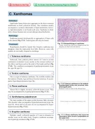
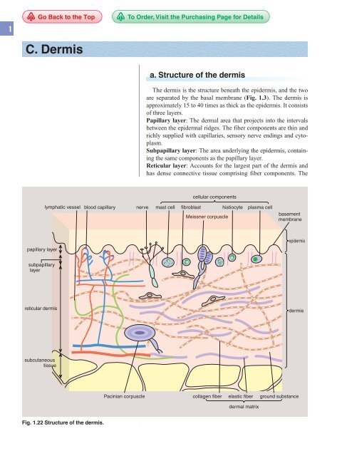
Fig. 1.22 <strong>Structure</strong> <strong>of</strong> <strong>the</strong> <strong>dermis</strong>.<br />
Pacinian corpuscle<br />
a. <strong>Structure</strong> <strong>of</strong> <strong>the</strong> <strong>dermis</strong><br />
The <strong>dermis</strong> is <strong>the</strong> structure beneath <strong>the</strong> epi<strong>dermis</strong>, and <strong>the</strong> two<br />
are separated by <strong>the</strong> basal membrane (Fig. 1.3). The <strong>dermis</strong> is<br />
approximately 15 to 40 times as thick as <strong>the</strong> epi<strong>dermis</strong>. It consists<br />
<strong>of</strong> three layers.<br />
Papillary layer: The dermal area that projects into <strong>the</strong> intervals<br />
between <strong>the</strong> epidermal ridges. The fiber components are thin and<br />
richly supplied with capillaries, sensory nerve endings and cytoplasm.<br />
Subpapillary layer: The area underlying <strong>the</strong> epi<strong>dermis</strong>, containing<br />
<strong>the</strong> same components as <strong>the</strong> papillary layer.<br />
Reticular layer: Accounts for <strong>the</strong> largest part <strong>of</strong> <strong>the</strong> <strong>dermis</strong> and<br />
has dense connective tissue comprising fiber components. The<br />
mast cell fibroblast<br />
cellular components<br />
Meissner corpuscle<br />
histiocyte<br />
collagen fiber elastic fiber<br />
dermal matrix<br />
plasma cell<br />
basement<br />
membrane<br />
epi<strong>dermis</strong><br />
<strong>dermis</strong><br />
ground substance
1<br />
14 1 <strong>Structure</strong> and Function <strong>of</strong> <strong>the</strong> Skin<br />
Table 1.2 Types <strong>of</strong> collagen fibers.<br />
Fiber Type<br />
Fibrillar<br />
I, II, III, V, XI<br />
Basement membrane IV<br />
Anchoring fibril VII<br />
Network forming VIII, X<br />
FACIT<br />
IX, XII, XIV, XIX, XX<br />
Micr<strong>of</strong>ibril<br />
VI<br />
Multiplexin<br />
XV, XVIII<br />
O<strong>the</strong>r<br />
XIII, XVII<br />
FACIT: fibril associated collagen with interrupted<br />
triple helix<br />
Abnormality in elastic MEMO<br />
fibers or collagen fibers<br />
If elastic fibers are decreased, lost or denatured,<br />
dermatolysis or senile cutis rhomboidalis<br />
nuchae may occur. If fibrillin<br />
molecules are congenitally abnormal, Marfan<br />
syndrome results (Chapter 18). Abnormalities<br />
in collagens, which are components <strong>of</strong> collagen<br />
fibers, may lead to Ehlers-Danlos syndrome,<br />
which causes skin fragility.<br />
2. Elastic fibers<br />
The elastic fiber is not as tough as <strong>the</strong> collagen fiber; however,<br />
it is extremely elastic and found abundantly in <strong>the</strong> <strong>dermis</strong> <strong>of</strong> <strong>the</strong><br />
scalp, face and <strong>the</strong> extensible organs such as arteries and tendons.<br />
In <strong>the</strong> <strong>dermis</strong>, <strong>the</strong> deeper <strong>the</strong> elastic fiber, <strong>the</strong> thicker it is. In<br />
<strong>the</strong> reticular layers, elastic fibers are scattered among collagen<br />
bundles running parallel to <strong>the</strong> skin surface. The closer <strong>the</strong> elastic<br />
fiber is to <strong>the</strong> papillary layer, <strong>the</strong> thinner it is and <strong>the</strong> more perpendicular<br />
it is to <strong>the</strong> skin surface. It forms an arch shape in <strong>the</strong><br />
papillary layer from which thin fibers are produced that perpendicularly<br />
reach <strong>the</strong> lamina densa. Elastic fibers are also connected<br />
to <strong>the</strong> lamina densa <strong>of</strong> glands, sweat ducts, smooth muscle,<br />
nerves and blood vessels.<br />
Elastic fibers are 1 mm to 3 mm in diameter. They cannot be<br />
differentiated from collagen fibers by HE staining. Elastic fibers<br />
stain dark blue to black in Weigert resorcin fuchsin, red-violet in<br />
aldehyde fuchsin, and brownish black in orcein. In elastic fibers,<br />
<strong>the</strong> characteristic striped pattern <strong>of</strong> collagen fibers is not<br />
observed by electron microscopy (Fig. 1.23). A skeletal fiber is<br />
10 nm to 15 nm in diameter and its main content is fibrillin. The<br />
homogeneous substances are highly elastic structural proteins<br />
called elastins.<br />
3. Ground substance, Matrix<br />
Ground substance, a gelatinous amorphous substance <strong>of</strong> sugar<br />
and proteins, is observed in between fibers and between cells in<br />
<strong>the</strong> <strong>dermis</strong>.<br />
The components <strong>of</strong> ground substance are principally proteoglycans<br />
and glycoproteins whose molecular weight is 150,000 to<br />
250,000 and whose sugar content is 2% to 15%. The molecules<br />
stabilize <strong>the</strong> fibers to give flexibility to <strong>the</strong> skin. Fibronectin, one<br />
kind <strong>of</strong> glycoprotein, contains a domain that connects fibrin,<br />
heparin and collagen; and binds integrin receptors on <strong>the</strong> cell surface<br />
are involved in cell proliferation, differentiation and wound<br />
healing. Besides <strong>the</strong>se components, blood and lymph-derived tissue<br />
fluid forms <strong>the</strong> remainder <strong>of</strong> <strong>the</strong> ground substance that is<br />
involved in <strong>the</strong> transport <strong>of</strong> substances essential to cellar activities<br />
and metabolism.<br />
Proteoglycans are a massive molecules with a molecular<br />
weight <strong>of</strong> 10 5 to 10 6 or more and a composition <strong>of</strong> multiple glycosaminoglycans<br />
(mucopolysaccharides) connecting with backbone<br />
proteins. Glycosamine, mostly produced by fibroblasts in<br />
<strong>the</strong> <strong>dermis</strong>, is rich in hyaluronic acids, which are associated with<br />
moisture retention.
c. Cellular components<br />
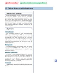
1. Fibroblast<br />
Fibroblast differentiates from <strong>the</strong> mesenchymal cells and produces<br />
collagen fibers, elastic fibers, and glycosaminoglycans.<br />
Fibroblasts appear as thin spindle-shaped cells sparse in collagen<br />
fibers (Fig. 1.25).<br />
By electron microscopy, multiple Golgi apparatus and granular<br />
endoplasmic reticuli are seen in <strong>the</strong> fibroblast. When collagen<br />
fibers are produced and <strong>the</strong> <strong>dermis</strong> matures, fibroblasts stop <strong>the</strong>ir<br />
activities and become fibrocytes. At this point, <strong>the</strong> cell nuclei<br />
shrink and have fewer endoplasmic reticuli. Adrenal cortex hormones<br />
and thyroid hormones are involved in this process.<br />
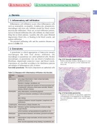
2. Histiocyte<br />
The histiocyte, a kind <strong>of</strong> macrophage, is broadly distributed in<br />
<strong>the</strong> connective tissue and intermingles with fibroblasts on <strong>the</strong><br />
outside <strong>of</strong> endocapillary cells (Fig. 1.26). A small circular nucleus<br />
and a large spindle- or star-shaped cell structure is seen in a<br />
histiocyte by light microscopy; fur<strong>the</strong>rmore, concave nuclei and<br />
<strong>the</strong> formation <strong>of</strong> psudopodial protrusions are observed by electron<br />
microscopy. Histiocytes contain Golgi apparatus, smooth<br />
and rough endoplasmic reticuli, and lysosomes. Lysosomes contain<br />
hydrolases and active acid phosphatases. The histiocyte<br />
releases collagenase and lysosomal enzymes containing elastase<br />
to digest <strong>the</strong> interstitium. It is involved in organ repair. The histiocyte<br />
degrades and phagocytoses mainly foreign substances and<br />
presents <strong>the</strong>m as antigens to T cells (Chapter 3).<br />
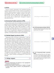
3. Mast cell<br />
The mast cell is found in <strong>the</strong> <strong>dermis</strong> around capillaries and in<br />
<strong>the</strong> periphery <strong>of</strong> subcutaneous tissues. The shape is roundish or<br />
spindled, and <strong>the</strong> diameter is 10 mm (Fig. 1.27). Mast cells produce<br />
and maintain various vasodilatory and hyperlucent chemical<br />
mediators. Mast cell granules stain red-violet in toluidine blue<br />
and methylene blue, and present with metachromasia. Cutaneous<br />
mast cells resemble basophils in form and function; however,<br />
<strong>the</strong>ir characteristics differ slightly from those <strong>of</strong> o<strong>the</strong>r organs,<br />
because <strong>the</strong>y differentiate in skin during intrauterine life.<br />
A mast cell intracytoplasmic granule appears as a circular<br />
structure with a diameter <strong>of</strong> 0.3 mm to 0.5 mm under electron<br />
microscopy. Multiple mast cell granules are evenly distributed in<br />
<strong>the</strong> cytoplasm. Chemotransmitters in <strong>the</strong> granules are released<br />
outside <strong>the</strong> cell under various stimuli, such as in type I allergic<br />
reactions (Fig. 1.28, Chapter 3). The main components <strong>of</strong> <strong>the</strong><br />
released substances are histamines and heparins, followed by<br />
Fig. 1.25 Fibroblasts (arrows).<br />
Fig. 1.26 Histiocytes (arrows)<br />
C. Dermis 15<br />
20 mm<br />
20 mm<br />
Histiocyte, Monocyte, MEMO<br />
Macrophage<br />
Large phagocytic cells in <strong>the</strong> living body are<br />
called macrophages. There are two kinds.<br />
-- Free macrophages: e.g., monocytes in <strong>the</strong><br />
blood, migrating macrophages in granuloma<br />
-- Fixed macrophages: e.g., histiocytes in <strong>the</strong><br />
<strong>dermis</strong> and subcutaneous tissues, Kupffer<br />
cells<br />
Histiocytes and monocytes are kinds <strong>of</strong><br />
macrophages.<br />
Melanophages<br />
MEMO<br />
Melanin granules that have exfoliated from<br />
<strong>the</strong> epi<strong>dermis</strong> to <strong>the</strong> <strong>dermis</strong> are <strong>of</strong>ten phagocytosed<br />
by histiocytes. Histiocytes that become<br />
brownish-red after repeatedly phagocytosing<br />
melanin granules are called melanophages.<br />
20 mm<br />
Histiocytes, <strong>the</strong> key to MEMO<br />
pathological diagnosis<br />
Epi<strong>the</strong>lioid cells, Touton giant cells, xanthoma<br />
cells and foreign-body giant cells in an<br />
epi<strong>the</strong>lioid cell granuloma are histiocytes that<br />
pathologically present in an atypical form.<br />
1
1<br />
16 1 <strong>Structure</strong> and Function <strong>of</strong> <strong>the</strong> Skin<br />
20 mm<br />
20 mm<br />
various enzymes, including neutrophil chemotactic factors<br />
(NCF), eosinophil chemotactic factors <strong>of</strong> anaphylaxis (ECF-A),<br />
tryptase, chymase and tumor necrosis factor (TNF)-like substances.<br />
The mast cell may produce and release inflammatory<br />
substances such as prostaglandins, leukotorienes and plateletactivating<br />
factors.<br />
4. Plasma cell<br />
a b c d e f g h i j k l m n o p q r<br />
The plasma cell is a differentiated B cell that has been stimulated<br />
by an antigen. It produces antibodies and is involved in<br />
humoral immunity. The shape <strong>of</strong> <strong>the</strong> plasma cell varies from circular<br />
to pear-shaped, and <strong>the</strong> diameter ranges from 8 mm to 14<br />
mm, which is twice as large as a leukocyte. It has a wheel-shaped<br />
nucleus with peripheral chromatin (Fig. 1.29).<br />
5. Dermal dendrocyte<br />
a b c d e f g h i The dermal j kdendrocyte l is mfound in n <strong>the</strong> dermal o upper p layer q (inr<br />
Fig. 1.27 Mast cells.<br />
a: Hematoxylin and eosin staining. b: Metachro-<br />
and between <strong>the</strong> papillary layer and <strong>the</strong> reticular layer). It is<br />
thought to be an immunocompetent cell (Chapter 3), and it is<br />
masia is seen by toluidine blue staining.<br />
characterized by containing clotting factor XIIIa.<br />
second exposure<br />
<strong>of</strong> <strong>the</strong> antigen<br />
nucleus nucleus<br />
sensitized<br />
mast cell<br />
antigen<br />
histamine<br />
vasodilation<br />
vascular<br />
permeability<br />
itch<br />
Fig. 1.28 Sensitization by mast cells.<br />
Fig. 1.29 Plasma cells (arrows).<br />
20 mm<br />
d. Vascular channels and nerves<br />
1. Blood vessels<br />
Multiple branches <strong>of</strong> arteries distributed in skin (Figs. 1.30 and<br />
1.31) are connected with each o<strong>the</strong>r in <strong>the</strong> dermal deep layer to<br />
form a horizontal network (subcutaneous plexus). With numerous<br />
branches ascending from <strong>the</strong> subcutaneous plexuses, <strong>the</strong><br />
arteries form a second network in <strong>the</strong> papillary lower layer (subpapillary<br />
plexus). The arterioles ascend through <strong>the</strong> papillary<br />
layer, forming capillary loops in <strong>the</strong> dermal papillaries before<br />
moving to venules that connect to each o<strong>the</strong>r to form two kinds<br />
<strong>of</strong> plexuses, whereby <strong>the</strong> blood flows into <strong>the</strong> cutaneous veins<br />
(Fig. 1.30). There are also characteristic plexuses in <strong>the</strong> periphery<br />
<strong>of</strong> <strong>the</strong> cutaneous appendages. The peripheral regions <strong>of</strong> <strong>the</strong><br />
eccrine glands are particularly rich in vascular networks, which<br />
control blood flow volume and body temperature by perspiration.<br />
Moreover, hair follicles in <strong>the</strong> anagen (growth) stage are also<br />
richly supplied with blood vessels, present in <strong>the</strong> surrounding<br />
dermal tissue.<br />
There is ano<strong>the</strong>r apparatus that circulates <strong>the</strong> blood directly<br />
from arteries to blood vessels: This is <strong>the</strong> arteriovenous anastomosis,<br />
which is controlled by sympa<strong>the</strong>tic nerves. The arteriovenous<br />
anastomosis controls <strong>the</strong> peripheral blood flow and is<br />
involved in body temperature regulation. Glomus apparatuses,<br />
which have spherical anastomotic branches, are seen everywhere<br />
in <strong>the</strong> skin. They are particularly well developed in <strong>the</strong> fingers, at<br />
apical ends <strong>of</strong> <strong>the</strong> toes, and below <strong>the</strong> nails. Many layers <strong>of</strong><br />
Go Back to <strong>the</strong> Top To Order, Visit <strong>the</strong> Purchasing Page for Details