1) Photo test 2) Photo-patch test 3) Photo-drug test 2 ...
1) Photo test 2) Photo-patch test 3) Photo-drug test 2 ...
1) Photo test 2) Photo-patch test 3) Photo-drug test 2 ...

Create successful ePaper yourself
Turn your PDF publications into a flip-book with our unique Google optimized e-Paper software.
Go Back to the Top To Order, Visit the Purchasing Page for Details<br />
2. <strong>Photo</strong>sensitivity <strong>test</strong> (Chapter 13)<br />
Various photosensitivity <strong>test</strong>s are conducted by examining the<br />
reaction to irradiation. Ultraviolet radiation is divided into ultraviolet<br />
A (UVA) (operative wavelength of 320 to 400 nm), ultraviolet<br />
B (UVB) (operative wavelength of 290 to 320 nm) and<br />
ultraviolet C (UVC) (operative wavelength of 100 to 290 nm).<br />
UVA, UVB and visible light are most frequently used.<br />
1) <strong>Photo</strong> <strong>test</strong><br />
The degree of photosensitivity and suitable operative wavelength<br />
can be measured and determined by the amount of radiation<br />
that causes cutaneous reactions such as pigmentation and<br />
erythema. By <strong>test</strong>ing the operative wavelength on patients, it is<br />
possible to know which particular radiation exposure should be<br />
eliminated.<br />
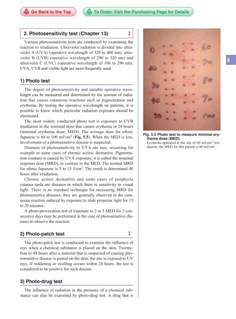
The most widely conducted photo <strong>test</strong> is exposure to UVB<br />
irradiation in the minimal dose that causes erythema in 24 hours<br />
(minimal erythema dose; MED). The average dose for ethnic<br />
Japanese is 60 to 100 mJ/cm 2 (Fig. 5.5). When the MED is low,<br />
involvement of a photosensitive disease is suspected.<br />
Diseases of photosensitivity to UVA are rare, occurring for<br />
example in some cases of chronic actinic dermatitis. Pigmentation<br />
(suntan) is caused by UVA exposure; it is called the minimal<br />
response dose (MRD), in contrast to the MED. The normal MRD<br />
for ethnic Japanese is 5 to 15 J/cm 2 . The result is determined 48<br />
hours after irradiation.<br />
Chronic actinic dermatitis and some cases of porphyria<br />
cutanea tarda are diseases in which there is sensitivity to visual<br />
light. There is no standard technique for measuring MRD for<br />
photosensitive diseases; they are generally observed in the cutaneous<br />
reaction induced by exposure to slide projector light for 15<br />
to 20 minutes.<br />
A photo-provocation <strong>test</strong> of exposure to 2 to 3 MED for 3 consecutive<br />
days may be performed in the case of photosensitive diseases<br />
to observe the reaction.<br />
2) <strong>Photo</strong>-<strong>patch</strong> <strong>test</strong><br />
The photo-<strong>patch</strong> <strong>test</strong> is conducted to examine the influence of<br />
rays when a chemical substance is placed on the skin. Twentyfour<br />
to 48 hours after a material that is suspected of causing photosensitive<br />
disease is pasted on the skin, the site is exposed to UV<br />
rays. If reddening or swelling occurs within 24 hours, the <strong>test</strong> is<br />
considered to be positive for such disease.<br />
3) <strong>Photo</strong>-<strong>drug</strong> <strong>test</strong><br />
The influence of radiation in the presence of a chemical substance<br />
can also be examined by photo-<strong>drug</strong> <strong>test</strong>. A <strong>drug</strong> that is<br />
Fig. 5.5 <strong>Photo</strong> <strong>test</strong> to measure minimal erythema<br />
dose (MED).<br />
Erythema appeared at the site of 60 mJ/cm 2 irradiation;<br />
the MED for this patient is 60 mJ/cm 2 .<br />
5
5<br />
68 5 Diagnosis of Skin Diseases<br />
Table 5.4 Typical type IV allergy <strong>test</strong>s.<br />
Tuberculin skin <strong>test</strong> (PPD skin <strong>test</strong>)<br />
A tuberculin skin <strong>test</strong> is used to detect delayed<br />
hypersensitivity to tuberculosis, by injecting<br />
tuberculosis antigen intradermally. 0.1 ml of<br />
tuberculosis antigen (purified protein derivative;<br />
PPD, 0.05 mg/ml) is injected into the inner side<br />
of the forearm. The long diameter of the<br />
erythema 48 hours after injection is used for<br />
interpretation: less than 10 mm is negative, and<br />
more than 10 mm as positive. Positive reaction<br />
is sometimes categorized into weak (only<br />
erythema), moderate (erythema and induration),<br />
and strong (erythema with vesicles and necrosis).<br />
Tuberculin skin <strong>test</strong> is specific to tuberculosis.<br />
However, patients with measles, sarcoidosis,<br />
Hodgkin’s disease, severe tuberculosis, and<br />
serious malignancies may show weak reaction<br />
or false negative.<br />
Trichophytin reaction<br />
An antigen derived from trichophyton<br />
(trichophytin antigen) may be used to <strong>test</strong><br />
intradermally for trichophytid and tinea profunda.<br />
Sporotrichin reaction<br />
Sporotrichin antigen is injected intradermally for<br />
diagnosis of sporotrichosis.<br />
Ito’s reaction<br />
Haemophilus ducreyi antigen is used for<br />
diagnosis of chancroid.<br />
Frei reaction<br />
Frei reaction is an intradermal <strong>test</strong> for<br />
lymphogranuloma venereum.<br />
Lepromin reaction (Mitsuda reaction)<br />
Lepromin reaction (Mitsuda reaction).<br />
Antigen derived from leproma is intradermally<br />
injected for diagnosis and classification of leprosy.<br />
Kveim <strong>test</strong><br />
Diagnosis of sarcoidosis used to be done by a<br />
skin <strong>test</strong> whose antigen is derived from another<br />
patient’s spleen and lymph nodes. Kveim <strong>test</strong> is<br />
rarely done today.<br />
5) Drug challenge <strong>test</strong><br />
The <strong>drug</strong> suspected of causing allergy is administered to the<br />
patient to determine whether the eruptions will recur. One onehundredth<br />
to one tenth of the usual dosage is given orally. In serious<br />
<strong>drug</strong> eruptions, there is a high risk that a <strong>drug</strong> challenge <strong>test</strong><br />
will cause anaphylactic shock. The <strong>drug</strong> challenge <strong>test</strong> is the<br />
most reliable allergy <strong>test</strong>.<br />
4. Skin function <strong>test</strong><br />
Tests for measuring various skin function, such as temperature<br />
control, secretion, and vascular regulation, are as follows.<br />
1) Measurement of skin temperature<br />
and thermography<br />
Thermography, which uses an infrared-camera-equipped emission<br />
pyrometer to express the distribution of skin temperature<br />
two-dimensionally, has become widely used for diagnosing diseases<br />
of the blood vessels, and nervous system disorders, inflammations,<br />
tumors, and other disorders.<br />
2) Transepidermal water loss (TEWL)<br />
Transepidermal water loss (TEWL) from the skin surface is<br />
measured by electric hygrometer (Fig. 5.8). This <strong>test</strong> is effective<br />
in determining the clinical condition of keratinization. The<br />
TEWL value usually is elevated in dyskeratoses, such as in<br />
ichthyosis.<br />
3) Skin capillary resistance <strong>test</strong><br />
The fragility of skin capillaries can be determined by measuring<br />
ecchymosis produced in artificially pressured blood vessels.<br />
In the Rumpel-Leede <strong>test</strong>, the upper arm is pressed by a blood<br />
pressure manchette to congest the blood vessels. Two minutes<br />
after pressure between the systolic and diastolic pressures is<br />
applied to the patient’s upper arm for 5 minutes to constrict the<br />
blood vessels, hemorrhagic spots occur. When 10 hemorrhagic<br />
spots or more produced, the <strong>test</strong> is positive for dysfunction of<br />
vascular regulation. It may also be positive if there is abnormality<br />
in the capillaries or platelets, such as Henoch-Schönlein purpura<br />
or thrombopenic purpura.<br />
5. Fungal examination<br />
Potassium hydroxide (KOH) is used for observation and detection<br />
of fungi and mites. Scales or blister contents are swabbed<br />
(Fig. 5.9) and applied to a glass slide onto which 20% KOH solution<br />
is dripped, and a slide cover is placed on top. The slide is<br />
Go Back to the Top To Order, Visit the Purchasing Page for Details