LYMPHATIC SYSTEM consists of: 1. lymph capillaries 2. collecting ...
LYMPHATIC SYSTEM consists of: 1. lymph capillaries 2. collecting ...
LYMPHATIC SYSTEM consists of: 1. lymph capillaries 2. collecting ...
You also want an ePaper? Increase the reach of your titles
YUMPU automatically turns print PDFs into web optimized ePapers that Google loves.
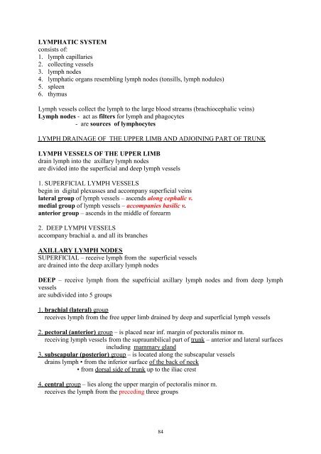
<strong>LYMPHATIC</strong> <strong>SYSTEM</strong><br />
<strong>consists</strong> <strong>of</strong>:<br />
<strong>1.</strong> <strong>lymph</strong> <strong>capillaries</strong><br />
<strong>2.</strong> <strong>collecting</strong> vessels<br />
3. <strong>lymph</strong> nodes<br />
4. <strong>lymph</strong>atic organs resembling <strong>lymph</strong> nodes (tonsills, <strong>lymph</strong> nodules)<br />
5. spleen<br />
6. thymus<br />
Lymph vessels collect the <strong>lymph</strong> to the large blood streams (brachiocephalic veins)<br />
Lymph nodes - act as filters for <strong>lymph</strong> and phagocytes<br />
- are sources <strong>of</strong> <strong>lymph</strong>ocytes<br />
LYMPH DRAINAGE OF THE UPPER LIMB AND ADJOINING PART OF TRUNK<br />
LYMPH VESSELS OF THE UPPER LIMB<br />
drain <strong>lymph</strong> into the axillary <strong>lymph</strong> nodes<br />
are divided into the superficial and deep <strong>lymph</strong> vessels<br />
<strong>1.</strong> SUPERFICIAL LYMPH VESSELS<br />
begin in digital plexusses and accompany superficial veins<br />
lateral group <strong>of</strong> <strong>lymph</strong> vessels – ascends along cephalic v.<br />
medial group <strong>of</strong> <strong>lymph</strong> vessels – accompanies basilic v.<br />
anterior group – ascends in the middle <strong>of</strong> forearm<br />
<strong>2.</strong> DEEP LYMPH VESSELS<br />
accompany brachial a. and all its branches<br />
AXILLARY LYMPH NODES<br />
SUPERFICIAL – receive <strong>lymph</strong> from the superficial vessels<br />
are drained into the deep axillary <strong>lymph</strong> nodes<br />
DEEP – receive <strong>lymph</strong> from the supefricial axillary <strong>lymph</strong> nodes and from deep <strong>lymph</strong><br />
vessels<br />
are subdivided into 5 groups<br />
<strong>1.</strong> brachial (lateral) group<br />
receives <strong>lymph</strong> from the free upper limb drained by deep and superficial <strong>lymph</strong> vessels<br />
<strong>2.</strong> pectoral (anterior) group – is placed near inf. margin <strong>of</strong> pectoralis minor m.<br />
receiving <strong>lymph</strong> vessels from the supraumbilical part <strong>of</strong> trunk – anterior and lateral surfaces<br />
including mammary gland<br />
3. subscapular (posterior) group – is located along the subscapular vessels<br />
drains <strong>lymph</strong> • from the inferior surface <strong>of</strong> the back <strong>of</strong> neck<br />
• from dorsal side <strong>of</strong> trunk up to the iliac crest<br />
4. central group – lies along the upper margin <strong>of</strong> pectoralis minor m.<br />
receives the <strong>lymph</strong> from the preceding three groups<br />
84
5. apical group (infraclavicular) – surrounds subclavian a.<br />
receives <strong>lymph</strong> vessels arising from the central nodes<br />
efferent vessels arising from apical axillary <strong>lymph</strong> nodes join to form subclavian trunk<br />
Right subclavian trunk – opens into the right brachiocephalic v. (angulus venosus)<br />
Left subclavian trunk – terminates into the thoracic duct which opens into the left<br />
brachiocephalic v. (angulus venosus)<br />
LYMPH DRAINAGE OF LOWER LIMB AND ADJOINING PART OF TRUNK<br />
the <strong>lymph</strong> from the lower limb and lower part <strong>of</strong> trunk is drained into the inguinal <strong>lymph</strong><br />
nodes<br />
INGUINAL LYMPH NODES<br />
are divided into the superficial and deep groups<br />
SUPERFICIAL INGUINAL LYMPH NODES<br />
Receive <strong>lymph</strong> from:<br />
•free lower limb – superficial structures (skin, subcutaneous tissue)<br />
•gluteal region<br />
• lateral abdominal wall<br />
• anterior abdominal wall<br />
• external genital organas<br />
• uterus!<br />
• lower part <strong>of</strong> the anal canal<br />
The <strong>lymph</strong> from the superficial <strong>lymph</strong> nodes is drained into the external iliac <strong>lymph</strong> nodes<br />
and into the deep inguinal <strong>lymph</strong> nodes<br />
DEEP INGUINAL LYMPH NODES<br />
are placed around femoral vessels in the femoral triangle near the inguinal ligament<br />
receive • deep <strong>lymph</strong> vessels <strong>of</strong> lower limb (accompanying blood vessels)<br />
efferent <strong>lymph</strong> vessels arising from the deep <strong>lymph</strong> nodes terminate in the external iliac<br />
<strong>lymph</strong> nodes<br />
LYMPH VESSELS OF FREE LOWER LIMB<br />
Deep – accompany femoral a. and its branches<br />
popliteal <strong>lymph</strong> nodes are placed around popliteal a.<br />
Superficial <strong>lymph</strong> vessels – accompany saphenous veins<br />
medial group – ascends along the great saphenous v. (to terminate into the inferior<br />
superficial inguinal nodes)<br />
lateral group – ascends along m the small saphenous v. (to terminate partly into the<br />
popliteal, partly into the inferior superficial inguinal nodes)<br />
85
LYMPH ORGANS<br />
THYMUS<br />
<strong>lymph</strong>oid organ and endocrine gland<br />
main function:<br />
- differentiation <strong>of</strong> <strong>lymph</strong>ocytes into the different classes<br />
- production <strong>of</strong> various factors and hormones which regulate <strong>lymph</strong>ocytes production,<br />
differentiation and activities within the thymus, peripheral <strong>lymph</strong>oid tissue and<br />
elsewhere<br />
located in the anterior mediastinum<br />
extends into the neck<br />
may reach lower part <strong>of</strong> thyroid gland (in children)<br />
relations:<br />
anteriorly – sternum and costal cartilages<br />
- infrahyoid muscles<br />
posteriorly – heart in pericardium<br />
- brachicephalic veins and sup. v. cava<br />
- aortic arch and its branches<br />
- trachea<br />
laterally – pleurae and lungs<br />
varies in size with age<br />
at birth: 10 – 15 gm<br />
grows up to the puberty: 30 – 40 gm<br />
progressively diminishes – atrophy, replacement by fat<br />
mid-adult life: 10 gm<br />
childhood<br />
pinkish-grey in colour<br />
lobulated<br />
right and left lobes connected by fibroareolar tissue<br />
Structure:<br />
fibrous capsule<br />
septa → lobules<br />
lobule – cortex, medulla (fewer <strong>lymph</strong>ocytes than the cortex)<br />
Blood supply:<br />
arteries<br />
Thymic branches <strong>of</strong> inf. thyroid a. and int. thoracic a.<br />
Veins – drained into the inf. thyroid, brachiocephalic and<br />
int. thoracic veins<br />
Nerve supply: sympathetic nerves – cervicothoracic ganglion<br />
parasympathetic fibres (vagus n.)<br />
86
SPLEEN<br />
- placed in the left hypochondriac region<br />
between the gastric fundus and diaphragm<br />
- long axis lies in the plane <strong>of</strong> the 10th rib<br />
- posterior extremity lies 4 cm far from the median plane<br />
- anterior extremity reaches mid-axillay line<br />
- it is s<strong>of</strong>t, friable in consistence, dark red in colour<br />
- irregularly ovoid – 12 cm long, 7 cm wide, 3-4 cm thick<br />
- has diaphragmatic and visceral surfaces<br />
superior and inferior margins<br />
anterior and posterior extremities<br />
relations:<br />
diaphragmatic surface – diaphragm which separates it from<br />
- pleural cavity - costodiaphragmatic recess<br />
visceral surface – stomach (gastric impression)<br />
- L kedney (renal impression)<br />
- pancreas (pancreatic impresiion)<br />
- left colic flexure (colic impression )<br />
hilum – splenic a. and v. enter the spleen<br />
spleen – intraperitoneal organ<br />
(develops in the posterior mezogastrium)<br />
gastrosplenic and lienorenal ligaments<br />
accessory spleens – in the gastrosplenic lig. and greater omentum<br />
function:<br />
phagocytosis<br />
immune responses due to <strong>lymph</strong>ocytes<br />
cytopoiesis (haemopoiesis and <strong>lymph</strong>opoiesis)<br />
storage place <strong>of</strong> blood<br />
Blood supply: splenic a. (coeliac a.)<br />
splenic v. (portal v.)<br />
Nerve supply: abdominal autonomic plexus<br />
87
THE LYMPH VESSELS AND LYMPH NODES OF THE HEAD, NECK AND TRUNK<br />
THE <strong>LYMPHATIC</strong> DRAINAGE OF THE HEAD AND NECK<br />
Lymph drainage <strong>of</strong> the superficial tissues <strong>of</strong> the head and neck<br />
Most <strong>lymph</strong> vessels accompany branches <strong>of</strong> external carotid artery<br />
Terminate in small groups <strong>of</strong> <strong>lymph</strong> nodes<br />
•Occipital nodes (occipital vessels)<br />
• Retroauricular (posterior auricular vessels)<br />
• Parotid ( temporal superficial vessels)<br />
• Buccal (facial vessels)<br />
• Submandibular (facial vessels in digastric triangle)<br />
Receives afferents from parotid and buccal nodes<br />
and <strong>lymph</strong> from the oral cavity, tonsils, tongue, teeth)<br />
• Anterior cervical (along the anterior jugular vein)<br />
• Superficial cervical (along the external jugular vein)<br />
nodes represent outlying nodes <strong>of</strong> deep cervical <strong>lymph</strong> nodes<br />
the <strong>lymph</strong> is drained into the deep cervical <strong>lymph</strong> nodes<br />
Lymph drainage <strong>of</strong> deep tissues <strong>of</strong> the head and neck<br />
Directs to the deep cervical <strong>lymph</strong> nodes<br />
Outlying grops <strong>of</strong> nodes:<br />
Retropharyngeal – receive <strong>lymph</strong> from nasopharynx, nasal cavity, auditory tube<br />
Paratracheal nodes – (around trachea and oesophagus) receive the <strong>lymph</strong> from the larynx,<br />
trachea, oesophagus<br />
the deep cervical <strong>lymph</strong> nodes<br />
are regional <strong>lymph</strong> nodes <strong>of</strong> the head and neck<br />
receive <strong>lymph</strong> from the superficial and deep structures <strong>of</strong> the head and neck<br />
lie along the carotid sheath (internal jugular vein, carotid arteries)<br />
subdivided into the superior and inferior group<br />
■ Superior deep cervical <strong>lymph</strong> nodes<br />
Lie close to the upper part <strong>of</strong> the internal jugular vein<br />
this group contains jugulodigastric node - near the tendon <strong>of</strong> digastricus m.- associated<br />
with <strong>lymph</strong> drainage <strong>of</strong> the tongue<br />
efferents terminate into the inferior deep cervical <strong>lymph</strong> nodes<br />
■ inferior deep cervical <strong>lymph</strong> nodes<br />
around lower part <strong>of</strong> the internal jugular vein<br />
contain juguloomohyoid node (near the tendon <strong>of</strong> omohyoid m.)<br />
efferents unite to form<br />
jugular trunk<br />
88
THE THORACIC DUCT<br />
main <strong>lymph</strong> vessel conveying the <strong>lymph</strong> to the blood stream<br />
arises in the abdomen (at the level <strong>of</strong> the 12th thoracic vertebra)<br />
by union <strong>of</strong> right and left lumbar trunks and intestinal trunk – here dilated – cisterna chyli<br />
here it receives the <strong>lymph</strong> from<br />
• abdominal walls and organs<br />
• pelvic walls and organs<br />
• lower limbs<br />
traverses the diaphragm through the aortic hiatus<br />
ascends in the posterior mediastinum - behind the oesophagus on right side <strong>of</strong> aorta<br />
turns to the left behind aortic arch and subclavian a.<br />
leaves the thorax (3 – 4 cm above the clavicle)<br />
arches above the subclavian a.<br />
opens into the left brachiocephalic v. (angulus venosus)<br />
here it receives the tributaries<br />
• left subclavian trunk (left upper limb )<br />
• left jugular trunk ( left half <strong>of</strong> head and neck )<br />
• left bronchopulmonary trunk (left half <strong>of</strong> the thorax – the walls and organs)<br />
In conclusion:<br />
Thoracic duct drains the <strong>lymph</strong> from the lower limbs, pelvis (walls, organs), abdomen walls,<br />
organs), left half <strong>of</strong> the thorax (walls, organs), left upper limb, left half <strong>of</strong> the neck and head<br />
--------------------------------------------------------------------------<br />
<strong>lymph</strong> from the right half <strong>of</strong> head and neck, right upper limb and right half <strong>of</strong> the thorax is<br />
drained into the right <strong>lymph</strong>atic trunk – terminates into the right angulus venosus<br />
right <strong>lymph</strong>atic trunk arises by union <strong>of</strong> right jugular, right subclavian and right<br />
bronchomediastinal trunks<br />
LYMPH DRAINAGE OF THE THORAX<br />
superficial tissues - skin, mammary gland, muscles connecting upper limb to the thorax-<br />
are drained to the axillary <strong>lymph</strong> nodes (see over there)<br />
deep structures – thoracic walls - ribs, intercostal muscles, diaphragm<br />
and thoracic organs are drained into<br />
the deep <strong>lymph</strong> nodes <strong>of</strong> the thorax<br />
<strong>lymph</strong> drainage <strong>of</strong> the thoracic walls<br />
parasternal, intercostal, diaphragmatic <strong>lymph</strong> nodes<br />
89
■ parasternal <strong>lymph</strong> nodes<br />
on sides <strong>of</strong> internal thoracic vessels<br />
receive <strong>lymph</strong> from<br />
• anterior thoracic and abdominal wall<br />
• mammary gland<br />
• liver (upper surface)<br />
efferents terminate in the bronchomediastinal trunk<br />
■ intercostal <strong>lymph</strong> nodes<br />
located near the necks <strong>of</strong> ribs<br />
receive <strong>lymph</strong> from<br />
• the posterolateral thoracic wall (ribs, intercostal muscles)<br />
efferents open into the thoracic duct and right <strong>lymph</strong>atic trunk<br />
■ diaphragmatic <strong>lymph</strong> nodes<br />
lie around the periphery <strong>of</strong> the diaphragm<br />
receive <strong>lymph</strong> from<br />
• diaphragm<br />
• superior surface <strong>of</strong> the liver<br />
efferents pass into the parasternal nodes, anterior mediastinal and posterior mediastinal<br />
<strong>lymph</strong> nodes<br />
<strong>lymph</strong> drainage <strong>of</strong> the thoracic organs<br />
anterior mediastinal , posterior mediastinal and tracheobronchial <strong>lymph</strong> nodes<br />
efferents empty into the thoracic duct and right <strong>lymph</strong>atic duct<br />
■ anterior mediastinal <strong>lymph</strong> nodes (brachiocephalic l. n.)<br />
are placed in front <strong>of</strong> the brachicephalic veins and branches <strong>of</strong> aortic arch<br />
receive <strong>lymph</strong> from the anterior mediastinum<br />
• thymus<br />
• thyroid gland<br />
• pericardium<br />
• diaphragmatic <strong>lymph</strong> nodes<br />
efferents terminate in the thoracic duct (and tracheobronchial <strong>lymph</strong> nodes)<br />
■ tracheobronchial <strong>lymph</strong> nodes<br />
located near the trachea and tracheal bifurcation<br />
subdivided into<br />
the paratracheal – on sides <strong>of</strong> trachea<br />
superior tracheobronchial - above bifurcation<br />
inferior tracheobronchial – below the bifurcation<br />
outlying <strong>lymph</strong> nodes - bronchopulmonary – in the pulmonary hilum<br />
- pulmonary – in the lung substance<br />
receive <strong>lymph</strong> from<br />
• lungs and bronchi<br />
• trachea<br />
• heart<br />
efferent vessels unite to form bronchomediastinal trunks<br />
90
■ posterior mediastinal <strong>lymph</strong> nodes<br />
around oesophagus and thoracic aorta<br />
drain <strong>lymph</strong> from<br />
• oesophagus<br />
• heart<br />
• diaphragm<br />
• liver<br />
efferents terminate into the thoracic duct<br />
THE <strong>LYMPHATIC</strong> DRAINAGE OF THE ABDOMEN<br />
the <strong>lymph</strong> from the abdominal walls and abdominal organs is drained into<br />
the lumbar and pre-aortic nodes<br />
■ lumbar <strong>lymph</strong> nodes (lateral aortic)<br />
lie on sides <strong>of</strong> the abdominal aorta and inf. vena cava<br />
receive <strong>lymph</strong> from<br />
• the abdominal walls<br />
• suprerenal glands<br />
• kidneys, ureters<br />
• gonads<br />
efferents unite to form lumbar trunk – right and left<br />
outlying <strong>lymph</strong> nodes in the pelvis - common iliac)- drain<br />
• pelvic walls and pelvic organs<br />
• lower limbs<br />
■ pre-aortic nodes – coeliac, superior mesenteric, inferior mesenteric<br />
receive <strong>lymph</strong> from the alimentary canal, liver, pancreas, spleen<br />
efferents form intestinal trunk<br />
coeliac <strong>lymph</strong> nodes<br />
near origin <strong>of</strong> coeliac artery<br />
receive <strong>lymph</strong> from the organs supplied by coeliac a. (liver, gall bladder, stomach, duodenum,<br />
pancreas, spleen)<br />
outlying nodes:<br />
hepatic - along common and proper hepatic a.<br />
gastric – near lesser and greater curvatures and around the pylorus<br />
pancreaticosplenic – along splenic a.<br />
superior and inferior mesenteric <strong>lymph</strong> nodes<br />
lie close to the origin <strong>of</strong> sup. and inf. mesenteric arteries<br />
receive <strong>lymph</strong> from the small and large intestines<br />
outlying nodes:<br />
<strong>lymph</strong> nodes <strong>of</strong> the mesentery – drain small intestine<br />
ileocolic nodes – terminal part <strong>of</strong> ileum, appendix<br />
nodes <strong>of</strong> the colon – large intestine<br />
superior rectal nodes – upper part <strong>of</strong> rectum<br />
91
THE <strong>LYMPHATIC</strong> DRAINAGE OF THE PELVIS<br />
■ common iliac <strong>lymph</strong> nodes<br />
along common iliac vessels<br />
receive <strong>lymph</strong> from the external and internal iliac <strong>lymph</strong> nodes<br />
■ external iliac <strong>lymph</strong> nodes<br />
along external iliac vessels<br />
receive <strong>lymph</strong> from:<br />
• inguinal <strong>lymph</strong> nodes<br />
• infraumbilical part <strong>of</strong> anterior abdominal wall<br />
• adductor region (along obturator vessels)<br />
• penis<br />
• prostate<br />
• urinary bladder, urethra<br />
• uterus, vagina<br />
efferents pass to the common iliac nodes<br />
■ internal iliac <strong>lymph</strong> nodes<br />
lie along internal iliac vessels<br />
receive <strong>lymph</strong> from:<br />
• pelvic organs<br />
• perineum (along the internal pudendal vessels)<br />
• muscles <strong>of</strong> gluteal region (along the gluteal vessels)<br />
efferents pass to the common iliac nodes<br />
92
IN SUM<br />
lower limbs (and adjoing part <strong>of</strong> trunk)<br />
regional <strong>lymph</strong> nodes – inginal<br />
efferents → external iliac → common iliac → lumbar → lumbar trunk → thoracic duct<br />
pelvis:<br />
regional <strong>lymph</strong> nodes – internal iliac <strong>lymph</strong> nodes<br />
efferents → common iliac nodes→ lumbar nodes → lumbar trunk → thoracic duct<br />
abdomen:<br />
regional <strong>lymph</strong> nodes<br />
- lumbar nodes →lumbar trunk → thoracic duct<br />
- coeliac, superior and inferior mesenteric → intestinal trunk → thoracic duct<br />
thorax:<br />
regional <strong>lymph</strong> nodes<br />
- parasternal, intercostal, diaphragmatic (for the walls) → thoracic duct<br />
- anterior mediastinal, posterior mediastinal → thoracic duct,<br />
→ right lymf. duct<br />
- bronchomediastinal → throacic duct<br />
→ right lymf duct<br />
head and neck:<br />
regional <strong>lymph</strong> nodes – deep cervical nodes<br />
efferents → jugular trunk → thoracic duct,<br />
→ right <strong>lymph</strong>. duct<br />
upper limb (and adjoing part <strong>of</strong> trunk)<br />
regional <strong>lymph</strong> nodes – axillary <strong>lymph</strong> nodes<br />
efferents → subclavian trunk → thoracic duct,<br />
→ right <strong>lymph</strong>. duct<br />
93


