Chapter 20: Lymphatic System
Chapter 20: Lymphatic System
Chapter 20: Lymphatic System

Create successful ePaper yourself
Turn your PDF publications into a flip-book with our unique Google optimized e-Paper software.
<strong>Chapter</strong> <strong>20</strong>: <strong>Lymphatic</strong> <strong>System</strong><br />
Copyright © <strong>20</strong>06 Pearson Education, Inc., publishing as Benjamin Cummings
<strong>Lymphatic</strong> <strong>System</strong>: Overview<br />
Consists of two semi-independent parts:<br />
A network of lymphatic vessels<br />
Lymphoid tissues and organs scattered throughout the body<br />
Returns interstitial fluid and leaked plasma proteins back<br />
to the blood<br />
Lymphoid organs house phagocytic cells and lymphocytes<br />
Lymph – interstitial fluid once it has entered lymphatic<br />
vessels<br />
Copyright © <strong>20</strong>06 Pearson Education, Inc., publishing as Benjamin Cummings
<strong>Lymphatic</strong> <strong>System</strong>: Overview<br />
Copyright © <strong>20</strong>06 Pearson Education, Inc., publishing as Benjamin Cummings<br />
Figure <strong>20</strong>.2a
<strong>Lymphatic</strong> <strong>System</strong>: Overview<br />
Copyright © <strong>20</strong>06 Pearson Education, Inc., publishing as Benjamin Cummings<br />
Figure <strong>20</strong>.1a
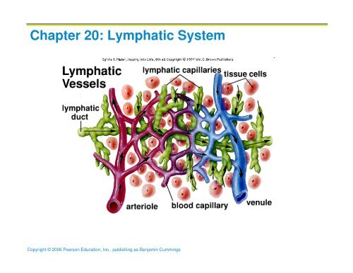
<strong>Lymphatic</strong> Vessels<br />
Fluid & plasma proteins are not all resorbed at the<br />
capillary beds and must be returned to the blood to<br />
maintain blood volume<br />
…lymphatic vessels accomplish this<br />
One-way system, lymph flows toward the heart<br />
Lymph vessels include:<br />
Microscopic, permeable, blind-ended capillaries<br />
<strong>Lymphatic</strong> collecting vessels<br />
Trunks and ducts<br />
Copyright © <strong>20</strong>06 Pearson Education, Inc., publishing as Benjamin Cummings
<strong>Lymphatic</strong> Vessels<br />
<strong>Lymphatic</strong> vessels begin at the blind-ended capillaries that<br />
weave between the tissue of the body<br />
<strong>Lymphatic</strong> capillaries are widespread, but are absent in:<br />
bones, bone marrow, teeth, CNS<br />
<strong>Lymphatic</strong> capillaries are incredibly permeable, much<br />
more so than blood capillaries<br />
This is due to:<br />
Loose fitting endothelial cells with weak cell-cell<br />
junctions thus forming minivalves<br />
Collagen filaments preventing vessels from collapsing<br />
Thus they form a one-way corridor<br />
Copyright © <strong>20</strong>06 Pearson Education, Inc., publishing as Benjamin Cummings
<strong>Lymphatic</strong> Capillaries<br />
Similar to blood capillaries, with modifications:<br />
Very permeable<br />
Loosely joined endothelial minivalves<br />
Withstand interstitial pressure and remain open<br />
The minivalves function as one-way gates:<br />
Greater interstitial fluid pressure, gates open<br />
Greater internal lymph vessel fluid pressure, gates<br />
close preventing back-flow<br />
Copyright © <strong>20</strong>06 Pearson Education, Inc., publishing as Benjamin Cummings
<strong>Lymphatic</strong> Capillaries<br />
Copyright © <strong>20</strong>06 Pearson Education, Inc., publishing as Benjamin Cummings<br />
Figure <strong>20</strong>.1b
<strong>Lymphatic</strong> Capillaries<br />
Inflammation results in the lymph capillary valves to open<br />
even wider to allow the following items to be absorbed:<br />
Cell debris<br />
Pathogens<br />
Cancer cells<br />
Cells in the lymph nodes cleanse and “examine” this debris<br />
Lacteals – specialized lymph capillaries present in<br />
intestinal mucosa<br />
Absorb digested fat and deliver chyle (white lymph) to the<br />
blood<br />
Copyright © <strong>20</strong>06 Pearson Education, Inc., publishing as Benjamin Cummings
<strong>Lymphatic</strong> Collecting Vessels<br />
From the lymph capillaries, lymph flows to<br />
collecting vessels<br />
Collecting vessels have the same three tunics as<br />
veins, but have thinner walls, with more internal<br />
valves and anastomose more frequently<br />
Collecting vessels (lymphatics) in the skin travel<br />
with superficial veins<br />
<strong>Lymphatic</strong>s of the trunk and digestive viscera<br />
travel with arteries<br />
Copyright © <strong>20</strong>06 Pearson Education, Inc., publishing as Benjamin Cummings
<strong>Lymphatic</strong> Trunks<br />
From the lymphatics (collecting vessels), lymph<br />
travels to lymphatic trunks<br />
<strong>Lymphatic</strong> trunks are formed by the union of the<br />
largest collecting vessels<br />
Major trunks include:<br />
Paired lumbar, bronchomediastinal, subclavian,<br />
and jugular<br />
and a single intestinal trunk<br />
Copyright © <strong>20</strong>06 Pearson Education, Inc., publishing as Benjamin Cummings
<strong>Lymphatic</strong> Ducts<br />
From the lymphatic trunks, lymph is delivered to<br />
one of two lymphatic ducts<br />
Right lymphatic duct – drains the right upper arm<br />
and the right side of the head and thorax<br />
Thoracic duct – arises from the cisterna chyli and<br />
drains the rest of the body<br />
Both empty into the venous circulation at the<br />
junction of the internal jugular vein and subclavian<br />
vein on its respective side<br />
Copyright © <strong>20</strong>06 Pearson Education, Inc., publishing as Benjamin Cummings
Lymph Transport<br />
The lymphatic system lacks a pumping organ<br />
Vessels are low-pressure conduits<br />
Uses the same methods as veins to propel lymph:<br />
Contraction of skeletal muscles<br />
Thoracic contraction during respiration<br />
Pulsations of nearby arteries<br />
Contractions of smooth muscle in the walls of the<br />
lymphatics<br />
Copyright © <strong>20</strong>06 Pearson Education, Inc., publishing as Benjamin Cummings
Lymphoid Cells & Lymphocytes<br />
Lymphocytes are the main cells involved in the<br />
immune response<br />
They mature into T cells & B cells<br />
T cells and B cells protect the body against<br />
antigens<br />
Antigen – anything the body perceives as foreign<br />
Bacteria and their toxins; viruses<br />
Mismatched RBCs or cancer cells<br />
Copyright © <strong>20</strong>06 Pearson Education, Inc., publishing as Benjamin Cummings
Lymphocytes<br />
T cells (Thymus)<br />
Manage the immune response<br />
Attack and destroy foreign cells<br />
B cells (Bone Marrow)<br />
Produce plasma cells, which secrete antibodies<br />
Antibodies immobilize antigens and “tag” them for<br />
destruction by leukocytes<br />
Copyright © <strong>20</strong>06 Pearson Education, Inc., publishing as Benjamin Cummings
Other Lymphoid Cells<br />
Macrophages – phagocytize foreign substances and<br />
help activate T cells<br />
Dendritic cells – capture antigens and bring them<br />
back to the lymph node<br />
Reticular cells – fibroblast–like cells that produce a<br />
stroma, or network, that supports other cell types<br />
in lymphoid organs<br />
Copyright © <strong>20</strong>06 Pearson Education, Inc., publishing as Benjamin Cummings
Lymphoid Tissue<br />
Composed of loose reticular tissue<br />
Functions to:<br />
House and provide proliferation site for lymphocytes<br />
Surveillance:<br />
Macrophages & lymphocytes live on the fibrous<br />
tissue<br />
Lymphocytes cycle between circulatory vessels,<br />
lymphoid tissue, and loose connective tissue of<br />
the body<br />
can move quickly from one to the other<br />
Copyright © <strong>20</strong>06 Pearson Education, Inc., publishing as Benjamin Cummings
Lymphoid Tissue<br />
Diffuse lymphatic tissue – scattered reticular tissue<br />
elements in every body organ<br />
Larger collections appear in the lamina propria of<br />
mucous membranes and lymphoid organs<br />
<strong>Lymphatic</strong> follicles (nodules) – solid, spherical<br />
bodies consisting of tightly packed reticular<br />
elements and cells<br />
Germinal center composed of dendritic and B cells<br />
Found in isolation and as part of larger lymphoid<br />
organs<br />
Copyright © <strong>20</strong>06 Pearson Education, Inc., publishing as Benjamin Cummings
Lymph Nodes<br />
Principal lymphoid organs of the body<br />
Embedded in connective tissue and clustered along lymphatic<br />
vessels<br />
Aggregations of these nodes occur near the body surface in<br />
inguinal, axillary, and cervical regions of the body<br />
Copyright © <strong>20</strong>06 Pearson Education, Inc., publishing as Benjamin Cummings
Lymph Nodes<br />
Two basic functions:<br />
Filtration – macrophages in the nodes<br />
remove/destroy microorganisms and debris<br />
preventing its delivery to the blood<br />
Immune system activation – lymphocytes in the<br />
nodes monitor lymph for antigens and mount an<br />
attack against them<br />
Copyright © <strong>20</strong>06 Pearson Education, Inc., publishing as Benjamin Cummings
Structure of a Lymph Node<br />
Nodes are
Structure of a Lymph Node<br />
Cortex contains follicles with germinal centers, heavy with<br />
dividing B cells<br />
Dendritic cells nearly encapsulate the follicles<br />
Deep cortex houses T cells in transit<br />
T cells circulate continuously among the blood, lymph nodes,<br />
and lymphatic stream<br />
Copyright © <strong>20</strong>06 Pearson Education, Inc., publishing as Benjamin Cummings
Structure of a Lymph Node<br />
Medullary cords extend from the cortex and contain B cells, T<br />
cells, and plasma cells<br />
Throughout the node are lymph sinuses crisscrossed by reticular<br />
fibers<br />
Macrophages reside on these fibers and phagocytize foreign<br />
matter<br />
Copyright © <strong>20</strong>06 Pearson Education, Inc., publishing as Benjamin Cummings
Circulation in the Lymph Nodes<br />
Lymph enters via afferent lymphatic vessels<br />
It then enters a large subcapsular sinus and travels into smaller sinuses of<br />
the cortex and medulla<br />
It meanders through these sinuses and exits the node at the hilum (hilus)<br />
via efferent lymphatic vessels<br />
Because there are fewer efferent vessels, lymph stagnates somewhat in the<br />
node<br />
This allows lymphocytes and macrophages time to carry out protective<br />
functions<br />
Copyright © <strong>20</strong>06 Pearson Education, Inc., publishing as Benjamin Cummings
Other Lymphoid Organs<br />
Copyright © <strong>20</strong>06 Pearson Education, Inc., publishing as Benjamin Cummings<br />
Figure <strong>20</strong>.5
Other Lymphoid Organs<br />
The spleen, thymus gland, and tonsils<br />
Peyer’s patches and bits of lymphatic tissue<br />
scattered in connective tissue<br />
All are composed of reticular connective tissue,<br />
except the thymus<br />
All help protect the body<br />
Only lymph nodes filter lymph<br />
Copyright © <strong>20</strong>06 Pearson Education, Inc., publishing as Benjamin Cummings
Spleen<br />
Largest lymphoid organ (fist-sized), located on the left side<br />
of the abdominal cavity beneath the diaphragm<br />
Blood-rich<br />
It is served by the splenic artery and vein, which enter and<br />
exit at the hilum<br />
Functions:<br />
Site of lymphocyte proliferation<br />
Immune surveillance and response<br />
Cleanses the blood: extracts aged and defective blood cells<br />
and platelets. Macrophages remove debris and foreign<br />
matter from blood flowing thru its sinuses<br />
Copyright © <strong>20</strong>06 Pearson Education, Inc., publishing as Benjamin Cummings
Additional Spleen Functions<br />
Stores breakdown products of RBCs for later reuse<br />
Spleen macrophages salvage and store iron for<br />
later use by bone marrow<br />
Site of fetal erythrocyte production (normally<br />
ceases after birth)<br />
Stores blood platelets<br />
Copyright © <strong>20</strong>06 Pearson Education, Inc., publishing as Benjamin Cummings
Structure of the Spleen<br />
Surrounded by a fibrous capsule, it has trabeculae that extend inward and<br />
contains lymphocytes, macrophages, and huge numbers of erythrocytes<br />
Two distinct areas:<br />
White pulp – containing mostly lymphocytes suspended on reticular<br />
fibers and involved in immune functions. Forms a “cuff’ around central<br />
arteries forming islands in a sea of…<br />
Red pulp – all remaining splenic tissue concerned with disposing of<br />
worn-out RBCs and bloodborne pathogens. Rich in macrophages<br />
Copyright © <strong>20</strong>06 Pearson Education, Inc., publishing as Benjamin Cummings
Copyright © <strong>20</strong>06 Pearson Education, Inc., publishing as Benjamin Cummings
Thymus<br />
A bilobed organ that secretes hormones (thymosin<br />
and thymopoietin) that cause T lymphocytes (T<br />
cells) to become immunocompetent (functional)<br />
Size of the thymus varies with age:<br />
In infants, it is found in the inferior neck and<br />
extends into the mediastinum where it partially<br />
overlies the heart<br />
It increases in size and is most active during<br />
childhood<br />
It stops growing during adolescence and then<br />
gradually atrophies<br />
Copyright © <strong>20</strong>06 Pearson Education, Inc., publishing as Benjamin Cummings
Internal Anatomy of the Thymus<br />
Thymic lobes contain an outer cortex and inner medulla<br />
Cortex contains densely packed lymphocytes and scattered<br />
macrophages<br />
Medulla contains fewer lymphocytes and thymic (Hassall’s)<br />
corpuscles<br />
Copyright © <strong>20</strong>06 Pearson Education, Inc., publishing as Benjamin Cummings
Thymus<br />
The thymus differs from other lymphoid organs in<br />
important ways<br />
It functions strictly in T lymphocyte maturation<br />
It does not directly fight antigens<br />
The stroma of the thymus consists of star-shaped<br />
epithelial cells (not reticular fibers)<br />
These thymocytes secrete the hormones that<br />
stimulate lymphocytes to become<br />
immunocompetent<br />
Copyright © <strong>20</strong>06 Pearson Education, Inc., publishing as Benjamin Cummings
Tonsils<br />
Simplest lymphoid organs; form a ring of<br />
lymphatic tissue around the pharynx<br />
Location:<br />
Palatine tonsils – either side of the posterior end of<br />
the oral cavity<br />
Lingual tonsils – lie at the base of the tongue<br />
Pharyngeal tonsil (adenoid)– posterior wall of the<br />
nasopharynx<br />
Tubal tonsils – surround the openings of the<br />
auditory tubes into the pharynx<br />
Copyright © <strong>20</strong>06 Pearson Education, Inc., publishing as Benjamin Cummings
Tonsils<br />
Palatine tonsils<br />
Copyright © <strong>20</strong>06 Pearson Education, Inc., publishing as Benjamin Cummings
Tonsils<br />
Lymphoid tissue of tonsils contains<br />
follicles with germinal centers<br />
Tonsil masses are not fully encapsulated<br />
Epithelial tissue overlying tonsil masses<br />
invaginates, forming blind-ended crypts<br />
Function in gathering/removing pathogens<br />
entering the pharynx from food and<br />
inhaled air<br />
Crypts trap and destroy bacteria and<br />
particulate matter<br />
Copyright © <strong>20</strong>06 Pearson Education, Inc., publishing as Benjamin Cummings
Aggregates of Lymphoid Follicles<br />
Peyer’s patches – isolated clusters of lymphoid<br />
tissue, similar to tonsils<br />
Found in the wall of the distal portion of the small<br />
intestine<br />
Similar structures are found in the appendix<br />
Peyer’s patches and the appendix:<br />
Destroy bacteria, preventing them from breaching<br />
the intestinal wall<br />
Generate “memory” lymphocytes for long-term<br />
immunity<br />
Copyright © <strong>20</strong>06 Pearson Education, Inc., publishing as Benjamin Cummings
MALT<br />
MALT – mucosa-associated lymphatic tissue:<br />
Peyer’s patches, tonsils, and the appendix<br />
(digestive tract)<br />
Lymphoid nodules in the walls of the bronchi<br />
(respiratory tract)<br />
MALT protects the digestive and respiratory<br />
systems from foreign matter<br />
Copyright © <strong>20</strong>06 Pearson Education, Inc., publishing as Benjamin Cummings
KU Game Day!!<br />
Copyright © <strong>20</strong>06 Pearson Education, Inc., publishing as Benjamin Cummings