Cervical lymph node dissection in papillary thyroid cancer: Current ...
Cervical lymph node dissection in papillary thyroid cancer: Current ...
Cervical lymph node dissection in papillary thyroid cancer: Current ...

Create successful ePaper yourself
Turn your PDF publications into a flip-book with our unique Google optimized e-Paper software.
Surgical Oncology (2010) 19, e57ee70<br />
REVIEW<br />
<strong>Cervical</strong> <strong>lymph</strong> <strong>node</strong> <strong>dissection</strong> <strong>in</strong> <strong>papillary</strong> <strong>thyroid</strong><br />
<strong>cancer</strong>: <strong>Current</strong> trends, persist<strong>in</strong>g controversies,<br />
and unclarified uncerta<strong>in</strong>ties<br />
George H. Sakorafas*, Dimitrios Sampanis, Michael Safioleas<br />
4th Department of Surgery, Athens University, Medical School Attikon University Hospital,<br />
Arkadias 19-21, GR-115 26, Athens, Greece<br />
Accepted 1 April 2009<br />
KEYWORDS<br />
Thyroid;<br />
Surgery;<br />
Papillary;<br />
Lymph <strong>node</strong><br />
metastases;<br />
Node <strong>dissection</strong>;<br />
Recurrence;<br />
Differentiated <strong>thyroid</strong><br />
<strong>cancer</strong>;<br />
Survival;<br />
Micrometastases;<br />
Complications;<br />
Thyroidectomy<br />
Abbreviations: PTC, Papillary <strong>thyroid</strong> <strong>cancer</strong>; CLND, <strong>Cervical</strong> <strong>lymph</strong><br />
<strong>node</strong> <strong>dissection</strong>; PTMC, Papillary <strong>thyroid</strong> microcarc<strong>in</strong>oma; RLN,<br />
Recurrent laryngeal nerve.<br />
* Correspond<strong>in</strong>g author. Tel./fax: þ30 (210) 74 87 192.<br />
E-mail address: georgesakorafas@yahoo.com (G.H. Sakorafas).<br />
0960-7404/$ - see front matter ª 2009 Published by Elsevier Ltd.<br />
doi:10.1016/j.suronc.2009.04.002<br />
available at www.sciencedirect.com<br />
journal homepage: www.elsevier.com/locate/suronc<br />
Abstract<br />
<strong>Cervical</strong><strong>lymph</strong><strong>node</strong>metastasesareverycommon<strong>in</strong>patientswith<strong>papillary</strong><strong>thyroid</strong><strong>cancer</strong><br />
(PTC). Despite that PTC has an excellent prognosis, <strong>lymph</strong>atic spread is associated with<br />
<strong>in</strong>creased risk of loco-regional recurrence, which significantly impairs quality-of-life and can<br />
alter prognosis of the patient. Therefore, the identification of <strong>lymph</strong> <strong>node</strong> metastases preoperatively<br />
is very important for the surgeon to plan the optimal surgical therapy for the <strong>in</strong>dividualpatient.Inmostwesterncountries,cervical<strong>lymph</strong><strong>node</strong><strong>dissection</strong>(CLND)is<br />
performed <strong>in</strong> the presence of cervical <strong>lymph</strong>adenopathy (therapeutic CLND). In contrast, <strong>in</strong><br />
eastern countries (ma<strong>in</strong>ly <strong>in</strong> Japan, where the use of postoperative radioiod<strong>in</strong>e adjuvant<br />
therapy is restricted by law), most surgeons perform prophylactic CLND (i.e., CLND <strong>in</strong> the<br />
absence of cervical <strong>lymph</strong>adenopathy). CLND is performed on a compartment-oriented basis.<br />
<strong>Current</strong>ly, given the very high <strong>in</strong>cidence of cervical <strong>lymph</strong> <strong>node</strong> metastases <strong>in</strong> PTC, there is<br />
a clear trend eeven <strong>in</strong> western countriese <strong>in</strong> favor of central (level IV) <strong>node</strong> <strong>dissection</strong>, even<br />
<strong>in</strong> patients without cl<strong>in</strong>ically or ultrasonographically evident <strong>node</strong> disease. This surgical<br />
strategy will prevent disease recurrence, which may require an additional and more morbid<br />
surgery. Experience is therefore required from the part of the operat<strong>in</strong>g surgeon, who should<br />
be able to perform safely CLND at the time of <strong>in</strong>itial surgery (<strong>thyroid</strong>ectomy), to m<strong>in</strong>imize<br />
surgical morbidity.<br />
ª 2009 Published by Elsevier Ltd.<br />
Contents<br />
Introduction . . ........................e58<br />
The extend of the problem . . . .............e59
e58 G.H. Sakorafas et al.<br />
Biologic behavior and cl<strong>in</strong>ical significance of cervical <strong>lymph</strong> <strong>node</strong> metastases/micrometastases <strong>in</strong> PTC . . .....e59<br />
Applied surgical anatomy; <strong>lymph</strong> <strong>node</strong> compartments and pattern of <strong>lymph</strong> <strong>node</strong> metastasis ..............e60<br />
Types of cervical <strong>lymph</strong> <strong>node</strong> <strong>dissection</strong> .....................................................e61<br />
Radical neck <strong>dissection</strong> .............................................................e61<br />
Extended radical neck <strong>dissection</strong> . . ....................................................e61<br />
Modified radical neck <strong>dissection</strong> . . ....................................................e61<br />
Selective neck <strong>dissection</strong> . . .........................................................e62<br />
Berry pick<strong>in</strong>g ....................................................................e62<br />
Surgical strategy . .....................................................................e62<br />
Elective vs. rout<strong>in</strong>e CLND ................................................................e62<br />
Elective or therapeutic CLND .........................................................e62<br />
Rout<strong>in</strong>e or prophylactic CLND ........................................................e62<br />
Practical considerations . ................................................................e63<br />
Preoperative <strong>in</strong>vestigation/documentation ...............................................e63<br />
Incision and access . ...............................................................e63<br />
Management of para<strong>thyroid</strong>s .........................................................e63<br />
Extent of CLND ...................................................................e63<br />
The role of contralateral neck <strong>dissection</strong> . ...............................................e64<br />
What’s the role of CLND <strong>in</strong> microscopic metastatic spread? ...................................e64<br />
Complications . . . .....................................................................e64<br />
Hypopara<strong>thyroid</strong>ism ...............................................................e65<br />
Recurrent laryngeal nerve <strong>in</strong>jury . . ....................................................e65<br />
Thoracic duct <strong>in</strong>jury ...............................................................e65<br />
Neck anesthesia/neuropathic pa<strong>in</strong>/decreased shoulder mobility . ..............................e65<br />
Other complications ...............................................................e65<br />
Newer surgical approaches ...............................................................e65<br />
Video-assisted technique . . . .........................................................e65<br />
Sent<strong>in</strong>el <strong>lymph</strong> <strong>node</strong> <strong>dissection</strong> . . . ....................................................e66<br />
Postoperative management . . . ...........................................................e66<br />
TSH suppression . . . ...............................................................e66<br />
Radioactive iod<strong>in</strong>e therapy (RIT) . . ....................................................e67<br />
Conflict of <strong>in</strong>terest statement . ...........................................................e67<br />
Authorship ...........................................................................e67<br />
References ..........................................................................e67<br />
Introduction<br />
Papillary <strong>thyroid</strong> <strong>cancer</strong> (PTC) is the most common type of<br />
<strong>thyroid</strong> <strong>cancer</strong>, represent<strong>in</strong>g about 75% of all <strong>thyroid</strong><br />
malignancies and more than 90% of differentiated <strong>thyroid</strong><br />
<strong>cancer</strong> [1,2]. The optimal strategy for treatment of patients<br />
with PTC comb<strong>in</strong>es complete surgical resection of cl<strong>in</strong>ically<br />
and radiologically evident disease with<strong>in</strong> the neck, appropriate<br />
use of radioiod<strong>in</strong>e ablation (RIA) (when <strong>in</strong>dicated),<br />
and postoperative TSH suppression. PTC shows a mild biological<br />
behavior and has an excellent prognosis. Adequate<br />
management leads to a survival rate of excess of 90%.<br />
Death by PTC is very rare [3]. However, cervical <strong>lymph</strong> <strong>node</strong><br />
metastases are common <strong>in</strong> PTC and are associated with<br />
a significant probability for loco-regional recurrence of the<br />
disease, even <strong>in</strong> low-risk patients. As a result, a rapid shift<br />
<strong>in</strong> patient care from a focus on overall survival to a focus on<br />
recurrence-free survival has recently noted. These considerations<br />
generated a strong <strong>in</strong>terest <strong>in</strong> a more comprehensive<br />
preoperative evaluation of the neck and renewed<br />
the controversy about the role and the extent of <strong>lymph</strong>adenectomy<br />
at the time of <strong>thyroid</strong>ectomy [4]. Preoperative<br />
identification of cervical <strong>lymph</strong> <strong>node</strong> metastases may be<br />
a problem, despite recent progress and cont<strong>in</strong>uous<br />
improvement of diagnostic modalities used <strong>in</strong> the preoperative<br />
<strong>in</strong>vestigation of <strong>thyroid</strong> diseases. Moreover, many<br />
questions rema<strong>in</strong> unanswered regard<strong>in</strong>g the optimal<br />
management of patients with cervical <strong>lymph</strong> <strong>node</strong> metastases.<br />
In select<strong>in</strong>g the optimal management, an <strong>in</strong>-depth<br />
understand<strong>in</strong>g of the biological behavior of cervical <strong>lymph</strong><br />
<strong>node</strong> metastases is required. Ideally, surgical treatment<br />
should be radical enough <strong>in</strong> order to achieve complete<br />
eradication of the disease, while eat the same timee<br />
m<strong>in</strong>imiz<strong>in</strong>g treatment and disease-related morbidity. To<br />
elim<strong>in</strong>ate the probability of leav<strong>in</strong>g beh<strong>in</strong>d residual<br />
disease, rout<strong>in</strong>e total <strong>thyroid</strong>ectomy with cervical <strong>lymph</strong><br />
<strong>node</strong> <strong>dissection</strong> (CLND) would be theoretically the ideal<br />
operation. However, such an aggressive surgical approach<br />
will represent over-treatment <strong>in</strong> a large percentage of<br />
patients, associated with an unjustified <strong>in</strong>crease of surgical<br />
morbidity.<br />
The aim of this paper is to critically summarize currently<br />
available data regard<strong>in</strong>g the optimal treatment of patients<br />
with PTC with a particular emphasis on the role of CLND.
<strong>Cervical</strong> <strong>node</strong> <strong>dissection</strong> <strong>in</strong> <strong>papillary</strong> <strong>thyroid</strong> <strong>cancer</strong> e59<br />
<strong>Current</strong> controversies and recent trends are presented and<br />
extensively discussed <strong>in</strong> order to help the practic<strong>in</strong>g<br />
surgeon to select the ideal operation for the <strong>in</strong>dividual<br />
patient with PTC.<br />
The extend of the problem<br />
In patients with PTC there is a high <strong>in</strong>cidence of cervical<br />
<strong>lymph</strong> <strong>node</strong> metastasis at the time of primary diagnosis,<br />
depend<strong>in</strong>g not only on the actual pathological stage of the<br />
disease, but also on which diagnostic modalities are<br />
employed to assess the potential metastases [5e7]. Cl<strong>in</strong>ical<br />
exam<strong>in</strong>ation may detect <strong>lymph</strong> <strong>node</strong> <strong>in</strong>volvement <strong>in</strong> 15e<br />
30% of patients [8]. However, data from centers where<br />
rout<strong>in</strong>e CLND or sent<strong>in</strong>el <strong>lymph</strong> <strong>node</strong> biopsy (SLNB, see<br />
below) are practiced showed that occult metastases may<br />
be observed <strong>in</strong> up to 90% of patients [9e14]. Interest<strong>in</strong>gly,<br />
for cl<strong>in</strong>ically <strong>node</strong>-negative PTC, <strong>lymph</strong> <strong>node</strong> metastases<br />
are found <strong>in</strong> 50e60% of the central <strong>lymph</strong> <strong>node</strong>s [15].<br />
Lymph <strong>node</strong> metastases occur <strong>in</strong> a significant percentage<br />
(rang<strong>in</strong>g from 15% to 65%) of patients with <strong>papillary</strong> <strong>thyroid</strong><br />
microcarc<strong>in</strong>oma (tumor 5 mm), extracapsular<br />
<strong>in</strong>vasion, and multifocality [19,20e22].<br />
Micrometastases (def<strong>in</strong>ed as the presence of metastatic<br />
deposits with<strong>in</strong> a <strong>lymph</strong> <strong>node</strong> of less than 2 mm <strong>in</strong> diameter)<br />
are a very common and particular problem [23]. Unfortunately,<br />
many authors do not dist<strong>in</strong>guish macro-and micrometastatic<br />
disease <strong>in</strong> their reports and this complicates the<br />
estimation of the true <strong>in</strong>cidence of micrometastatic<br />
spread. Obviously, the reported rates of micrometastases <strong>in</strong><br />
PTC vary with the technique used to detect them. Data<br />
specifically regard<strong>in</strong>g micrometastatic spread are limited,<br />
and come ma<strong>in</strong>ly from centers where rout<strong>in</strong>e CLND is performed;<br />
these reports describe that <strong>lymph</strong> <strong>node</strong> micrometastases<br />
are observed <strong>in</strong> 53e66% of patients [24,25]. In<br />
an <strong>in</strong>terest<strong>in</strong>g study, Quba<strong>in</strong> et al. [24] described the<br />
pattern of micrometastatic spread <strong>in</strong> a cohort of 80<br />
patients who underwent rout<strong>in</strong>e central and ipsilateral<br />
modified radical neck <strong>dissection</strong>s. They exam<strong>in</strong>ed a total of<br />
2551 <strong>lymph</strong> <strong>node</strong>s with immunohistochemistry (IHC) to<br />
identify all micrometastases and describe their distribution.<br />
In their study, even PTMC was associated with<br />
micrometastatic disease <strong>in</strong> 26%, while tumors greater than<br />
10 mm were associated with micrometastases 66% of the<br />
time. Others, however, have reported that only 12% of<br />
patients had purely micrometastatic disease <strong>in</strong> isolation<br />
[26]. These authors have reported that the rate of patients<br />
with macroscopic metastases was more than double that of<br />
those with solely micrometastases (29% vs. 12%) [25].<br />
Biologic behavior and cl<strong>in</strong>ical significance of<br />
cervical <strong>lymph</strong> <strong>node</strong> metastases/<br />
micrometastases <strong>in</strong> PTC<br />
Despite the very high <strong>in</strong>cidence of cervical <strong>lymph</strong> <strong>node</strong><br />
metastases <strong>in</strong> PTC, the reported rates of loco-regional<br />
recurrence range between 3% and 30% for low-risk PTC<br />
(Table 1) [27,28]. Even for high-risk cases (Table 1), the<br />
rates are only 59% often <strong>in</strong> patients with evidence of<br />
macroscopically <strong>in</strong>volved <strong>node</strong>s. These data <strong>in</strong>dicate that<br />
the majority of <strong>lymph</strong> <strong>node</strong> metastases do not progress<br />
follow<strong>in</strong>g <strong>in</strong>itial treatment whether they are micrometastases<br />
or macrometastases. However, the presence of<br />
macrometastatic disease has been widely recognized as an<br />
<strong>in</strong>dependent risk factor for loco-regional recurrence <strong>in</strong> PTC<br />
[6,15,29e37]. <strong>Cervical</strong> recurrence occurs <strong>in</strong> up to 20% of<br />
patients with low-risk PTC and up to 60% of those with highrisk<br />
disease [29,32,38,39]. Prognosis for the development of<br />
loco-regional recurrences relies on other variables<br />
comb<strong>in</strong>ed <strong>in</strong> a variety of prognostic algorithms [38,40,41].<br />
Massive extra<strong>thyroid</strong> extension, male gender, and age 55<br />
years or older have been associated with <strong>in</strong>creased probability<br />
of disease recurrence [42]. Large tumor size is<br />
<strong>in</strong>versely associated with disease-free survival. Indeed, Ito<br />
et al. [42] recently reported that the 10-year <strong>lymph</strong> <strong>node</strong>sdisease-free<br />
survival rate of patients with carc<strong>in</strong>oma larger<br />
than 3 cm was low at 87%, whereas that of patients with<br />
carc<strong>in</strong>oma
e60 G.H. Sakorafas et al.<br />
[5,36,43,44,50e54]. Most authors agree that extended<br />
CLND does not significantly reduce overall survival of<br />
patients with PTC.<br />
Despite that the role of <strong>lymph</strong> <strong>node</strong> metastases <strong>in</strong><br />
def<strong>in</strong><strong>in</strong>g prognosis (i.e., overall survival) currently rema<strong>in</strong>s<br />
relatively limited, the surgeon should recognize that local<br />
nodal recurrence is a significant problem for patients,<br />
associated with a poor prognosis and high morbidity and<br />
mortality rates, usually due to <strong>in</strong>vasion of the trachea or<br />
the great vessels or to recurrent laryngeal nerve <strong>in</strong>volvement<br />
[55]. Therefore, the impact of local recurrence on<br />
patient’s quality-of-life is tremendous. Moreover, about<br />
10% of patients with local recurrence and 50% of those with<br />
distant metastasis will die of the disease [39]. In these<br />
patients, reoperation is a traumatic event and may be<br />
associated with unacceptably high complication rates, such<br />
as <strong>in</strong>jury to the recurrent laryngeal nerve, hypopara<strong>thyroid</strong>ism,<br />
palsy of the sp<strong>in</strong>al accessory nerve, and unsightly<br />
surgical scars (see below) [30,56]. For these reasons and<br />
because death is very uncommon <strong>in</strong> PTC [3], survival is no<br />
longer the outcome of <strong>in</strong>terest <strong>in</strong> PTC; <strong>in</strong>stead, locoregional<br />
recurrence is used as a valid endpo<strong>in</strong>t to evaluate<br />
the effectiveness of therapy for PTC. These data renewed<br />
the <strong>in</strong>terest regard<strong>in</strong>g the rates of local recurrence <strong>in</strong> PTC<br />
dur<strong>in</strong>g the last decade.<br />
Data regard<strong>in</strong>g the biological behavior of micrometastatic<br />
disease are few. There is evidence that only<br />
a small proportion of microscopic metastases <strong>in</strong> PTC will<br />
become cl<strong>in</strong>ically apparent even over many years<br />
[15,16,18e22]. Survival is not affected by the presence of<br />
micrometastases, and even rates of loco-regional recurrence<br />
are low, suggest<strong>in</strong>g that micrometastatic deposits<br />
have little prognostic significance [25]. Therefore, patients<br />
with micrometastatic disease seem to have the same<br />
prognosis as patients without any metastatic disease.<br />
Applied surgical anatomy; <strong>lymph</strong> <strong>node</strong><br />
compartments and pattern of <strong>lymph</strong> <strong>node</strong><br />
metastasis<br />
The neck conta<strong>in</strong>s a very rich <strong>lymph</strong>atic network. Indeed,<br />
two-fifths of the body’ <strong>lymph</strong> <strong>node</strong>s are located <strong>in</strong> the head<br />
and neck region [57,58]. The <strong>thyroid</strong> gland has an extensive<br />
<strong>lymph</strong>atic dra<strong>in</strong>age, which may follow a number of directions.<br />
Until the early 1990s, cervical <strong>lymph</strong> <strong>node</strong>s classification<br />
was based on anatomic location, with the anterior<br />
nodal groups labeled as submental, submandibular,<br />
<strong>in</strong>ternal jugular, supraclavicular, posterior triangle, and<br />
parotid [59]. This classification was cumbersome and<br />
a better topographic classification was adapted to aid <strong>in</strong><br />
mapp<strong>in</strong>g nodal surgical <strong>in</strong>tervention. Nowadays, the most<br />
widely used classification system is based on recommendations<br />
by the American Jo<strong>in</strong>t Committee on Cancer (AJCC)<br />
and the American Academy of Otolaryngology e Head and<br />
Neck Surgery and uses landmarks from cross-sectional<br />
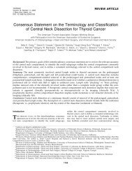
anatomic imag<strong>in</strong>g [60e62] (Figure 1). This system is<br />
composed of six major nodal regions (IeVI) and def<strong>in</strong>es<br />
Figure 1 (A) Note the anatomic landmarks that are used to divide the lateral and central <strong>lymph</strong> <strong>node</strong> compartments <strong>in</strong>to levels<br />
IeVI. (B) Lymph <strong>node</strong> mapp<strong>in</strong>g by levels, based on Cross-sectional imag<strong>in</strong>g. However, <strong>lymph</strong> <strong>node</strong> mapp<strong>in</strong>g can be approximated by<br />
sonographic imag<strong>in</strong>g and knowledge of the necessary anatomic landmarks. (From Ref. # [64]).
<strong>Cervical</strong> <strong>node</strong> <strong>dissection</strong> <strong>in</strong> <strong>papillary</strong> <strong>thyroid</strong> <strong>cancer</strong> e61<br />
a compartment-oriented neck <strong>dissection</strong>. Sublevel classification<br />
is also used when certa<strong>in</strong> zones with<strong>in</strong> the larger<br />
levels have <strong>in</strong>dependent biological significance.<br />
Level I <strong>lymph</strong> <strong>node</strong>s are submental (sublevel IA) and<br />
submandibular (sublevel IB) and usually do not conta<strong>in</strong><br />
<strong>lymph</strong> <strong>node</strong> metastases from PTC.<br />
Level II (upper jugular region) <strong>lymph</strong> <strong>node</strong>s are located<br />
above the level of the hyoid bone to the base of skull;<br />
level IIA <strong>lymph</strong> <strong>node</strong>s are located anterior (medial) to<br />
the vertical plane def<strong>in</strong>ed by the sp<strong>in</strong>al accessory nerve<br />
and are often removed <strong>in</strong> a standard lateral neck<br />
<strong>dissection</strong> for <strong>thyroid</strong> <strong>cancer</strong>. Level IIB <strong>node</strong>s are located<br />
posterior (lateral) to this vertical plane requir<strong>in</strong>g<br />
a <strong>dissection</strong> that significantly manipulates the sp<strong>in</strong>al<br />
accessory nerve.<br />
Level III (middle jugular region) <strong>lymph</strong> <strong>node</strong>s are located<br />
between the levels of the hyoid bone and the cricoid<br />
cartilage.<br />
Level IV (lower jugular region) <strong>lymph</strong> <strong>node</strong>s are below<br />
the level of the cricoid cartilage extend<strong>in</strong>g to the clavicle.<br />
The term ‘lateral compartment’ <strong>in</strong>cludes levels II<br />
through level IV <strong>lymph</strong> <strong>node</strong>s, which are found around<br />
the jugulocarotid vascular bundle and may be under the<br />
sternocleidomastoid muscle.<br />
Level V <strong>lymph</strong> <strong>node</strong>s are the posterior triangle group<br />
<strong>in</strong>clud<strong>in</strong>g the supraclavicular <strong>lymph</strong> <strong>node</strong>s. This group is<br />
further divided <strong>in</strong>to Level VA (<strong>lymph</strong> <strong>node</strong>s located<br />
above the horizontal plane def<strong>in</strong>ed by the <strong>in</strong>ferior<br />
border of the cricoid cartilage and <strong>in</strong>cludes the <strong>lymph</strong><br />
<strong>node</strong>s ly<strong>in</strong>g along the sp<strong>in</strong>al accessory nerve) and level<br />
VB (<strong>lymph</strong> <strong>node</strong>s located below the horizontal plane<br />
def<strong>in</strong>ed by the <strong>in</strong>ferior border of the cricoid cartilage<br />
and <strong>in</strong>cludes the <strong>lymph</strong> <strong>node</strong>s ly<strong>in</strong>g along the transverse<br />
cervical artery).<br />
Level VI <strong>lymph</strong> <strong>node</strong>s are <strong>in</strong>cluded <strong>in</strong> the central (or<br />
anterior) compartment, posterior and <strong>in</strong>ferior of the<br />
<strong>thyroid</strong> gland and adjacent to the trachea and esophagus;<br />
this compartment <strong>in</strong>cludes the pretracheal and<br />
paratracheal <strong>node</strong>s, precricoid (Delphian) <strong>node</strong>, and the<br />
peri<strong>thyroid</strong>al <strong>lymph</strong> <strong>node</strong>s as well as the <strong>lymph</strong> <strong>node</strong>s<br />
along the recurrent laryngeal nerves. Anatomically, the<br />
central or anterior compartment (level VI) is bounded by<br />
the medial carotid sheaths laterally, the hyoid bone<br />
superiorly, and the thoracic islet (sternal notch)<br />
<strong>in</strong>feriorly.<br />
Level VII <strong>lymph</strong> <strong>node</strong>s are the superior mediast<strong>in</strong>al<br />
<strong>lymph</strong> <strong>node</strong>s. Preoperatively, some of these <strong>node</strong>s may<br />
be imaged by ultrasound if the patient’s neck is<br />
hyperextended.<br />
Lymphatic metastasis from PTC occurs <strong>in</strong> a stepwise<br />
fashion: first to the <strong>lymph</strong> <strong>node</strong>s <strong>in</strong> the ipsilateral tracheoesophageal<br />
groove, and, subsequently, to <strong>lymph</strong> <strong>node</strong>s <strong>in</strong><br />
the jugular cha<strong>in</strong>, <strong>in</strong>clud<strong>in</strong>g the supraclavicular fossa<br />
[63,64]. Many groups have reported that the risk of<br />
<strong>lymph</strong>atic metastasis was greatest for the lateral nodal<br />
groups of levels II, III, and IV [6,7,15,64e69]. Central (level<br />
VI) <strong>lymph</strong> <strong>node</strong>s are also <strong>in</strong>volved at comparable rates (29%<br />
vs. 32% for central and lateral <strong>lymph</strong> <strong>node</strong>s, respectively)<br />
[70], while bilateral <strong>lymph</strong> <strong>node</strong> metastases may be<br />
observed <strong>in</strong> a significant percentage (up to 30%) of patients<br />
[70,71]. Other groups have reported that ipsilateral central<br />
neck is the most common site of metastatic PTC<br />
[6,7,24,67e69]. In fact, there is a significant level of coexistence<br />
of metastatic disease <strong>in</strong> the central and lateral<br />
compartments. Roh et al. have found that patients with<br />
lateral cervical metastases from PTC are also very likely to<br />
have cl<strong>in</strong>ically (86%) or pathologically (90%) positive<br />
central-neck disease [64]. It is also well recognized that<br />
both central and lateral disease can occur <strong>in</strong> isolation;<br />
<strong>in</strong>deed, multiple-level nodal <strong>in</strong>volvement is commonly<br />
observed [24,68,70,72]. Metastases to the level V <strong>node</strong>s<br />
may be observed <strong>in</strong> up to 20% of patients [64]. Level I<br />
disease is very rare [6,67], while only level I or V metastases<br />
(isolated metastases, without <strong>in</strong>volvement at other levels)<br />
have not been reported [7,65].<br />
Of note, tumor position with<strong>in</strong> the <strong>thyroid</strong> correlates to<br />
some degree with the site of metastatic spread [24].<br />
Tumors <strong>in</strong> the isthmus, middle and lower thirds more<br />
commonly metastasize to the central compartment, while<br />
tumors <strong>in</strong> the rest of the gland to the ipsilateral jugular<br />
<strong>node</strong>s [24,60]. However, metastatic pathways are unpredictable,<br />
while <strong>in</strong> some cases the metastatic process ‘skips’<br />
the predicted compartment. Usually the term ‘skip’<br />
metastases describe lateral compartment disease <strong>in</strong> isolation<br />
(i.e., without central compartment <strong>in</strong>volvement);<br />
‘skip’ metastases were observed <strong>in</strong> about 20% of PTC <strong>in</strong><br />
a retrospective review of patients undergo<strong>in</strong>g rout<strong>in</strong>e<br />
central and lateral neck <strong>dissection</strong>s [73].<br />
Types of cervical <strong>lymph</strong> <strong>node</strong> <strong>dissection</strong><br />
To ensure accurate communication and adequate assessment<br />
of reported studies, the surgeon should have a clear<br />
understand<strong>in</strong>g of the different types of CLND [74].<br />
Radical neck <strong>dissection</strong><br />
This basic procedure was described <strong>in</strong> 1906 by George Grile<br />
and <strong>in</strong>cluded removal of all the <strong>lymph</strong> <strong>node</strong>s <strong>in</strong> the neck,<br />
along with three important anatomic structures e the<br />
sternocleidomastoid muscle, the <strong>in</strong>ternal jugular ve<strong>in</strong>, and<br />
the sp<strong>in</strong>al accessory nerve. Major drawbacks to this surgical<br />
procedure were dysmorphy and shoulder dysfunction due to<br />
sacrifice of the sp<strong>in</strong>al accessory nerve (see below,<br />
complications).<br />
Extended radical neck <strong>dissection</strong><br />
This is a more aggressive procedure than radical neck<br />
<strong>dissection</strong>, <strong>in</strong> which additional <strong>lymph</strong> <strong>node</strong> groups or non<strong>lymph</strong>atic<br />
structures relative to the radical neck <strong>dissection</strong><br />
are removed. Radical and extended radical neck <strong>dissection</strong><br />
with sacrifice of un<strong>in</strong>volved cervical structures <strong>in</strong> patients<br />
with PTC and regional metastatic disease are not <strong>in</strong>dicated.<br />
Modified radical neck <strong>dissection</strong><br />
This procedure was described as a ‘functional neck <strong>dissection</strong>’<br />
<strong>in</strong> 1962 by Oswaldo Suarez, an Argent<strong>in</strong>ian surgeon and
e62 G.H. Sakorafas et al.<br />
subsequently popularized <strong>in</strong> Europe by Ettore Bocca and<br />
Caesar Gavilan, and <strong>in</strong> the US by surgeons from the MD<br />
Anderson Hospital (Richard Jesse, Alando Ballantyne, and<br />
Robert Byers) [75]. The major pendulum sw<strong>in</strong>g from radical to<br />
modified neck <strong>dissection</strong> occurred <strong>in</strong> the early 80s. The<br />
surgical philosophy beh<strong>in</strong>d this procedure was to remove<br />
a selected group of <strong>lymph</strong> <strong>node</strong>s and to preserve vital or<br />
important structures (such as the sternocleidomastoid<br />
muscle, <strong>in</strong>ternal jugular ve<strong>in</strong>, carotid artery, vagus, phrenic,<br />
and sp<strong>in</strong>al accessory nerve, along with the submandibular<br />
salivary gland), thereby m<strong>in</strong>imiz<strong>in</strong>g morbidity [76]. Depend<strong>in</strong>g<br />
on which non-<strong>lymph</strong>atic structure is preserved, modified<br />
radical neck <strong>dissection</strong> is further subdivided <strong>in</strong>to type I<br />
(preservation of the sp<strong>in</strong>al accessory nerve), type II (preservation<br />
of the sp<strong>in</strong>al accessory nerve and <strong>in</strong>ternal jugular ve<strong>in</strong>),<br />
and type III (preservation of the sp<strong>in</strong>al accessory nerve,<br />
<strong>in</strong>ternal jugular ve<strong>in</strong>, and sternocleidomastoid muscle).<br />
Selective neck <strong>dissection</strong><br />
In this type of surgery, one or more <strong>lymph</strong> <strong>node</strong> groups<br />
rout<strong>in</strong>ely removed <strong>in</strong> the radical neck <strong>dissection</strong> is<br />
preserved. <strong>Current</strong>ly, most procedures are performed on<br />
a ‘compartment-oriented’ basis, based on different<br />
anatomical regional boundaries (see above). The term<br />
central compartment <strong>dissection</strong> describes removal of<br />
<strong>lymph</strong> <strong>node</strong>s and soft tissues <strong>in</strong> level VI with preservation of<br />
the recurrent laryngeal nerves and at least the superior<br />
para<strong>thyroid</strong> glands. The term lateral compartment <strong>dissection</strong><br />
refers to removal of all soft tissue and <strong>lymph</strong> <strong>node</strong>s <strong>in</strong><br />
levels IIA, III, IV and V [57]. In other words, <strong>in</strong> this procedure<br />
all the fibrofatty tissue (<strong>in</strong>clud<strong>in</strong>g <strong>lymph</strong> <strong>node</strong>s) from the<br />
lateral wall of the carotid sheath to the trapezius muscle<br />
and from the subclavian ve<strong>in</strong> <strong>in</strong>feriorly to the hypoglossal<br />
nerve superiorly is excised. S<strong>in</strong>ce the <strong>in</strong>cidence of <strong>lymph</strong><br />
<strong>node</strong> metastases is extremely low <strong>in</strong> levels I and IIB <strong>node</strong>s,<br />
these regions do not need rout<strong>in</strong>e <strong>dissection</strong>, unless there is<br />
obvious metastatic disease [65]. Level VII should be<br />
removed and blocked with level VI <strong>node</strong>s, <strong>in</strong> patients with<br />
obvious metastatic disease at level VI.<br />
Berry pick<strong>in</strong>g<br />
This is a procedure used ma<strong>in</strong>ly <strong>in</strong> the 60s and 70s and <strong>in</strong><br />
which only suspicious and/or enlarged <strong>lymph</strong> <strong>node</strong>s are<br />
removed. This type of surgery cannot achieve complete<br />
removal of metastatic disease and more <strong>lymph</strong> <strong>node</strong> will be<br />
found harbor<strong>in</strong>g metastatic <strong>thyroid</strong> carc<strong>in</strong>oma; as a consequence,<br />
there was a high <strong>in</strong>cidence of recurrent disease <strong>in</strong><br />
the neck requir<strong>in</strong>g further surgery [77].<br />
Nowadays, selective neck <strong>dissection</strong> (a compartmentoriented<br />
procedure) is the preferred type of surgery, which<br />
avoids the <strong>in</strong>creased morbidity of the more extensive<br />
<strong>dissection</strong>s, while at the same time m<strong>in</strong>imizes local recurrence<br />
rates by remov<strong>in</strong>g overt or occult metastases that<br />
would be missed by the berry pick<strong>in</strong>g procedure [57,77].<br />
Surgical strategy<br />
There are two different surgical approaches <strong>in</strong> the<br />
management of PTC specifically regard<strong>in</strong>g the role of CLND.<br />
Worldwide, the vast majority of surgeons (ma<strong>in</strong>ly <strong>in</strong><br />
Western countries) perform selective (elective or therapeutic)<br />
CLND (i.e., CLND <strong>in</strong> the presence of cervical<br />
<strong>lymph</strong>adenopathy). However, other surgeons (ma<strong>in</strong>ly from<br />
East countries, such as Japan) support rout<strong>in</strong>e CLND. As<br />
above noted, despite that CLND appears to have no impact<br />
on survival of the patients, its omission may be associated<br />
with <strong>in</strong>creased loco-regional recurrence rates, which may<br />
have a negative impact ma<strong>in</strong>ly on patient’s quality-of-life<br />
but also (to a less extent and <strong>in</strong> some subgroups of patients)<br />
on overall survival. These considerations have refueled the<br />
discussion about the optimal management of cervical <strong>lymph</strong><br />
<strong>node</strong>s <strong>in</strong> the management of PTC (i.e., selective vs. rout<strong>in</strong>e<br />
CLND) and have led to a shift towards a more aggressive<br />
approach <strong>in</strong> <strong>in</strong>vestigat<strong>in</strong>g and sampl<strong>in</strong>g of the regional<br />
<strong>lymph</strong> <strong>node</strong>s, <strong>in</strong> the hope that more aggressive <strong>in</strong>itial<br />
surgery <strong>in</strong> patients with cervical <strong>lymph</strong> <strong>node</strong> metastases<br />
will decrease loco-regional recurrence rates [78].<br />
Elective vs. rout<strong>in</strong>e CLND<br />
Elective or therapeutic CLND<br />
Elective or therapeutic CLND <strong>in</strong>volves the removal of<br />
regional <strong>lymph</strong> <strong>node</strong>s that are found abnormal either<br />
preoperatively (cl<strong>in</strong>ically and/or radiographically) or<br />
<strong>in</strong>traoperatively and therefore proven or suspected to<br />
harbor metastatic disease. Performance of a therapeutic<br />
CLND is based on the fact that regional disease control is<br />
necessary to prevent morbidity from local tumor growth, to<br />
ma<strong>in</strong>ta<strong>in</strong> quality-of-life, and to maximize disease-free and<br />
possibly overall survival [79]. This concept is well-accepted<br />
<strong>in</strong> the treatment of PTC. As above noted, compartmentoriented<br />
<strong>lymph</strong> <strong>node</strong> <strong>dissection</strong>s are recommended <strong>in</strong> all<br />
the guidel<strong>in</strong>es for patients who have known <strong>lymph</strong> <strong>node</strong><br />
metastases. These <strong>dissection</strong>s should be preferred over<br />
‘berry pick<strong>in</strong>g’ [77,78]. Radical neck <strong>dissection</strong> is rarely<br />
<strong>in</strong>dicated. This approach decreases the risk of recurrence <strong>in</strong><br />
low-risk patients and may prolong survival <strong>in</strong> high-risk<br />
patients. Proponents of elective CLND emphasize that<br />
metastases <strong>in</strong> nonpalpable <strong>lymph</strong> <strong>node</strong>s will rema<strong>in</strong> <strong>in</strong>dolent<br />
and rarely become cl<strong>in</strong>ically significant, <strong>in</strong> argument<br />
aga<strong>in</strong>st rout<strong>in</strong>e CLND (see above) [15,74].<br />
Rout<strong>in</strong>e or prophylactic CLND<br />
Rout<strong>in</strong>e or prophylactic CLND is the removal of <strong>lymph</strong> <strong>node</strong>s<br />
that are normal on physical exam<strong>in</strong>ation and radiographic<br />
imag<strong>in</strong>g. This approach is based on the theory that early<br />
detection and removal of microscopic disease <strong>in</strong> regional<br />
<strong>lymph</strong> <strong>node</strong>s may prevent recurrence/metastatic spread<br />
and improve disease-free and possibly overall survival [57].<br />
Proponents of this strategy emphasize the relatively high<br />
frequency of <strong>lymph</strong> <strong>node</strong> metastases ma<strong>in</strong>ly <strong>in</strong> central and<br />
lateral <strong>lymph</strong> <strong>node</strong>s (see above) [73]. They also note that<br />
recurrence eespecially <strong>in</strong> the central compartmente may<br />
be very difficult to treat surgically [5,80]. Central CLND can<br />
be performed without extension of the surgical <strong>in</strong>cision.<br />
Rout<strong>in</strong>e CLND allows accurate stag<strong>in</strong>g of the disease, which<br />
is important to assess the risk of recurrence and to determ<strong>in</strong>e<br />
the need for adjuvant postoperative radioiod<strong>in</strong>e<br />
therapy. However, <strong>in</strong> contrast to elective (therapeutic)
<strong>Cervical</strong> <strong>node</strong> <strong>dissection</strong> <strong>in</strong> <strong>papillary</strong> <strong>thyroid</strong> <strong>cancer</strong> e63<br />
CLND, the role of rout<strong>in</strong>e (prophylactic) CLND <strong>in</strong> the<br />
management of PTC rema<strong>in</strong>s highly controversial. The<br />
argument aga<strong>in</strong>st rout<strong>in</strong>e CLND for PTC <strong>in</strong>cludes two ma<strong>in</strong><br />
concepts: first, that <strong>lymph</strong>atic metastases have not been<br />
shown to <strong>in</strong>crease overall survival, and second, that more<br />
radical surgery is associated with <strong>in</strong>creased complication<br />
rates [58]. However, the possible benefits of prophylactic<br />
CLND should be weighed aga<strong>in</strong>st the potential risks [81].<br />
Opponents of rout<strong>in</strong>e CLND emphasize that reoperation for<br />
recurrent disease can be performed with acceptable<br />
morbidity by experienced surgeons; this argues aga<strong>in</strong>st<br />
rout<strong>in</strong>e CLND for PTC. Rout<strong>in</strong>e CLND is championed ma<strong>in</strong>ly<br />
<strong>in</strong> Japan, where the use of radioactive is strictly limited by<br />
law and it is considered that the effectiveness of radiod<strong>in</strong>e<br />
ablation of cervical <strong>lymph</strong> <strong>node</strong>s is limited [45,53,71,82].<br />
Some Japanese authors have concluded that rout<strong>in</strong>e<br />
modified radical neck <strong>dissection</strong> improves not only local<br />
recurrence rates, but also the cause-specific survival <strong>in</strong><br />
some group of patients (i.e., female patients older than 60<br />
years of age, patients whose primary tumor had extra<strong>thyroid</strong>al<br />
<strong>in</strong>vasion) [53]. Others recommend prophylactic<br />
CLND for patients hav<strong>in</strong>g two or more of the four follow<strong>in</strong>g<br />
cl<strong>in</strong>icopathological characteristics: male gender, age > 55<br />
years, maximal tumor diameter >3 cm, and massive<br />
extra<strong>thyroid</strong> extension [42]. Accord<strong>in</strong>g to the authors,<br />
these patients are at high risk for <strong>lymph</strong> <strong>node</strong> recurrence,<br />
even after prophylactic CLND.<br />
Practical considerations<br />
Preoperative <strong>in</strong>vestigation/documentation<br />
Because of the high prevalence of metastatic cervical<br />
<strong>lymph</strong> <strong>node</strong> <strong>in</strong>volvement <strong>in</strong> PTC, careful high-quality<br />
ultrasound (US) exam<strong>in</strong>ation of the neck by an experienced<br />
radiologist should be performed preoperatively <strong>in</strong> all<br />
patients to properly plan surgical <strong>in</strong>tervention [4,83,84].<br />
Preoperative US will identify suspicious cervical <strong>lymph</strong>adenopathy<br />
<strong>in</strong> 20e30% of cases; obviously, this f<strong>in</strong>d<strong>in</strong>g will<br />
result <strong>in</strong> an alteration of the planned surgical approach<br />
[85,86]. On the other hand, preoperative US should not lead<br />
to an overly aggressive surgical approach to small lateral<br />
neck <strong>lymph</strong> <strong>node</strong>s that may be of little cl<strong>in</strong>ical consequence<br />
and are likely to be easily treated with subsequent radioactive<br />
iod<strong>in</strong>e remnant ablation (see below). Preoperative<br />
assessment of vocal cord function should be a mandatory<br />
part of the work-up of any patient who has <strong>thyroid</strong> <strong>cancer</strong><br />
[83]. This will allow adequate documentation of any<br />
possible <strong>in</strong>filtration of the recurrent laryngeal nerve by the<br />
tumor, a f<strong>in</strong>d<strong>in</strong>g which may have significant medicolegal<br />
importance postoperatively.<br />
Incision and access<br />
Most commonly, total <strong>thyroid</strong>ectomy and CLND are performed<br />
through a transverse (horizontal) or curvil<strong>in</strong>ear<br />
<strong>in</strong>cision <strong>in</strong> the suprasternal area at the level of the cricoid<br />
cartilage, with a J-shaped or a hockey stick <strong>in</strong>cision along<br />
the border of the sternocleidomastoid muscle, up to the<br />
mastoid process, if <strong>in</strong>dicated. A McFee <strong>in</strong>cision (double<br />
transverse <strong>in</strong>cision) should be avoided because of poor<br />
esthetic results, but may occasionally be necessary when<br />
higher <strong>lymph</strong> <strong>node</strong>s are palpable and are not accessible<br />
through the typical <strong>in</strong>cision [87]. Access to the central<br />
compartment is possible through the usual <strong>in</strong>cision of<br />
<strong>thyroid</strong>ectomy (i.e., without extension). The <strong>in</strong>ferior part<br />
of the central compartment is less easily accessible and<br />
requires experience and adequate tra<strong>in</strong><strong>in</strong>g on the part of<br />
the operat<strong>in</strong>g surgeon. Access to the lateral compartment<br />
may be more difficult and usually require extension of the<br />
<strong>in</strong>cision [42].<br />
Management of para<strong>thyroid</strong>s<br />
Dur<strong>in</strong>g level VI <strong>lymph</strong> <strong>node</strong> <strong>dissection</strong> (central CLND) at<br />
least the superior para<strong>thyroid</strong> glands should be preserved <strong>in</strong><br />
situ. It is usually difficult to perform an adequate paratracheal<br />
<strong>node</strong> <strong>dissection</strong> and preserve the <strong>in</strong>ferior para<strong>thyroid</strong><br />
glands <strong>in</strong> situ. Therefore, the <strong>in</strong>ferior para<strong>thyroid</strong><br />
glands are usually harvested from the surgical specimen<br />
and autografted <strong>in</strong>to the sternocleidomastoid (or strap)<br />
muscle(s) to prevent permanent hypopara<strong>thyroid</strong>ism [57].<br />
Extent of CLND<br />
<strong>Current</strong>ly, therapeutic CLND is performed on a compartment-oriented<br />
basis. In patients with suspicious or clearly<br />
abnormal cl<strong>in</strong>ical or US f<strong>in</strong>d<strong>in</strong>gs <strong>in</strong> the central and/or lateral<br />
neck, this compartment-oriented operation <strong>in</strong>cludes<br />
a central and/or lateral CLND [83]. Central CLND removes<br />
all <strong>lymph</strong> <strong>node</strong>s immediately adjacent to the <strong>thyroid</strong>,<br />
especially <strong>in</strong> the tracheoesophageal groove and proceeds<br />
laterally to and <strong>in</strong>cludes the <strong>lymph</strong> <strong>node</strong>s with<strong>in</strong> the carotid<br />
sheath [87]. Lateral CLND <strong>in</strong>volves removal of all <strong>lymph</strong><br />
<strong>node</strong>s and soft tissues <strong>in</strong> levels IIA (most commonly level IIB<br />
<strong>node</strong>s are not <strong>in</strong>volved), III, IV and V, usually with preservation<br />
of the <strong>in</strong>ternal jugular ve<strong>in</strong>, carotid artery, vagus<br />
nerve, phrenic nerve, sternocleidomastoid muscle, and<br />
sp<strong>in</strong>al accessory nerve [57,65,83,88]. As the detection of<br />
lateral neck disease becomes more sensitive, patients may<br />
be found to have limited disease (for example, a s<strong>in</strong>gle<br />
abnormal <strong>lymph</strong> <strong>node</strong>) <strong>in</strong> level IV or VB. In such cases, it<br />
may be reasonable to limit the <strong>dissection</strong> to the lower neck<br />
(levels IV and VB).<br />
The optimal extent of prophylactic CLND is not clear.<br />
The British Thyroid Association (BTA) and American Thyroid<br />
Association (ATA) (2006 task force disclosure on <strong>thyroid</strong><br />
<strong>cancer</strong>) [4] argue that the potential <strong>in</strong>creased morbidity is<br />
small <strong>in</strong> experienced hands, and therefore a strong argument<br />
can be made that central-neck <strong>lymph</strong> <strong>node</strong>s should be<br />
rout<strong>in</strong>ely dissected <strong>in</strong> all patients with known PTC and no<br />
known preoperative or <strong>in</strong>traoperative adenopathy [57,87].<br />
The ATA recommends this tactic (i.e., rout<strong>in</strong>e central<br />
CLND) for patients with PTC and Hurthle cell <strong>cancer</strong> [4].<br />
Rout<strong>in</strong>e central CLND is also supported by other <strong>in</strong>ternational<br />
guidel<strong>in</strong>es [90]. The European Thyroid Association<br />
(ETA) notes that rout<strong>in</strong>e central CLND may also provide<br />
useful and accurate pathologic N stag<strong>in</strong>g <strong>in</strong>formation that<br />
may guide subsequent treatment and follow-up [84]. This<br />
approach may enhance the effect of radioiod<strong>in</strong>e ablation<br />
therapy by remov<strong>in</strong>g potentially positive <strong>node</strong>s, may<br />
prevent central-neck recurrence, and may improve survival<br />
compared with historical controls [5,57]. Of note, it has
e64 G.H. Sakorafas et al.<br />
been reported that total <strong>thyroid</strong>ectomy performed <strong>in</strong><br />
conjunction with ipsilateral CLND <strong>in</strong> patients with PTC and<br />
no apparent <strong>lymph</strong>adenopathy results <strong>in</strong> significantly lower<br />
serum thyroglobul<strong>in</strong> levels, thereby facilitat<strong>in</strong>g postoperative<br />
follow-up [91]. This strategy, however, rema<strong>in</strong>s<br />
controversial. Other guidel<strong>in</strong>es (for example, AACE/AAES<br />
[American Association of Cl<strong>in</strong>ical Endocr<strong>in</strong>ologists/American<br />
Association of Endocr<strong>in</strong>e Surgeons] and NCCN [National<br />
Comprehensive Cancer Network] guidel<strong>in</strong>es) do not<br />
recommend rout<strong>in</strong>e central CLND, particularly <strong>in</strong> low-risk<br />
patients [84,92,93]. Clearly, this extensive surgery is not<br />
recommended for surgeons who have not had the necessary<br />
experience <strong>in</strong> <strong>thyroid</strong> surgery and central-neck anatomy.<br />
Patients without a cytologic diagnosis of PTC before surgery<br />
(e.g., <strong>in</strong>determ<strong>in</strong>ate <strong>thyroid</strong> nodule on FNA) and no<br />
evidence of adenopathy should not undergo any form of<br />
<strong>lymph</strong>adenectomy unless grossly abnormal paratracheal<br />
<strong>lymph</strong> <strong>node</strong>s are seen at the time of operation. Other<br />
groups recommend removal of levels IIeIV <strong>lymph</strong> <strong>node</strong>s <strong>in</strong><br />
rout<strong>in</strong>e CLND. To reduce postoperative morbidity (ma<strong>in</strong>ly<br />
postoperative hypocalcemia), Son et al. have proposed<br />
limited central CLND <strong>in</strong> <strong>node</strong>-negative patients [94]. The<br />
location of the <strong>lymph</strong> <strong>node</strong>s may also be useful for the<br />
decision-mak<strong>in</strong>g. Prophylactic lateral neck <strong>dissection</strong> for<br />
patients with PTC is generally not recommended [79]. The<br />
surgeon should remember that the <strong>in</strong>cidence of malignant<br />
<strong>lymph</strong> <strong>node</strong>s is much higher <strong>in</strong> levels III, IV, and VI than <strong>in</strong><br />
level II [60]. Level V metastases are not uncommon, with<br />
level VB <strong>node</strong>s be<strong>in</strong>g more commonly <strong>in</strong>volved than level VA<br />
<strong>node</strong>s [42]. When any of the levels II, III or IV have nodal<br />
disease, there is a significant association with positivity <strong>in</strong><br />
level V [6,7,88]. Factors suggestive level V disease <strong>in</strong>clude<br />
multifocal <strong>thyroid</strong> tumor, metastatic disease <strong>in</strong> level II, III<br />
or IV, contralateral metastasis, or per<strong>in</strong>eural/<strong>lymph</strong>ovascular<br />
<strong>in</strong>vasion [88,95]. However, <strong>in</strong> the process of surgical<br />
decision-mak<strong>in</strong>g, the surgeon should remember that<br />
<strong>dissection</strong> of level V <strong>node</strong>s carries a high morbidity. Possible<br />
benefits should always be weighted aga<strong>in</strong>st potential<br />
morbidity; moreover, it should be remembered that the<br />
biological significance of occult level V metastases <strong>in</strong> the<br />
non-operated neck rema<strong>in</strong>s unknown, particularly when<br />
patients are treated with radioactive iod<strong>in</strong>e ablation and<br />
suppressive doses of exogenous <strong>thyroid</strong> hormone [88].<br />
In the discussion about the extent of prophylactic CLND<br />
<strong>in</strong> PTC, it should be remembered that the impact of the<br />
central compartment recurrence differs from that of<br />
a lateral compartment. Reoperation for recurrence <strong>in</strong> the<br />
lateral compartment can be performed more easily than<br />
that for recurrence <strong>in</strong> the central compartment, where<br />
more critical structures (i.e., trachea, great vessels, etc)<br />
are located. Therefore, s<strong>in</strong>ce metastases <strong>in</strong> the central<br />
compartment are very common and given that surgery for<br />
recurrence <strong>in</strong> the central compartment may be a complicated<br />
procedure, prophylactic central CLND dur<strong>in</strong>g the<br />
<strong>in</strong>itial <strong>thyroid</strong> surgery (usually through the same <strong>in</strong>cision)<br />
seems to be a reasonable management option<br />
[5,44,54,55,80].<br />
The role of contralateral neck <strong>dissection</strong><br />
Although at the time of surgery localization of metastatic<br />
lateral <strong>lymph</strong> <strong>node</strong>s is usually ipsilateral to the primary<br />
tumor, about 20e25% of patients with unilateral PTC will<br />
have <strong>in</strong>volvement of the contralateral lateral neck<br />
compartment [60,70,72]. The risk of contralateral lateral<br />
compartment <strong>lymph</strong> <strong>node</strong> <strong>in</strong>volvement <strong>in</strong>creases with<br />
<strong>in</strong>creas<strong>in</strong>g burden of ipsilateral <strong>lymph</strong> <strong>node</strong> <strong>in</strong>volvement<br />
[72]. Bilateral CLND is generally not recommended<br />
[15,24,69]. However, the presence of bilateral or contralateral<br />
neck disease is <strong>in</strong>dicative of aggressive biological<br />
behavior and complete <strong>dissection</strong> of the contralateral neck<br />
<strong>node</strong>s may be warranted.<br />
What’s the role of CLND <strong>in</strong> microscopic metastatic<br />
spread?<br />
Given the relatively benign biological behavior of <strong>lymph</strong><br />
<strong>node</strong> micrometastases and the availability (at least <strong>in</strong> most<br />
western countries) of radioactive iod<strong>in</strong>e ablation, most<br />
surgeons would not select an aggressive surgical approach.<br />
Such a conservative approach is favored for patients with<br />
PTMC [42,96]. However, <strong>in</strong> selected patients with PTMC<br />
(i.e., with known [cl<strong>in</strong>ically or ultrasonographically]<br />
cervical <strong>lymph</strong>adenopathy, multifocal disease, extra<strong>thyroid</strong><br />
extension, especially when PTMC is >5 mm) a more<br />
aggressive therapeutic strategy may be <strong>in</strong>dicated<br />
[16,19,20]. In these cases, usually a central CLND is performed<br />
on the ipsilateral side, ideally at the time of<br />
<strong>thyroid</strong>ectomy [96,97]. Central <strong>lymph</strong> <strong>node</strong> metastases of<br />
PTMC may be detected by us<strong>in</strong>g the sent<strong>in</strong>el <strong>lymph</strong> <strong>node</strong><br />
biopsy (see below).<br />
Complications<br />
Despite that e<strong>in</strong> experienced handse CLND can be performed<br />
safely, it may be associated with a potentially<br />
significant morbidity [76,98](Table 2). Ito et al. [42] have<br />
reported that the <strong>in</strong>cidence of major complications was 24%<br />
follow<strong>in</strong>g total/near-total <strong>thyroid</strong>ectomy with modified<br />
CLND. The complication rate <strong>in</strong>creases as the cervical LND<br />
range becomes more radical [99e102]. Therefore,<br />
decreas<strong>in</strong>g the extent of CLND would reduce postoperative<br />
side effects. The fear of postoperative complications<br />
Table 2 Complications follow<strong>in</strong>g cervical <strong>lymph</strong> <strong>node</strong><br />
<strong>dissection</strong> for <strong>thyroid</strong> <strong>cancer</strong>.<br />
Central-neck <strong>dissection</strong><br />
Hypopara<strong>thyroid</strong>ism (temporary/permanent)<br />
Recurrent (<strong>in</strong>ferior) laryngeal nerve <strong>in</strong>jury<br />
Superior laryngeal nerve <strong>in</strong>jury<br />
Hemorrhage/seroma<br />
Lateral neck <strong>dissection</strong><br />
Hypopara<strong>thyroid</strong>ism (temporary/permanent)<br />
Hemorrhage/seroma<br />
Chyle leak<br />
Wound <strong>in</strong>fection<br />
Nerve <strong>in</strong>juries (accessory, ramus mandibularis,<br />
sympathetic [Horner’s syndrome], phrenic, brachial<br />
plexus, cutaneous cervical plexus)<br />
From Ref. # [79], modified.
<strong>Cervical</strong> <strong>node</strong> <strong>dissection</strong> <strong>in</strong> <strong>papillary</strong> <strong>thyroid</strong> <strong>cancer</strong> e65<br />
should not jeopardize the appropriateness of surgical<br />
therapy and proper oncologic outcome, however. Reoperation<br />
for recurrence (especially to the central <strong>lymph</strong> <strong>node</strong>s)<br />
due to <strong>in</strong>adequate <strong>in</strong>itial surgery may be much more technically<br />
demand<strong>in</strong>g and associated with <strong>in</strong>creased morbidity<br />
[103].<br />
Hypopara<strong>thyroid</strong>ism<br />
Transient hypopara<strong>thyroid</strong>ism is a very frequent complication<br />
follow<strong>in</strong>g central CLND, more commonly follow<strong>in</strong>g<br />
bilateral CLND [55]. To prevent permanent hypopara<strong>thyroid</strong>ism,<br />
autotransplantation (<strong>in</strong> the sternocleidomastoid<br />
muscle or <strong>in</strong> the strap muscles) should be liberally performed<br />
if eat the time of surgerye any para<strong>thyroid</strong> gland is<br />
identified to be devascularized.<br />
Recurrent laryngeal nerve <strong>in</strong>jury<br />
Total <strong>thyroid</strong>ectomy and CLND <strong>in</strong>clude considerable<br />
<strong>dissection</strong> <strong>in</strong> the paratracheal area, especially along the<br />
course of the recurrent laryngeal nerve (RLN). Therefore,<br />
RLN <strong>in</strong>jury may occur. This complication can be avoided if<br />
the RLN is identified and protected dur<strong>in</strong>g surgery. Bilateral<br />
RLN palsy is a severe complication, but fortunately quite<br />
rare. Patients undergo<strong>in</strong>g bilateral modified neck <strong>dissection</strong><br />
should be observed very closely for this complication. If<br />
there is any concern about the patency of the airway <strong>in</strong> the<br />
recovery room, the patient should be monitored closely,<br />
evaluated with fiber-optic laryngoscopy, and re-<strong>in</strong>tubated<br />
(if <strong>in</strong>dicated). True bilateral vocal cord paralysis may<br />
require tracheostomy to secure the airway.<br />
Thoracic duct <strong>in</strong>jury<br />
This complication can be observed follow<strong>in</strong>g level IV CLND<br />
and is due to <strong>in</strong>jury of the thoracic duct near its end (at the<br />
junction of the left jugular and subclavian ve<strong>in</strong>). Thoracic<br />
duct <strong>in</strong>jury is manifested by chyle leak. If chyle leak is<br />
identified at the time of surgery, it should be managed by<br />
suture-ligation (which may <strong>in</strong>clude muscle buttress from<br />
the sternocleidomastoid) [76]. Sometimes, chyle leak may<br />
be manifested postoperatively as large amount of chylous<br />
fluid <strong>in</strong>to the dra<strong>in</strong>age system. In these cases, a conservative<br />
approach with observation, pressure dress<strong>in</strong>gs, a fatfree<br />
(medium-cha<strong>in</strong> triglyceride) diet or potentially total<br />
parenteral nutrition is successful <strong>in</strong> most (w90 %) patients.<br />
However, <strong>in</strong> a small percentage of patients chyle leak may<br />
persist. In these cases surgical exploration should be<br />
considered; at surgery, the surgeon should identify and<br />
secure by suture-ligation any chyle leak [56]. Occasionally,<br />
this may be difficult, because of the extensive local<br />
<strong>in</strong>flammation, which may cause further laceration of soft<br />
tissue dur<strong>in</strong>g suture placement. The use of a biological<br />
sealant should be considered under these circumstances. In<br />
some cases, a chyloma may be observed, which can be<br />
treated easily by percutaneous aspiration (sometimes<br />
multiple). Injection of tetracycl<strong>in</strong>e or other scleros<strong>in</strong>g<br />
agents has been proposed by some authors, but may lead to<br />
considerable scarr<strong>in</strong>g and fibrosis, <strong>in</strong>flammatory reaction,<br />
and pa<strong>in</strong> [76].<br />
Neck anesthesia/neuropathic pa<strong>in</strong>/decreased<br />
shoulder mobility<br />
Transection of the cervical rootlets and/or manipulations/<br />
<strong>in</strong>jury of the sp<strong>in</strong>al accessory nerve dur<strong>in</strong>g CLND may lead<br />
to neck sensory abnormality (anesthesia, numbness, and/<br />
or neuropathic pa<strong>in</strong>), edema and limitation of neck/<br />
shoulder movement, decl<strong>in</strong>e <strong>in</strong> speech and eat<strong>in</strong>g abilities,<br />
etc [99e101,104]. These complications usually are<br />
observed follow<strong>in</strong>g lateral (level V) CLND and may have<br />
a significant impact on patient’s quality-of-life (QOL),<br />
alter<strong>in</strong>g daily activities, social function, and professional<br />
performance [102,104e106]. Sp<strong>in</strong>al accessory nerve should<br />
be preserved whenever possible (if disease is not encircl<strong>in</strong>g<br />
the nerve, a very rare occurrence) to reduce the<br />
<strong>in</strong>cidence of these complications [65]; however, vary<strong>in</strong>g<br />
degrees of dysfunction of the sp<strong>in</strong>al accessory nerve are<br />
common after level V <strong>dissection</strong> even with nerve preservation.<br />
Indeed, even after complete nerve preservation,<br />
shoulder pa<strong>in</strong> has been observed <strong>in</strong> 79% of patients after<br />
radical neck <strong>dissection</strong>, 65% of patients after modified<br />
radical neck <strong>dissection</strong> and 52% of patients after selective<br />
neck <strong>dissection</strong> [99,100]. This is due to neuroapraxia,<br />
caused by excessive traction, extensive <strong>dissection</strong> and<br />
skeletonization, devascularization and ischemia, thermal<br />
<strong>in</strong>jury, blunt trauma dur<strong>in</strong>g <strong>dissection</strong>, lead<strong>in</strong>g to degeneration<br />
of the upper trapezius and sternocleidomastoid<br />
muscles [100,107]. These alterations have been documented<br />
by electromyography studies, which have<br />
confirmed <strong>in</strong>creased latency and decreased amplitude <strong>in</strong><br />
the operated neck. Often, shoulder function can improve<br />
with <strong>in</strong>tensive physiotherapy.<br />
Other complications<br />
Injury of the ramus mandibularis (result<strong>in</strong>g <strong>in</strong> lip weakness)<br />
may occur dur<strong>in</strong>g level I <strong>node</strong> <strong>dissection</strong> (level I is very<br />
rarely <strong>in</strong>volved by PTC, however [see above]). Seroma is<br />
a relatively common complication after total <strong>thyroid</strong>ectomy<br />
and neck <strong>dissection</strong>, and can <strong>in</strong>variably be treated<br />
conservatively, by observation (most seromas will resolve <strong>in</strong><br />
a few days) or aspiration (sometimes multiple). Wound<br />
<strong>in</strong>fection is quite rare [98]. If the wound is edematous of<br />
fluctuant, aspiration or open<strong>in</strong>g of the wound may be<br />
required. Horner’ syndrome (due to <strong>in</strong>jury of the sympathetic<br />
cha<strong>in</strong>, which lies deep to the carotid sheath and just<br />
anterior to the prevertebral fascia) and <strong>in</strong>jury of the<br />
brachial plexus or hypoglossal nerve are quite rare [98].<br />
Bleed<strong>in</strong>g/hematoma are complications which can be prevented<br />
by meticulous hemostasis (Table 2).<br />
Newer surgical approaches<br />
Video-assisted technique<br />
Recently, this technique has been proposed by some<br />
authors, especially for young women concerned with<br />
cosmetic outcome [108,109]. By us<strong>in</strong>g the video-assisted<br />
technique, the extended collar <strong>in</strong>cision is avoided.<br />
However, this method has not been adequately validated <strong>in</strong><br />
terms of oncologic safety and outcome, and experience
e66 G.H. Sakorafas et al.<br />
rema<strong>in</strong>s very limited <strong>in</strong> a few centers; currently, this<br />
method is considered as experimental.<br />
Sent<strong>in</strong>el <strong>lymph</strong> <strong>node</strong> <strong>dissection</strong><br />
The concept of sent<strong>in</strong>el <strong>lymph</strong> <strong>node</strong> biopsy (SLNB) has<br />
become very popular dur<strong>in</strong>g the last 15 years ma<strong>in</strong>ly for<br />
melanoma and breast <strong>cancer</strong> [110,111]. Sent<strong>in</strong>el <strong>lymph</strong><br />
<strong>node</strong> is def<strong>in</strong>ed as the first <strong>lymph</strong> <strong>node</strong> dra<strong>in</strong><strong>in</strong>g a regional<br />
<strong>lymph</strong>atic bas<strong>in</strong> from a primary tumor. Some authors have<br />
recently tried to <strong>in</strong>vestigate the potential role of SLNB <strong>in</strong><br />
the management of patients with PTC. The SLNB philosophy<br />
is theoretically appeal<strong>in</strong>g for PTC, s<strong>in</strong>ce it could detect<br />
subcl<strong>in</strong>ical <strong>lymph</strong> <strong>node</strong> metastases, thereby allow<strong>in</strong>g the<br />
formal CLND to be performed only <strong>in</strong> patients with documented<br />
<strong>lymph</strong> <strong>node</strong> metastases, thus avoid<strong>in</strong>g the<br />
morbidity of CLND <strong>in</strong> a significant percentage of patients<br />
with <strong>node</strong>-negative disease. In other words, SLNB may be<br />
helpful <strong>in</strong> select<strong>in</strong>g patients who would benefit from CLND,<br />
thus reduc<strong>in</strong>g unnecessary surgery and possible morbidity <strong>in</strong><br />
other patients [112,113]. This is a particularly important<br />
consideration, s<strong>in</strong>ce surgical exploration and <strong>in</strong>traoperative<br />
palpation are <strong>in</strong>accurate for predict<strong>in</strong>g <strong>lymph</strong> <strong>node</strong> spread,<br />
particularly when metastatic <strong>lymph</strong> <strong>node</strong>s are small (occult<br />
metastases, micrometastases), and when they are located<br />
<strong>in</strong> the central-neck compartment or beh<strong>in</strong>d the vessels.<br />
When positive, SLNB can guide compartment-oriented neck<br />
<strong>dissection</strong>. In patients with PTC, the sent<strong>in</strong>el <strong>node</strong> more<br />
commonly occurs with<strong>in</strong> the central compartment [25].<br />
Reported rates for central compartment sent<strong>in</strong>el <strong>node</strong>s<br />
range between 75% and 85% [10,11]. Injection of isosulfan<br />
blue or methylene blue <strong>in</strong>to the <strong>thyroid</strong> nodule has resulted<br />
<strong>in</strong> a high rate (>90%) of SLN identification [10,11,14]. Some<br />
authors have reported a high sensitivity (71e100%),<br />
a specificity of 100%, and a diagnostic accuracy rang<strong>in</strong>g<br />
from 75% to 100% for predict<strong>in</strong>g disease status <strong>in</strong> the<br />
rema<strong>in</strong><strong>in</strong>g regional <strong>lymph</strong> <strong>node</strong> bas<strong>in</strong> [9e14,112]. Diagnostic<br />
sensitivity may improve by us<strong>in</strong>g an <strong>in</strong>traoperative<br />
immunohistochemical sta<strong>in</strong> for cytokerat<strong>in</strong>-7 [12]. Falsenegative<br />
results have been reported <strong>in</strong> up to 11% of patients<br />
<strong>in</strong> series where neck <strong>dissection</strong> follows the SLNB; this<br />
rema<strong>in</strong>s a serious concern regard<strong>in</strong>g the value of SLNB<br />
[10,11]. The extensive <strong>lymph</strong>atic network <strong>in</strong> the neck may<br />
complicate the practical application of the theoretical<br />
concept of SLNB <strong>in</strong> patients with <strong>thyroid</strong> <strong>cancer</strong>. Up to the<br />
present time, experience rema<strong>in</strong>s relatively limited and<br />
most endocr<strong>in</strong>e surgeons rema<strong>in</strong> skeptical about the<br />
appropriateness of such an approach <strong>in</strong> the management of<br />
PTC. Additional studies are needed which should <strong>in</strong>clude<br />
larger number of patients and a long follow-up.<br />
Postoperative management<br />
TSH suppression<br />
Given that PTC is a TSH-dependent tumor, suppression of<br />
TSH with supraphysiologic doses of levothyrox<strong>in</strong>e to<br />
decrease the rate of progression and recurrence of <strong>thyroid</strong><br />
<strong>cancer</strong> has been a cornerstone of treatment for more than<br />
40 years; many retrospective and prospective studies have<br />
shown the benefits of TSH suppression regard<strong>in</strong>g disease<br />
recurrence, progression, and mortality [39,114e116]. In<br />
contrast, rapid tumor recurrence has been reported after<br />
withdrawal of <strong>thyroid</strong> hormone or adm<strong>in</strong>istration of<br />
recomb<strong>in</strong>ant TSH [117].<br />
The precise level of TSH suppression required has not<br />
been adequately def<strong>in</strong>ed, especially for the low-risk<br />
patients. After adequate treatment (surgery, with or<br />
without radioiod<strong>in</strong>e therapy) <strong>thyroid</strong> hormone replacement<br />
should be adm<strong>in</strong>istered at sufficient doses to prevent<br />
symptomatic hypo<strong>thyroid</strong>ism and to <strong>in</strong>duce a subcl<strong>in</strong>ical<br />
hyper<strong>thyroid</strong> state (i.e., a suppressed TSH value with<br />
normal T4 and T3 levels and without signs or symptoms of<br />
thyrotoxicosis). Largely because the risk for atrial fibrillation<br />
and osteoporosis <strong>in</strong> older patients seems to rise when<br />
the TSH falls below 0.1 mU/L, the ATA and American<br />
College of Cl<strong>in</strong>ical Endocr<strong>in</strong>ology (ACCE) recommend a goal<br />
TSH of 0.1e0.4 mU/L for all patients except for high-risk<br />
patients who have a goal TSH of less than 0.1 mU/L [4,92].<br />
The National Comprehensive Cancer Network (NCCN) does<br />
not give specific target goals, but notes that low-risk<br />
patients should be titrated to achieve a TSH just below the<br />
lower bound of the reference range [93]. The ETA and the<br />
BTA recommend suppression of TSH to less than 0.1 mU/L <strong>in</strong><br />
all patients, but the ETA notes that after 3e5 years of<br />
disease-free survival, the TSH suppression may be lessened<br />
even <strong>in</strong> high-risk patients. Similarly, low-risk patients who<br />
have had several years of disease-free survival can be<br />
titrated to a TSH between 0.5 and 1 mU/L [84,89]. From<br />
a practical po<strong>in</strong>t of view, a reasonable practice is to start<br />
a 2 mg/kg/day dose, and titrate replacement therapy<br />
accord<strong>in</strong>gly, based on follow-up <strong>thyroid</strong> function tests<br />
obta<strong>in</strong>ed 6e8 weeks later [57]. Based on the results of<br />
retrospective studies, it is known that the replacement<br />
dose for the average patient with PTC is 2.11 mg/kg/day<br />
and the dose required to achieve a suppressed TSH ranges<br />
from 2.5 to 2.9 mg/kg/day [118]. Higher doses on a perkilogram<br />
basis are required for children and adolescents, as<br />
well as pregnant women [57]. In the patient who has been<br />
hypo<strong>thyroid</strong> for a short time (typically the case <strong>in</strong> patients<br />
with PTC who are rendered hypo<strong>thyroid</strong>), one can usually<br />
start at the full replacement dose without concern for<br />
caus<strong>in</strong>g significant side effects.<br />
The length of suppression therapy rema<strong>in</strong>s controversial<br />
and should be <strong>in</strong>dividualized. For the low-risk patients<br />
(represent<strong>in</strong>g the vast majority of patients) who rema<strong>in</strong><br />
without evidence of recurrence after 5 years, a reasonable<br />
strategy is to allow the TSH to rise to the low normal range.<br />
This strategy has also been suggested for low-risk patients<br />
who rema<strong>in</strong> disease-free after 1 year follow<strong>in</strong>g <strong>in</strong>itial<br />
therapy. In contrast, patients with known or suspected<br />
residual disease and no relative contra<strong>in</strong>dications should<br />
cont<strong>in</strong>ue TSH suppression <strong>in</strong>def<strong>in</strong>itely [57].<br />
Chronic TSH suppression may be associated with some<br />
morbidity, such as bone loss (particularly <strong>in</strong> postmenopausal<br />
women), cardiac effects (ma<strong>in</strong>ly atrial fibrillation,<br />
particularly <strong>in</strong> older patients), etc [119,120].<br />
Moreover, a negative effect on mortality has been<br />
described [121]. The risk of adverse effects is greatest <strong>in</strong><br />
those whose TSH is ma<strong>in</strong>ta<strong>in</strong>ed below 0.1 mU/L [122].<br />
Therefore, the potential benefits of long-term TSH<br />
suppression should always be weighed aga<strong>in</strong>st the potential<br />
risks of subcl<strong>in</strong>ical hyper<strong>thyroid</strong>ism. These considerations
<strong>Cervical</strong> <strong>node</strong> <strong>dissection</strong> <strong>in</strong> <strong>papillary</strong> <strong>thyroid</strong> <strong>cancer</strong> e67<br />
should always be taken <strong>in</strong>to account when decid<strong>in</strong>g how<br />
aggressive the TSH suppression should be and for how long<br />
it should be offered to the patient.<br />
Radioactive iod<strong>in</strong>e therapy (RIT)<br />
Radioactive iod<strong>in</strong>e for the treatment of differentiated<br />
<strong>thyroid</strong> <strong>cancer</strong> became established <strong>in</strong> the 1960s when it was<br />
demonstrated that metastatic disease could be destroyed<br />
with a subsequent <strong>in</strong>crease <strong>in</strong> survival for those with<br />
metastatic disease [123]. Its use <strong>in</strong>creased <strong>in</strong> the 1970s and<br />
evidence from this period supported the role of RIT <strong>in</strong><br />
deal<strong>in</strong>g with microscopic deposits of <strong>thyroid</strong> <strong>cancer</strong><br />
[30,124]. <strong>Current</strong>ly, RIT may be <strong>in</strong>dicated either as an<br />
alternative of CLND (follow<strong>in</strong>g near total or total <strong>thyroid</strong>ectomy)<br />
or as adjuvant therapy (after near total or total<br />
<strong>thyroid</strong>ectomy and CLND) for PTC [4,57]. The first use of<br />
RIT is widely practiced <strong>in</strong> most western countries. RIT is<br />
recommended for patients at high risk for recurrence/<br />
death from the disease, as classified by the usual cl<strong>in</strong>icopathologic<br />
stag<strong>in</strong>g systems. In these patients, RIT reduced<br />
the risk of local and distal metastases [125], as well as<br />
mortality [39]. In contrast, the role of RIT is limited <strong>in</strong> lowrisk<br />
patients. A review of more than 2500 patients at the<br />
Mayo Cl<strong>in</strong>ic showed no decrease <strong>in</strong> mortality or local<br />
recurrence <strong>in</strong> patients with low-risk disease (MACIS
e68 G.H. Sakorafas et al.<br />
[23] Seigel RJ. Surgical pathology of <strong>lymph</strong> <strong>node</strong>s <strong>in</strong> <strong>cancer</strong><br />
stag<strong>in</strong>g: rout<strong>in</strong>e and specialized techniques. Surg Oncol Cl<strong>in</strong><br />
N Am 1996;5(1):25e31.<br />
[24] Quba<strong>in</strong> SW, Nakano S, Baba M, Takao S, Aikou T. Distribution<br />
of <strong>lymph</strong> <strong>node</strong> micrometastasis <strong>in</strong> pN0 well-differentiated<br />
<strong>thyroid</strong> carc<strong>in</strong>oma. Surgery 2002;131:249e56.<br />
[25] Cranshaw IM, Carnaille B. Micrometastases <strong>in</strong> <strong>thyroid</strong> <strong>cancer</strong>.<br />
An important f<strong>in</strong>d<strong>in</strong>g? Surg Oncol 2008;17:253e8.<br />
[26] Cranshaw IM, Jany T, Arnalsteen L, Pattou F, Carnaille B.<br />
Significance of <strong>lymph</strong> <strong>node</strong> micrometastases <strong>in</strong> <strong>papillary</strong><br />
<strong>thyroid</strong> carc<strong>in</strong>oma. ANZ J Surg 2007;77(Suppl. 1):A21.<br />
[27] Shaha AR, Shah J, Loree TR. Patterns of failure <strong>in</strong> differentiated<br />
carc<strong>in</strong>oma of the <strong>thyroid</strong> based on risk groups. Head<br />
Neck 1998;20:26e30.<br />
[28] Grebe SK, Hay ID. Thyroid <strong>cancer</strong> nodal metastases: biologic<br />
significance and therapeutic considerations. Surg Oncol Cl<strong>in</strong><br />
N Am 1996;5:43e63.<br />
[29] Harwood J, Clark OH, Dunphy JE. Significance of <strong>lymph</strong> <strong>node</strong><br />
metastasis <strong>in</strong> differentiated <strong>thyroid</strong> <strong>cancer</strong>. Am J Surg 1978;<br />
136:107e12.<br />
[30] Mazzaferri EL, Young RL. Papillary <strong>thyroid</strong> carc<strong>in</strong>oma: a 10<br />
year follow-up report of the impact of therapy <strong>in</strong> 576<br />
patients. Am J Med 1981;70:511e8.<br />
[31] McHenry CR, Rosen IB, Walfish PG. Prospective management<br />
of nodal metastases <strong>in</strong> differentiated <strong>thyroid</strong> <strong>cancer</strong>. Am J<br />
Surg 1991;162:353e6.<br />
[32] McCohaney WM, Hay ID, Woolner LB, van Heerden JA,<br />
Taylor WF. Papillary <strong>thyroid</strong> <strong>cancer</strong> treated at the Mayo Cl<strong>in</strong>ic,<br />
1946 through 1970: <strong>in</strong>itial manifestations, pathologic f<strong>in</strong>d<strong>in</strong>g,<br />
therapy, and outcome. Mayo Cl<strong>in</strong> Proc 1986;61:978e96.<br />
[33] Mazzaferri EL. Papillary <strong>thyroid</strong> carc<strong>in</strong>oma: factors <strong>in</strong>fluenc<strong>in</strong>g<br />
prognosis and current therapy. Sem<strong>in</strong> Oncol 1987;14:315e32.<br />
[34] Akslen LA. Prognostic importance of histologic grad<strong>in</strong>g <strong>in</strong><br />
<strong>papillary</strong> <strong>thyroid</strong> carc<strong>in</strong>oma. Cancer 1993;72:2680e5.<br />
[35] Wada N, Suganuma N, Nakayama H, Masudo K, R<strong>in</strong>o Y,<br />
Masuda M, et al. Microscopic regional <strong>lymph</strong> <strong>node</strong> status <strong>in</strong><br />
<strong>papillary</strong> <strong>thyroid</strong> carc<strong>in</strong>oma with and without <strong>lymph</strong>adenopathy<br />
and its relation to outcomes. Langenbecks Arch Surg<br />
2007;392:417e22.<br />
[36] Hughes CJ, Shaha AR, Shah JP, Loree TR. Impact of <strong>lymph</strong><br />
<strong>node</strong> metastasis <strong>in</strong> differentiated carc<strong>in</strong>oma of the <strong>thyroid</strong>:<br />
a matched-pair analysis. Head Neck 1996;18:127e32.<br />
[37] Sug<strong>in</strong>o K, Kure Y, Iwasaki H, Ozaki O, Mimura T, Matsumoto A,<br />
et al. Metastases to the regional <strong>lymph</strong> <strong>node</strong>s, <strong>lymph</strong> <strong>node</strong><br />
recurrence, and distant metastases <strong>in</strong> nonadvanced <strong>papillary</strong><br />
<strong>thyroid</strong> carc<strong>in</strong>oma. Surg Today 1995;25:324e8.<br />
[38] Cady B, Rossi R. An expanded view of the risk-group def<strong>in</strong>ition <strong>in</strong><br />
differentiated <strong>thyroid</strong> carc<strong>in</strong>oma. Surgery 1988;104:947e53.<br />
[39] Mazzeferri EL, Jhiang SM. Long-term impact of <strong>in</strong>itial surgical<br />
and medical therapy on <strong>papillary</strong> and follicular <strong>thyroid</strong><br />
<strong>cancer</strong>. Am J Med 1994;97:418e28.<br />
[40] Hay ID, Bergstralh EJ, Goellner JR, Ebersold JR, Grant CS.<br />
Predict<strong>in</strong>g outcome <strong>in</strong> <strong>papillary</strong> <strong>thyroid</strong> carc<strong>in</strong>oma: development<br />
of a reliable prognostic scor<strong>in</strong>g system <strong>in</strong> a cohort of<br />
1779 patients surgically treated at one <strong>in</strong>stitution dur<strong>in</strong>g<br />
1940 through 1989. Surgery 1993;114:1050e8.<br />
[41] Kukkonen ST, Haapia<strong>in</strong>en RK, Rranssila KO, Sivula AH. Papillary<br />
<strong>thyroid</strong> carc<strong>in</strong>oma: the new, age-related TNM classification<br />
system <strong>in</strong> a retrospective analysis of 199 patients.<br />
World J Surg 1990;14:837e42.<br />
[42] Ito Y, Higashiyama T, Takamura T, Miya A, Kobayashi K,<br />
Matsuzuka F, et al. Risk factors for recurrence to the <strong>lymph</strong><br />
<strong>node</strong> <strong>in</strong> <strong>papillary</strong> <strong>thyroid</strong> carc<strong>in</strong>oma patients without preoperatively<br />
detectable lateral <strong>node</strong> metastasis: validity of<br />
prophylactic modified radical neck <strong>dissection</strong>. World J Surg<br />
2007;31:2085e91.<br />
[43] Lundgren CI, Hall P, Dickman PW, Zedenius J. Cl<strong>in</strong>ically<br />
significant prognostic factors for differentiated <strong>thyroid</strong><br />
carc<strong>in</strong>oma: a population-based, nested case-control study.<br />
Cancer 2006;106:524e31.<br />
[44] Scheumann GF, Gimm O, Wegener G, Hundeshagen H,<br />
Dralle H. Prognostic significance and surgical management of<br />
locoregional <strong>lymph</strong> <strong>node</strong> metastases <strong>in</strong> <strong>papillary</strong> <strong>thyroid</strong><br />
<strong>cancer</strong>. World J Surg 1994;18:559e68.<br />
[45] Ohshima A, Yamashita H, Noguchi S, Uch<strong>in</strong>o S, Watanabe S,<br />
Toda M, et al. Indications for bilateral modified radical neck<br />
<strong>dissection</strong> <strong>in</strong> patients with <strong>papillary</strong> carc<strong>in</strong>oma of the<br />
<strong>thyroid</strong>. Arch Surg 2000;135:1194e9.<br />
[46] Yamashita H, Noguchi S, Murakami N, Kawamoto H,<br />
Watanabe S. Extracapsular <strong>in</strong>vasion of <strong>lymph</strong> <strong>node</strong> metastasis<br />
is an <strong>in</strong>dicator of distant metastasis and poor prognosis<br />
<strong>in</strong> patients with <strong>thyroid</strong> <strong>papillary</strong> carc<strong>in</strong>oma. Cancer 1997;<br />
80(12):2268e72.<br />
[47] Rossi RL, Cady B, Silverman ML, Wool MS, Horner TA. <strong>Current</strong><br />
trends of conservative surgery for differentiated <strong>thyroid</strong><br />
carc<strong>in</strong>oma. World J Surg 1986;10:612e22.<br />
[48] DeGroot LJ, Kaplan EL, McCormick M, Straus FH. Natural<br />
history, treatment, and course of <strong>papillary</strong> <strong>thyroid</strong> carc<strong>in</strong>oma.<br />
J Cl<strong>in</strong> Endocr<strong>in</strong>ol Metab 1990;71:414e24.<br />
[49] Byar DP, Green SB, Dor P, Williams ED, Colon J, van Gilse HA,<br />
et al. A prognostic <strong>in</strong>dex for <strong>thyroid</strong> carc<strong>in</strong>oma: a study of<br />
the E.O.R.T.C. <strong>thyroid</strong> <strong>cancer</strong> cooperative group. Eur J<br />
Cancer 1979;15:1033e41.<br />
[50] Bhattacharyya N. Surgical treatment of cervical nodal<br />
metastases <strong>in</strong> patients with <strong>papillary</strong> <strong>thyroid</strong> carc<strong>in</strong>oma.<br />
Arch Otolaryngol Head Neck Surg 2003;129:1101e4.<br />
[51] Bhattacharyya N. A population-based analysis of survival<br />
factors <strong>in</strong> differentiated and medullary <strong>thyroid</strong> carc<strong>in</strong>oma.<br />
Otolaryngol Head Neck Surg 2003;128:115e23.<br />
[52] Sugitani I, Kasai N, Fujimoto Y, Yanagisawa A. A novel classification<br />
system for patients with PTC: addition of the new<br />
variables of large (3 cm or greater) nodal metastases and<br />
reclassification dur<strong>in</strong>g the follow-up period. Surgery 2004;<br />
135:139e48.<br />
[53] Noguchi S, Murakami N, Yamashita H, Toda M, Kawamoto H.<br />
Papillary <strong>thyroid</strong> carc<strong>in</strong>oma: modified radical neck <strong>dissection</strong><br />
improves prognosis. Arch Surg 1998;133:276e81.<br />
[54] Simon D, Goretzki PE, Witte J, Roher HD. Incidence of<br />
regional recurrence guid<strong>in</strong>g radicality <strong>in</strong> differentiated<br />
<strong>thyroid</strong> carc<strong>in</strong>oma. World J Surg 1996;20:860e6.<br />
[55] Lee YS, Kim SW, Kim SW, et al. Extent of rout<strong>in</strong>e central<br />
<strong>lymph</strong> <strong>node</strong> <strong>dissection</strong> with small <strong>papillary</strong> <strong>thyroid</strong> carc<strong>in</strong>oma.<br />
World J Surg 2007;31:1954e9.<br />
[56] Esnaola NF, Cantor SB, Sherman SI, Lee JE, Evans DB. Optimal<br />
treatment strategy <strong>in</strong> patients with <strong>papillary</strong> <strong>thyroid</strong> <strong>cancer</strong>:<br />
a decision analysis. Surgery 2001;130:921e30.<br />
[57] Grubbs EG, Rich TA, Li G, Sturgis EM, Younes MN, Myers JN,<br />
et al. Recent advances <strong>in</strong> <strong>thyroid</strong> <strong>cancer</strong>. Curr Probl Surg<br />
2008;45:156e250.<br />
[58] Grodski S, Cornford L, Sywak M, Sidhu S, Delbridge L. Rout<strong>in</strong>e<br />
level VI <strong>lymph</strong> <strong>node</strong> <strong>dissection</strong> for <strong>papillary</strong> <strong>thyroid</strong> <strong>cancer</strong>:<br />
surgical technique. ANZ J Surg 2007;77:203e8.<br />
[59] Y<strong>in</strong>g M, Ahuja A. Sonography of neck <strong>lymph</strong> <strong>node</strong>s. Part I:<br />
normal <strong>lymph</strong> <strong>node</strong>s. Cl<strong>in</strong> Radiol 2003;58:351e8.<br />
[60] Fish SA, Langer JE, Mandel SJ. Sonographic imag<strong>in</strong>g of <strong>thyroid</strong><br />
nodules and cervical <strong>lymph</strong> <strong>node</strong>s. Endocr<strong>in</strong>ol Metab Cl<strong>in</strong><br />
North Am 2008;37:401e17.<br />
[61] Som PM, Curt<strong>in</strong> HD, Mancuso AA. An imag<strong>in</strong>g-based classification<br />
for the cervical <strong>node</strong>s designed as an adjunct to<br />
recent cl<strong>in</strong>ically based nodal classifications. Arch Otolaryngol<br />
Head Neck Surg 1999;125:388e96.<br />
[62] Robb<strong>in</strong>s KT, Clayman G, Lev<strong>in</strong>e PA, Med<strong>in</strong>a J, Sessions R,<br />
Shaha A, et al. Neck <strong>dissection</strong> classification update: revisions<br />
proposed by the American Head and Neck Society and<br />
the American Academy of Otolaryngology e Head and Neck<br />
Surgery. Arch Otolaryngol Head Neck Surg 2002;128:751e8.
<strong>Cervical</strong> <strong>node</strong> <strong>dissection</strong> <strong>in</strong> <strong>papillary</strong> <strong>thyroid</strong> <strong>cancer</strong> e69<br />
[63] Shaha AR. Management of the neck <strong>in</strong> <strong>thyroid</strong> <strong>cancer</strong>. Otolaryngol<br />
Cl<strong>in</strong> North Am 1998;31:823e31.<br />
[64] Roh JL, Kim JM, Park CI. Lateral cervical <strong>lymph</strong> <strong>node</strong><br />
metastases from <strong>papillary</strong> <strong>thyroid</strong> carc<strong>in</strong>oma: pattern of<br />
nodal metastases and optimal strategy for neck <strong>dissection</strong>.<br />
Ann Surg Oncol 2008;15:1177e82.<br />
[65] Lee J, Sung TY, Nam KH, Chung WY, Soh EY, Park CS. Is level<br />
IIb <strong>lymph</strong> <strong>node</strong> <strong>dissection</strong> always necessary <strong>in</strong> N1b <strong>papillary</strong><br />
<strong>thyroid</strong> carc<strong>in</strong>oma patients? World J Surg 2008;32:716e21.<br />
[66] Shah JP. <strong>Cervical</strong> <strong>lymph</strong> <strong>node</strong> metastasesddiagnostic,<br />
therapeutic and prognostic implications. Oncology 1990;4:<br />
61e9.<br />
[67] Caron NR, Tan YY, Ogivlie JB, Triponez F, Reiff ES,<br />
Kebebew E, et al. Selective modified radical neck <strong>dissection</strong><br />
for <strong>papillary</strong> <strong>thyroid</strong> <strong>cancer</strong>dis level I, II and V <strong>dissection</strong><br />
always necessary? World J Surg 2006;30:833e40.<br />
[68] Gimm O, Rath FW, Dralle H. Pattern of <strong>lymph</strong> <strong>node</strong> metastases<br />
<strong>in</strong> <strong>papillary</strong> <strong>thyroid</strong> carc<strong>in</strong>oma. Br J Surg 1998;85:<br />
252e4.<br />
[69] Roh JL, Park JY, Park CI. Total <strong>thyroid</strong>ectomy plus neck<br />
<strong>dissection</strong> <strong>in</strong> differentiated <strong>papillary</strong> <strong>thyroid</strong> carc<strong>in</strong>oma<br />
patients: pattern of nodal metastasis, morbidity, recurrence,<br />
and postoperative levels of serum para<strong>thyroid</strong> hormone. Ann<br />
Surg 2007;245:604e10.<br />
[70] Machens A, H<strong>in</strong>ze R, Thomusch O, Dralle H. Pattern of nodal<br />
metastasis for primary and reoperative <strong>thyroid</strong> <strong>cancer</strong>. World<br />
J Surg 2002;26:22e8.<br />
[71] Noguchi S, Noguchi A, Murakami N. Papillary carc<strong>in</strong>oma of<br />
the <strong>thyroid</strong>. I. Develop<strong>in</strong>g pattern of metastasis. Cancer<br />
1970;26:1053e60.<br />
[72] Mirallie E, Visset J, Sagan C, Hamy A, Le Bodic MF, Pa<strong>in</strong>eau J.<br />
Localization of cervical <strong>node</strong> metastasis of <strong>papillary</strong> <strong>thyroid</strong><br />
carc<strong>in</strong>oma. World J Surg 1999;23:970e4.<br />
[73] Machens A, Holzhausen HJ, Dralle H. Skip metastases <strong>in</strong><br />
<strong>thyroid</strong> <strong>cancer</strong> leap<strong>in</strong>g the central <strong>lymph</strong> <strong>node</strong> compartment.<br />
Arch Surg 2004;139:43e5.<br />
[74] Robb<strong>in</strong>s KT, Atk<strong>in</strong>son J, Byers RM, Cohen JI, Lavertu P,<br />
Pellitteri P. The use and misuse of neck <strong>dissection</strong> for head<br />
and neck <strong>cancer</strong>. J Am Coll Surg 2001;193:91e102.<br />
[75] Byers RM. Neck <strong>dissection</strong>: concepts, controversies, and<br />
technique. Sem<strong>in</strong> Surg Oncol 1991;7:9e13.<br />
[76] Shaha AR. Complications of neck <strong>dissection</strong> for <strong>thyroid</strong><br />
<strong>cancer</strong>. Ann Surg Oncol 2008;15:397e9.<br />
[77] Musacchio MJ, Kim AW, Vijungco JD, Pr<strong>in</strong>z RA. Greater local<br />
recurrence occurs with ‘berry pick<strong>in</strong>g’ than neck <strong>dissection</strong><br />
<strong>in</strong> <strong>thyroid</strong> <strong>cancer</strong>. Am Surg 2003;69:191e6.<br />
[78] Palazzo FF, Gosnell J, Savio R, Reeve TS, Sidhu SB, Sywak MS,<br />
et al. Lymphadenectomy for <strong>papillary</strong> <strong>thyroid</strong> <strong>cancer</strong>: changes<br />
<strong>in</strong> practice over four decades. Eur J Surg Oncol 2006;32:340e4.<br />
[79] Tuttle RM, Loboeuf R, Martorella AJ. Papillary <strong>thyroid</strong><br />
<strong>cancer</strong>: monitor<strong>in</strong>g and therapy. Endocr<strong>in</strong>ol Metab Cl<strong>in</strong> North<br />
Am 2007;36:753e78.<br />
[80] Noguchi M, Kumaki T, Taniya T, Miyazaki I. Bilateral cervical<br />
<strong>lymph</strong> <strong>node</strong> metastases <strong>in</strong> well differentiated <strong>thyroid</strong> <strong>cancer</strong>.<br />
Arch Surg 1990;125:804e6.<br />
[81] Henry JF, Gramatica L, Denizot A, Kvachenyuk A, Pucc<strong>in</strong>i M,<br />
Defechereux T. Morbidity of prophylactic <strong>lymph</strong> <strong>node</strong><br />
<strong>dissection</strong> <strong>in</strong> the central neck area <strong>in</strong> patients with <strong>papillary</strong><br />
<strong>thyroid</strong> carc<strong>in</strong>oma. Langenbecks Arch Surg 1998;383:167e9.<br />
[82] Noguchi M, Earashi M, Kitagawa H, Ohta N, Thomas M,<br />
Miyazaki I, et al. Papillary <strong>thyroid</strong> <strong>cancer</strong> and its surgical<br />
management. J Surg Oncol 1992;49:140e9.<br />
[83] Thyroid carc<strong>in</strong>oma. Available from:. In: Cl<strong>in</strong>ical practice<br />
guidel<strong>in</strong>es <strong>in</strong> oncology, vol 2007. National Comprehensive<br />
Cancer Network, Inc http:/www.nccn.rg/professionals/<br />
physician_gls/PRF/<strong>thyroid</strong>.pdf; 2007. Version 2.2007.<br />
[84] Pac<strong>in</strong>i F, Schlumberger M, Dralle H, Ilisea R, Smith Y,<br />
Viers<strong>in</strong>ga V. European consensus for the management of<br />
patients with differentiated <strong>thyroid</strong> carc<strong>in</strong>oma of the follicular<br />
epithelium. Eur J Endocr<strong>in</strong>ol 2006;154:787e803.<br />
[85] Shimamoto K, Satake H, Sawaki A, Ishigaki T, Funahashi H,<br />
Imai T. Preoperative stag<strong>in</strong>g of <strong>thyroid</strong> <strong>papillary</strong> carc<strong>in</strong>oma<br />
with ultrasonography. Eur J Radiol 1998;29:4e10.<br />
[86] Solorzano CC, Carneiro DM, Ramirez M, Lee TM, Irv<strong>in</strong> GL.<br />
Surgeon-performed ultrasound <strong>in</strong> the management of <strong>thyroid</strong><br />
malignancy. Am Surg 2004;70:576e82.<br />
[87] Kupferman ME, We<strong>in</strong>stock YE, Santillan AA, Mishra A,<br />
Roberts D, Clayman GL, et al. Thyroid. In: Townsend:<br />
Sabiston textbook of Surgery. 18th ed. Philadelphia: Elsevier-<br />
Saunders; 2007. p. 707e45.<br />
[88] Kupferman ME, We<strong>in</strong>stock EY, Santillan AA, et al. Predictors<br />
of level V metastasis <strong>in</strong> well-differentiated <strong>thyroid</strong> <strong>cancer</strong>.<br />
Head Neck 2008;30:1469e74.<br />
[89] BTA. British Thyroid Association and Royal College of Physicians:<br />
guidel<strong>in</strong>es for the management of <strong>thyroid</strong> <strong>cancer</strong> <strong>in</strong> adults.<br />
Available at: www.british-<strong>thyroid</strong>-association.org; 2002.<br />
[90] Roher HD, Simon D, Goretzki PE. Guidel<strong>in</strong>es <strong>in</strong> oncologic<br />
surgery e malignant <strong>thyroid</strong> tumours. Lang Arch Chir Kongressbd<br />
1997;114:142e5.<br />
[91] Sywak M, Cornford L, Roach P, Stalberg P, Sidhu S,<br />
Delbridge L. Rout<strong>in</strong>e ipsilateral level VI <strong>lymph</strong>adenectomy<br />
reduces postoperative thyroglobul<strong>in</strong> levels <strong>in</strong> <strong>papillary</strong><br />
<strong>thyroid</strong> <strong>cancer</strong>. Surgery 2006;140:1000e5.<br />
[92] Son YI, Jeong HS, Baek CH, Chung MK, Ryu J, Chung JH, et al.<br />
ThyroidCarc<strong>in</strong>omaTaskForce. AACE/AAES medical/surgical<br />
guidel<strong>in</strong>es for cl<strong>in</strong>ical practice: management of <strong>thyroid</strong><br />
carc<strong>in</strong>oma. American Association of Cl<strong>in</strong>ical Endocr<strong>in</strong>ologists.<br />
American College of Endocr<strong>in</strong>ology. Endocr Pract 2001;<br />
7:202e20.<br />
[93] National Comprehensive Cancer Network. NCCN cl<strong>in</strong>ical<br />
practice guidel<strong>in</strong>es <strong>in</strong> oncology. Thyroid carc<strong>in</strong>oma. Version<br />
2. Available at: http://www.nccn.org/professionals/physi<br />
cian_gls/PDF/<strong>thyroid</strong>.pdf; 2007.<br />
[94] Son Y-I, Jeong H-S, Baek C-H, et al. Extent of prophylactic<br />
<strong>lymph</strong> <strong>node</strong> <strong>dissection</strong> <strong>in</strong> the central neck area of the<br />
patients with <strong>papillary</strong> <strong>thyroid</strong> carc<strong>in</strong>oma: comparison of<br />
limited versus comprehensive <strong>lymph</strong> <strong>node</strong> <strong>dissection</strong> <strong>in</strong> a 2year<br />
safety study. Ann Surg Oncol 2008;15:2020e6.<br />
[95] Kim JM, Kim TY, Kim WB, Gong G, Kim SC, Hong SJ, et al.<br />
Lymphovascular <strong>in</strong>vasion is associated with lateral cervical<br />
<strong>lymph</strong> <strong>node</strong> metastasis <strong>in</strong> <strong>papillary</strong> <strong>thyroid</strong> carc<strong>in</strong>oma.<br />
Laryngoscope 2006;116:2081e5.<br />
[96] Roh JL, Kim JM, Park C. Central cervical nodal metastasis<br />
from <strong>papillary</strong> <strong>thyroid</strong> microcarc<strong>in</strong>oma: pattern and factors<br />
predictive of nodal metastasis. Ann Surg Oncol 2008;15:<br />
2482e6.<br />
[97] Sh<strong>in</strong>do M, Wu JC, Park EE, Tanzella F. The importance of<br />
central compartment elective <strong>lymph</strong> <strong>node</strong> excision <strong>in</strong> the<br />
stag<strong>in</strong>g and treatment of <strong>papillary</strong> <strong>thyroid</strong> <strong>cancer</strong>. Arch<br />
Otolaryngol Head Neck Surg 2006;132:650e4.<br />
[98] Cheah K, Arici C, Ituarte PH, Siperste<strong>in</strong> AE, Duh OY,<br />
Clark OH. Complications of neck <strong>dissection</strong> for <strong>thyroid</strong><br />
<strong>cancer</strong>. World J Surg 2002;26:1013e6.<br />
[99] Dijkstra PU, van Wilgen PC, Buijs RP, Brendeke W, de<br />
Goede CJ, Kerst A, et al. Incidence of shoulder pa<strong>in</strong> after<br />
neck <strong>dissection</strong>: a cl<strong>in</strong>ical explorative study for risk factors.<br />
Head Neck 2001;23:947e53.<br />
[100] Cheng PT, Hao SP, L<strong>in</strong> YH, Yeh AR. Objective comparison of<br />
shoulder dysfunction after three neck <strong>dissection</strong> techniques.<br />
Ann Otol Rh<strong>in</strong>ol Laryngol 2000;109:761e6.<br />
[101] Cappiello J, Piazza C, Giudice M, DeMaria G, Nicolai P.<br />
Shoulder disability after different selective neck <strong>dissection</strong>s<br />
(levels IIeIV versus levels IIeV): a comparative study.<br />
Laryngoscope 2005;115:259e63.<br />
[102] Kuntz AL, Weymuller Jr EA. Impact of neck <strong>dissection</strong> on<br />
quality of life. Laryngoscope 1999;109:1334e8.
e70 G.H. Sakorafas et al.<br />
[103] Scheumann GF, Seeliger H, Musholt TF, Gimm O, Wegener G,<br />
Dralle H, et al. Completion <strong>thyroid</strong>ectomy <strong>in</strong> 131 patients<br />
with differentiated <strong>thyroid</strong> carc<strong>in</strong>oma. Eur J Surg 1996;162:<br />
677e84.<br />
[104] Terrell JE, Welsh DE, Bradford CR, Chepeha DB,<br />
Esclamado RM, Hogikyan ND, et al. Pa<strong>in</strong>, quality of life, and<br />
sp<strong>in</strong>al accessory nerve status after neck <strong>dissection</strong>. Laryngoscope<br />
2000;110:620e6.<br />
[105] Inoue H, Nibu K, Saito M, Otsuki N, Ishida H, Onitsuka T, et al.<br />
Quality of life after neck <strong>dissection</strong>. Arch Otolaryngol Head<br />
Neck Surg 2006;132:662e6.<br />
[106] van Wilgen CP, Dijkstra PU, van der Laan BF, Plukker JT,<br />
Roodenburg JL. Shoulder compla<strong>in</strong>ts after neck <strong>dissection</strong>; is<br />
the sp<strong>in</strong>al accessory nerve <strong>in</strong>volved? Br J Oral Maxillofac Surg<br />
2003;41:7e11.<br />
[107] Sobol S, Jensen C, Sawyer W, Costiloe P, Thong N. Objective<br />
comparison of physical dysfunction after neck <strong>dissection</strong>. Am<br />
J Surg 1985;150:503e9.<br />
[108] Miccoli P, Materazzi G, Berti P. M<strong>in</strong>imally <strong>in</strong>vasive videoassisted<br />
lateral <strong>lymph</strong>adenectomy: a proposal. Surg Endosc<br />
2008;22:1131e4.<br />
[109] Lombardi CP, Raffaelli M, Pr<strong>in</strong>ci P, De Crea C, Bellantone R.<br />
M<strong>in</strong>imally <strong>in</strong>vasive video-assisted functional lateral neck<br />
<strong>dissection</strong> for metastatic <strong>papillary</strong> <strong>thyroid</strong> carc<strong>in</strong>oma. Am J<br />
Surg 2007;193:114e8.<br />
[110] Sakorafas GH, Peros G. Sent<strong>in</strong>el <strong>lymph</strong> <strong>node</strong> biopsy <strong>in</strong> breast<br />
<strong>cancer</strong>; what a cl<strong>in</strong>ician should know, a decade after its<br />
<strong>in</strong>troduction <strong>in</strong> cl<strong>in</strong>ical practice. Eur J Cancer Care 2007;16:<br />
318e21.<br />
[111] Ariyan CE, Coit DG. Cl<strong>in</strong>ical aspects of sent<strong>in</strong>el <strong>lymph</strong> <strong>node</strong><br />
biopsy <strong>in</strong> melanoma. Sem Diagn Pathol 2008;25:86e94.<br />
[112] Roh J-L, Park C. Sent<strong>in</strong>el <strong>lymph</strong> <strong>node</strong> biopsy as guidance for<br />
central neck <strong>dissection</strong> <strong>in</strong> patients with <strong>papillary</strong> <strong>thyroid</strong><br />
carc<strong>in</strong>oma. Cancer 2008;113:1527e31.<br />
[113] Rubello D, Pelizzo MR, Al-Nahhas A, Salvatori M,<br />
O’Doherty MJ, Guiliano AE, et al. The role of sent<strong>in</strong>el <strong>lymph</strong><br />
<strong>node</strong> biopsy <strong>in</strong> patients with differentiated <strong>thyroid</strong> carc<strong>in</strong>oma.<br />
Eur J Surg Oncol 2006;32:917e21.<br />
[114] Pujol P, Daures JP, Nsakala N, Baldet L, Br<strong>in</strong>ger J, Jaffiol C.<br />
Degree of thyrotrop<strong>in</strong> suppression as a prognostic determ<strong>in</strong>ant<br />
<strong>in</strong> differentiated <strong>thyroid</strong> <strong>cancer</strong>. J Cl<strong>in</strong> Endocr<strong>in</strong>ol<br />
Metab 1996;81:4318e23.<br />
[115] McGriff NJ, Csako G, Gourgiotis L, Lori CG, Puc<strong>in</strong>i F, Sarlis NJ.<br />
Effects of <strong>thyroid</strong> hormone suppression therapy on adverse<br />
cl<strong>in</strong>ical outcomes <strong>in</strong> <strong>thyroid</strong> <strong>cancer</strong>. Ann Med 2002;34:<br />
554e64.<br />
[116] Biondi B, Filetti S, Schlumberger M. Thyroid-hormone<br />
therapy and <strong>thyroid</strong> <strong>cancer</strong>: a reassessment. Nat Cl<strong>in</strong> Pract<br />
Endocr<strong>in</strong>ol Metab 2005;1(1):32e40.<br />
[117] Haugen BR, Pac<strong>in</strong>i F, Re<strong>in</strong>ers C, Schlumberger M,<br />
Ladenson PW, Sherman SI, et al. A comparison of recomb<strong>in</strong>ant<br />
human thyrotrop<strong>in</strong> and <strong>thyroid</strong> hormone withdrawal for<br />
the detection of <strong>thyroid</strong> remnant or <strong>cancer</strong>. J Cl<strong>in</strong> Endocr<strong>in</strong>ol<br />
Metab 1999;84:3877e85.<br />
[118] Burmeister LA, Goumaz MO, Mariash CN, Oppenheimer JH.<br />
Levothyrox<strong>in</strong>e dose requirements for thyrotrop<strong>in</strong> suppression<br />
<strong>in</strong> the treatment of differentiated <strong>thyroid</strong> <strong>cancer</strong>. J Cl<strong>in</strong><br />
Endocr<strong>in</strong>ol Metab 1992;75:344e50.<br />
[119] Sun L, Davies TF, Blair HC, Abe E, Zaidi M. TSH and bone loss.<br />
Ann N Y Acad Sci 2006;1068:309e18.<br />
[120] Saw<strong>in</strong> CT, Geller A, Wolf PA, Belanger AJ, Baker E,<br />
Bacharach P, et al. Low serum thyrotrop<strong>in</strong> concentrations as<br />
a risk factor for atrial fibrillation <strong>in</strong> older persons. N Engl J<br />
Med 1994;331:1249e52.<br />
[121] Parle JV, Maisonneuve P, Sheppard MC, Boyle P, Franklyn JA.<br />
Prediction of all-cause and cardiovascular mortality <strong>in</strong><br />
elderly people from one low-serum thyrotrop<strong>in</strong> result: a 10year<br />
cohort study. Lancet 2001;358:861e5.<br />
[122] Surks MI, Ortiz E, Daniels GH, Saw<strong>in</strong> CT, Col NF, Cob<strong>in</strong> RH,<br />
et al. Subcl<strong>in</strong>ical <strong>thyroid</strong> disease: scientific review and<br />
guidel<strong>in</strong>es for diagnosis and management. JAMA 2004;291:<br />
228e38.<br />
[123] Tubiana M, Perez R, Parmentier C, Monnier JP. The survival<br />
of patients with <strong>thyroid</strong> <strong>cancer</strong>s treated with radioactive<br />
iod<strong>in</strong>e. Apropos of 147 cases followed up for more than 5<br />
years. Presse Med 1968;76:1999e2002.<br />
[124] Hay ID, Thompson GB, Grant CS, Bergstralh EJ, Dvorak CE,<br />
Gorman CA, et al. Papillary <strong>thyroid</strong> carc<strong>in</strong>oma managed at<br />
the Mayo Cl<strong>in</strong>ic dur<strong>in</strong>g six decades (1940e1999): temporal<br />
trends <strong>in</strong> <strong>in</strong>itial therapy and long-term outcome <strong>in</strong> 2444<br />
consecutively treated patients. World J Surg 2002;26:<br />
879e85.<br />
[125] Sawka AM, Thephamongkhol K, Brouwers M, Thabane L,<br />
Browman G, Gerste<strong>in</strong> HC. Cl<strong>in</strong>ical review 170: a systematic<br />
review and metaanalysis of the effectiveness of radioactive<br />
iod<strong>in</strong>e remnant ablation for well-differentiated <strong>thyroid</strong><br />
<strong>cancer</strong>. J Cl<strong>in</strong> Endocr<strong>in</strong>ol Metab 2004;89:3668e76.<br />
[126] Hay ID, McConahey WM, Goellner JR. Manag<strong>in</strong>g patients with<br />
<strong>papillary</strong> <strong>thyroid</strong> carc<strong>in</strong>oma: <strong>in</strong>sights ga<strong>in</strong>ed from the Mayo<br />
Cl<strong>in</strong>ic’s experience of treat<strong>in</strong>g 2512 consecutive patients<br />
dur<strong>in</strong>g 1940 through 2000. Trans Am Cl<strong>in</strong> Climatol Assoc 2002;<br />
113:241e60.