Chapter 5 Maintenance of the Human Body
Chapter 5 Maintenance of the Human Body
Chapter 5 Maintenance of the Human Body

Create successful ePaper yourself
Turn your PDF publications into a flip-book with our unique Google optimized e-Paper software.
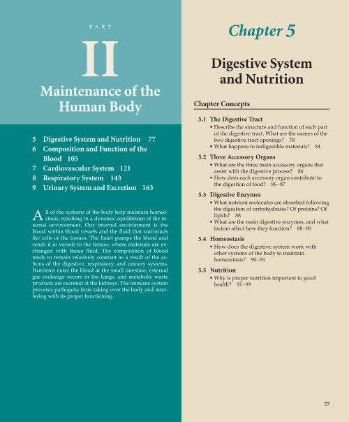
P A R T<br />
II<br />
<strong>Maintenance</strong> <strong>of</strong> <strong>the</strong><br />
<strong>Human</strong> <strong>Body</strong><br />
5 Digestive System and Nutrition 77<br />
6 Composition and Function <strong>of</strong> <strong>the</strong><br />
Blood 105<br />
7 Cardiovascular System 121<br />
8 Respiratory System 143<br />
9 Urinary System and Excretion 163<br />
A<br />
ll <strong>of</strong> <strong>the</strong> systems <strong>of</strong> <strong>the</strong> body help maintain homeostasis,<br />
resulting in a dynamic equilibrium <strong>of</strong> <strong>the</strong> internal<br />
environment. Our internal environment is <strong>the</strong><br />
blood within blood vessels and <strong>the</strong> fluid that surrounds<br />
<strong>the</strong> cells <strong>of</strong> <strong>the</strong> tissues. The heart pumps <strong>the</strong> blood and<br />
sends it in vessels to <strong>the</strong> tissues, where materials are exchanged<br />
with tissue fluid. The composition <strong>of</strong> blood<br />
tends to remain relatively constant as a result <strong>of</strong> <strong>the</strong> actions<br />
<strong>of</strong> <strong>the</strong> digestive, respiratory, and urinary systems.<br />
Nutrients enter <strong>the</strong> blood at <strong>the</strong> small intestine, external<br />
gas exchange occurs in <strong>the</strong> lungs, and metabolic waste<br />
products are excreted at <strong>the</strong> kidneys. The immune system<br />
prevents pathogens from taking over <strong>the</strong> body and interfering<br />
with its proper functioning.<br />
Digestive System<br />
and Nutrition<br />
<strong>Chapter</strong> Concepts<br />
<strong>Chapter</strong> 5<br />
5.1 The Digestive Tract<br />
• Describe <strong>the</strong> structure and function <strong>of</strong> each part<br />
<strong>of</strong> <strong>the</strong> digestive tract. What are <strong>the</strong> names <strong>of</strong> <strong>the</strong><br />
two digestive tract openings? 78<br />
•What happens to indigestible materials? 84<br />
5.2 Three Accessory Organs<br />
• What are <strong>the</strong> three main accessory organs that<br />
assist with <strong>the</strong> digestive process? 86<br />
•How does each accessory organ contribute to<br />
<strong>the</strong> digestion <strong>of</strong> food? 86–87<br />
5.3 Digestive Enzymes<br />
• What nutrient molecules are absorbed following<br />
<strong>the</strong> digestion <strong>of</strong> carbohydrates? Of proteins? Of<br />
lipids? 88<br />
• What are <strong>the</strong> main digestive enzymes, and what<br />
factors affect how <strong>the</strong>y function? 88–89<br />
5.4 Homeostasis<br />
•How does <strong>the</strong> digestive system work with<br />
o<strong>the</strong>r systems <strong>of</strong> <strong>the</strong> body to maintain<br />
homeostasis? 90–91<br />
5.5 Nutrition<br />
•Why is proper nutrition important to good<br />
health? 91–99<br />
77
78 Part II <strong>Maintenance</strong> <strong>of</strong> <strong>the</strong> <strong>Human</strong> <strong>Body</strong><br />
Enjoying <strong>the</strong> summer night at an outdoor cafe, Sam<br />
washes down his last piece <strong>of</strong> pizza with a sip <strong>of</strong> wine.<br />
Even before Sam swallows his food, <strong>the</strong> enzymes in his<br />
mouth’s saliva begin to break starch molecules apart. The<br />
wine’s alcohol is absorbed in <strong>the</strong> stomach, where <strong>the</strong><br />
process <strong>of</strong> transforming Sam’s meal into a nutrient-laden liquid<br />
begins. In <strong>the</strong> small intestine, wormlike projections from<br />
<strong>the</strong> intestinal wall absorb sugars, amino acids, and o<strong>the</strong>r<br />
needed molecules into Sam’s bloodstream. Even <strong>the</strong> large<br />
intestine contributes by taking in needed water and salts. His<br />
body now refueled, Sam heads <strong>of</strong>f for a night <strong>of</strong> dancing.<br />
In this chapter, you will learn how <strong>the</strong> body digests food,<br />
and <strong>the</strong> importance <strong>of</strong> proper nutrition. Science is beginning<br />
to find <strong>the</strong> cellular basis for believing that fruits and vegetables,<br />
and yes, especially broccoli, can ensure a brighter and<br />
healthier life. Sam—and all <strong>of</strong> us—can play a part by being<br />
aware <strong>of</strong> <strong>the</strong>se findings. Avoiding sugars and fats and consuming<br />
protein in moderate amounts can help us maintain a<br />
normal weight and avoid certain illnesses.<br />
5.1 The Digestive Tract<br />
Digestion takes place within a tube called <strong>the</strong><br />
digestive tract, which begins with <strong>the</strong> mouth<br />
and ends with <strong>the</strong> anus (Fig. 5.1). The functions<br />
<strong>of</strong> <strong>the</strong> digestive system are to ingest<br />
food, digest it to nutrients that can cross<br />
plasma membranes, absorb nutrients, and<br />
eliminate indigestible remains.<br />
The Mouth<br />
The mouth, which receives food, is bounded externally<br />
by <strong>the</strong> lips and cheeks. The lips extend<br />
from <strong>the</strong> base <strong>of</strong> <strong>the</strong> nose to <strong>the</strong> start <strong>of</strong> <strong>the</strong> chin.<br />
The red portion <strong>of</strong> <strong>the</strong> lips is poorly keratinized,<br />
and this allows blood to show through.<br />
Most people enjoy eating food, largely<br />
because <strong>the</strong>y like its texture and taste.<br />
Sensory receptors called taste buds occur primarily<br />
on <strong>the</strong> tongue. They communicate<br />
with <strong>the</strong> brain where <strong>the</strong> sensation <strong>of</strong> taste<br />
occurs. The tongue is composed <strong>of</strong> skeletal<br />
muscle whose contraction changes <strong>the</strong> shape<br />
<strong>of</strong> <strong>the</strong> tongue. Muscles exterior to <strong>the</strong> tongue<br />
cause it to move about. A fold <strong>of</strong> mucous<br />
membrane attaches <strong>the</strong> underside <strong>of</strong> <strong>the</strong><br />
tongue to <strong>the</strong> floor <strong>of</strong> <strong>the</strong> mouth.<br />
The mouth has a ro<strong>of</strong> that separates it<br />
from <strong>the</strong> nasal cavities. The ro<strong>of</strong> has<br />
two parts: an anterior (toward <strong>the</strong> front)<br />
hard palate and a posterior (toward <strong>the</strong> back)<br />
s<strong>of</strong>t palate (Fig. 5.2a). The hard palate<br />
contains several bones, while <strong>the</strong> s<strong>of</strong>t palate<br />
does not. The s<strong>of</strong>t palate ends in a fingershaped<br />
projection called <strong>the</strong> uvula.<br />
salivary glands<br />
esophagus<br />
diaphragm<br />
liver<br />
gallbladder<br />
common bile duct<br />
duodenum<br />
transverse colon<br />
ascending colon<br />
cecum<br />
appendix<br />
anus<br />
The tonsils are in <strong>the</strong> back <strong>of</strong> <strong>the</strong> mouth, on ei<strong>the</strong>r side <strong>of</strong><br />
<strong>the</strong> tongue and in <strong>the</strong> nasopharynx (where <strong>the</strong>y are called<br />
adenoids). If <strong>the</strong> tonsils become inflamed, <strong>the</strong> person has<br />
tonsillitis. If tonsillitis keeps on recurring, <strong>the</strong> tonsils may be<br />
surgically removed (called a tonsillectomy).<br />
Three pairs <strong>of</strong> salivary glands send juices (saliva) by<br />
way <strong>of</strong> ducts to <strong>the</strong> mouth. One pair <strong>of</strong> salivary glands lies at<br />
<strong>the</strong> sides <strong>of</strong> <strong>the</strong> face immediately below and in front <strong>of</strong><br />
<strong>the</strong> ears. These glands swell when a person has <strong>the</strong> mumps, a<br />
disease caused by a viral infection. Salivary glands have ducts<br />
that open on <strong>the</strong> inner surface <strong>of</strong> <strong>the</strong> cheek at <strong>the</strong> location <strong>of</strong><br />
pharynx<br />
mouth<br />
tongue<br />
stomach<br />
pancreas<br />
pancreatic duct<br />
small intestine<br />
descending colon<br />
sigmoid colon<br />
rectum<br />
anal canal<br />
Figure 5.1 Digestive system.<br />
Trace <strong>the</strong> path <strong>of</strong> food from <strong>the</strong> mouth to <strong>the</strong> anus. The large intestine consists <strong>of</strong> <strong>the</strong><br />
cecum, <strong>the</strong> colon (composed <strong>of</strong> <strong>the</strong> ascending, transverse, descending, and sigmoid<br />
colon), and <strong>the</strong> rectum and anal canal. Note also <strong>the</strong> location <strong>of</strong> <strong>the</strong> accessory organs <strong>of</strong><br />
digestion: <strong>the</strong> pancreas, <strong>the</strong> liver, and <strong>the</strong> gallbladder.
a.<br />
hard palate<br />
s<strong>of</strong>t palate<br />
uvula<br />
tonsil<br />
molars (3)<br />
premolars (2)<br />
canine (1)<br />
incisors (2)<br />
<strong>the</strong> second upper molar. Ano<strong>the</strong>r pair <strong>of</strong> salivary glands lies<br />
beneath <strong>the</strong> tongue, and still ano<strong>the</strong>r pair lies beneath <strong>the</strong><br />
floor <strong>of</strong> <strong>the</strong> mouth. The ducts from <strong>the</strong>se salivary glands<br />
open under <strong>the</strong> tongue. You can locate <strong>the</strong> openings if you<br />
use your tongue to feel for small flaps on <strong>the</strong> inside <strong>of</strong> your<br />
cheek and under your tongue. Saliva contains bicarbonate<br />
and an enzyme called salivary amylase that begins <strong>the</strong><br />
process <strong>of</strong> digesting starch.<br />
The Teeth<br />
With our teeth, we chew food into pieces convenient for<br />
swallowing. During <strong>the</strong> first two years <strong>of</strong> life, <strong>the</strong> smaller 20<br />
deciduous, or baby, teeth appear. These are eventually replaced<br />
by 32 adult teeth (Fig. 5.2a). The third pair <strong>of</strong> molars,<br />
called <strong>the</strong> wisdom teeth, sometimes fail to erupt. If <strong>the</strong>y<br />
push on <strong>the</strong> o<strong>the</strong>r teeth and/or cause pain, <strong>the</strong>y can be removed<br />
by a dentist or oral surgeon.<br />
Each tooth has two main divisions, a crown and a root<br />
(Fig. 5.2b). The crown has a layer <strong>of</strong> enamel, an extremely<br />
hard outer covering <strong>of</strong> calcium compounds; dentin, a thick<br />
layer <strong>of</strong> bonelike material; and an inner pulp, which contains<br />
<strong>the</strong> nerves and <strong>the</strong> blood vessels. Dentin and pulp are<br />
also found in <strong>the</strong> root.<br />
<strong>Chapter</strong> 5 Digestive System and Nutrition 79<br />
Figure 5.2 Adult mouth and teeth.<br />
a. The chisel-shaped incisors bite; <strong>the</strong> pointed canines tear; <strong>the</strong> fairly flat premolars grind; and <strong>the</strong> flattened molars crush food. The last molar,<br />
called a wisdom tooth, may fail to erupt, or if it does, it is sometimes crooked and useless. Often dentists recommend <strong>the</strong> extraction <strong>of</strong> <strong>the</strong><br />
wisdom teeth. b. Longitudinal section <strong>of</strong> a tooth. The crown is <strong>the</strong> portion that projects above <strong>the</strong> gum line and can be replaced by a dentist if<br />
damaged. When a “root canal” is done, <strong>the</strong> nerves are removed. When <strong>the</strong> periodontal membrane is inflamed, <strong>the</strong> teeth can loosen.<br />
crown<br />
root<br />
b.<br />
enamel<br />
dentin<br />
pulp<br />
gum<br />
Tooth decay, called dental caries, or cavities, occurs<br />
when bacteria within <strong>the</strong> mouth metabolize sugar and<br />
give <strong>of</strong>f acids, which erode teeth. Two measures can<br />
prevent tooth decay: eating a limited amount <strong>of</strong> sweets<br />
and daily brushing and flossing <strong>of</strong> teeth. Fluoride treatments,<br />
particularly in children, can make <strong>the</strong> enamel<br />
stronger and more resistant to decay. Gum disease is more<br />
apt to occur with aging. Inflammation <strong>of</strong> <strong>the</strong> gums (gingivitis)<br />
can spread to <strong>the</strong> periodontal membrane, which<br />
lines <strong>the</strong> tooth socket. A person <strong>the</strong>n has periodontitis,<br />
characterized by a loss <strong>of</strong> bone and loosening <strong>of</strong> <strong>the</strong> teeth<br />
so that extensive dental work may be required. Stimulation<br />
<strong>of</strong> <strong>the</strong> gums in a manner advised by your dentist is<br />
helpful in controlling this condition. Medications are also<br />
available.<br />
The tongue mixes <strong>the</strong> chewed food with saliva. It <strong>the</strong>n<br />
forms this mixture into a mass called a bolus in preparation<br />
for swallowing.<br />
The salivary glands send saliva into <strong>the</strong> mouth,<br />
where <strong>the</strong> teeth chew <strong>the</strong> food and <strong>the</strong> tongue<br />
forms it into a bolus for swallowing.<br />
root canal<br />
periodontal<br />
membrane<br />
jawbone<br />
cementum
80 Part II <strong>Maintenance</strong> <strong>of</strong> <strong>the</strong> <strong>Human</strong> <strong>Body</strong><br />
Table 5.1<br />
The Pharynx<br />
The pharynx is a region that receives air from <strong>the</strong> nasal cavities<br />
and food from <strong>the</strong> mouth. The s<strong>of</strong>t palate has a projection<br />
called <strong>the</strong> uvula, which projects into <strong>the</strong> pharynx and<br />
hard palate<br />
trachea<br />
Path <strong>of</strong> Food<br />
Organ Function <strong>of</strong> Organ Special Feature(s) Function <strong>of</strong> Special Feature(s)<br />
Mouth Receives food; starts Teeth Chew food<br />
digestion <strong>of</strong> starch Tongue Forms bolus<br />
Pharynx Passageway ——— ———<br />
Esophagus Passageway ——— ———<br />
Stomach Storage <strong>of</strong> food; acidity<br />
kills bacteria; starts<br />
digestion <strong>of</strong> protein<br />
Gastric glands Release gastric juices<br />
Small intestine Digestion <strong>of</strong> all foods; Intestinal glands Release intestinal juices<br />
absorption <strong>of</strong> nutrients Villi Absorb nutrients<br />
Large intestine Absorption <strong>of</strong> water;<br />
storage <strong>of</strong> indigestible remains<br />
——— ———<br />
s<strong>of</strong>t palate<br />
nasopharynx<br />
tonsil<br />
uvula<br />
tonsil<br />
bolus<br />
epiglottis<br />
covering<br />
glottis<br />
esophagus<br />
Figure 5.3 Swallowing.<br />
When food is swallowed, <strong>the</strong> s<strong>of</strong>t palate closes <strong>of</strong>f <strong>the</strong> nasopharynx,<br />
and <strong>the</strong> epiglottis covers <strong>the</strong> glottis, forcing <strong>the</strong> bolus to pass down <strong>the</strong><br />
esophagus. Therefore, a person does not brea<strong>the</strong> while swallowing.<br />
which people <strong>of</strong>ten confuse with <strong>the</strong> tonsils. The tonsils,<br />
however, are embedded in <strong>the</strong> mucous membrane <strong>of</strong> <strong>the</strong><br />
pharynx.<br />
Table 5.1 traces <strong>the</strong> path <strong>of</strong> food. From <strong>the</strong> mouth, food<br />
passes through <strong>the</strong> pharynx and esophagus to <strong>the</strong> stomach,<br />
small intestine, and large intestine. The food passage<br />
and air passage cross in <strong>the</strong> pharynx because <strong>the</strong> trachea<br />
(windpipe) is anterior to (in front <strong>of</strong>) <strong>the</strong> esophagus, a long<br />
muscular tube that takes food to <strong>the</strong> stomach. Swallowing,<br />
a process that occurs in <strong>the</strong> pharynx (Fig. 5.3), is a reflex<br />
action performed automatically, without conscious<br />
thought. Usually during swallowing, <strong>the</strong> s<strong>of</strong>t palate<br />
moves back to close <strong>of</strong>f <strong>the</strong> nasopharynx, and <strong>the</strong> trachea<br />
moves up under <strong>the</strong> epiglottis to cover <strong>the</strong> glottis. The<br />
glottis is <strong>the</strong> opening to <strong>the</strong> larynx (voice box) and <strong>the</strong>refore<br />
<strong>the</strong> air passage. During swallowing, food normally<br />
enters <strong>the</strong> esophagus because <strong>the</strong> air passages are blocked.<br />
We do not brea<strong>the</strong> when we swallow.<br />
Unfortunately, we have all had <strong>the</strong> unpleasant experience<br />
<strong>of</strong> having food “go <strong>the</strong> wrong way.” The wrong way<br />
may be ei<strong>the</strong>r into <strong>the</strong> nasal cavities or into <strong>the</strong> trachea. If it<br />
is <strong>the</strong> latter, coughing will most likely force <strong>the</strong> food up out<br />
<strong>of</strong> <strong>the</strong> trachea and into <strong>the</strong> pharynx again. The up-and-down<br />
movement <strong>of</strong> <strong>the</strong> Adam’s apple, <strong>the</strong> front part <strong>of</strong> <strong>the</strong> larynx,<br />
is easy to observe when a person swallows.<br />
The Esophagus<br />
The esophagus is a muscular tube that passes from <strong>the</strong> pharynx<br />
through <strong>the</strong> thoracic cavity and diaphragm into <strong>the</strong> abdominal<br />
cavity, where it joins <strong>the</strong> stomach. The esophagus is<br />
ordinarily collapsed, but it opens and receives <strong>the</strong> bolus<br />
when swallowing occurs.<br />
Arhythmic contraction called peristalsis pushes <strong>the</strong><br />
food along <strong>the</strong> digestive tract. Peristalsis begins in <strong>the</strong> esophagus<br />
and continues in all <strong>the</strong> organs <strong>of</strong> <strong>the</strong> digestive tract.
Occasionally, peristalsis begins even though <strong>the</strong>re is no food<br />
in <strong>the</strong> esophagus. This produces <strong>the</strong> sensation <strong>of</strong> a lump in<br />
<strong>the</strong> throat.<br />
The esophagus plays no role in <strong>the</strong> chemical digestion <strong>of</strong><br />
food. Its sole purpose is to conduct <strong>the</strong> food bolus from <strong>the</strong><br />
mouth to <strong>the</strong> stomach. Sphincters are muscles that encircle<br />
tubes and act as valves; tubes close when sphincters contract,<br />
and <strong>the</strong>y open when sphincters relax. The entrance <strong>of</strong><br />
<strong>the</strong> esophagus to <strong>the</strong> stomach is marked by a constriction,<br />
<strong>of</strong>ten called a sphincter, although <strong>the</strong> muscle is not as developed<br />
as in a true sphincter. Relaxation <strong>of</strong> <strong>the</strong> sphincter allows<br />
<strong>the</strong> bolus to pass into <strong>the</strong> stomach, while contraction<br />
prevents <strong>the</strong> acidic contents <strong>of</strong> <strong>the</strong> stomach from backing up<br />
into <strong>the</strong> esophagus.<br />
Heartburn, which feels like a burning pain rising up<br />
into <strong>the</strong> throat, occurs during reflux when some <strong>of</strong> <strong>the</strong> stomach<br />
contents escape into <strong>the</strong> esophagus. When vomiting occurs,<br />
a contraction <strong>of</strong> <strong>the</strong> abdominal muscles and diaphragm<br />
propels <strong>the</strong> contents <strong>of</strong> <strong>the</strong> stomach upward through <strong>the</strong><br />
esophagus.<br />
The air passage and food passage cross in <strong>the</strong><br />
pharynx, which takes food to <strong>the</strong> esophagus. The<br />
esophagus conducts <strong>the</strong> bolus <strong>of</strong> food from <strong>the</strong><br />
pharynx to <strong>the</strong> stomach. Peristalsis begins in <strong>the</strong><br />
esophagus and occurs along <strong>the</strong> entire length <strong>of</strong><br />
<strong>the</strong> digestive tract.<br />
muscularis<br />
adventitia<br />
lumen<br />
mucosa<br />
submucosa<br />
circular<br />
muscle<br />
longitudinal<br />
muscle<br />
serosa<br />
a. b.<br />
<strong>Chapter</strong> 5 Digestive System and Nutrition 81<br />
The Wall <strong>of</strong> <strong>the</strong> Digestive Tract<br />
The wall <strong>of</strong> <strong>the</strong> esophagus in <strong>the</strong> abdominal cavity is comparable<br />
to that <strong>of</strong> <strong>the</strong> digestive tract, which has <strong>the</strong>se layers<br />
(Fig. 5.4):<br />
Mucosa (mucous membrane layer) A layer <strong>of</strong><br />
epi<strong>the</strong>lium supported by connective tissue and<br />
smooth muscle lines <strong>the</strong> lumen (central cavity) and<br />
contains glandular epi<strong>the</strong>lial cells that secrete<br />
digestive enzymes and goblet cells that secrete<br />
mucus.<br />
Submucosa (submucosal layer) A broad band <strong>of</strong> loose<br />
connective tissue that contains blood vessels lies<br />
beneath <strong>the</strong> mucosa. Lymph nodules, called<br />
Peyer’s patches, are in <strong>the</strong> submucosa. Like <strong>the</strong><br />
tonsils, <strong>the</strong>y help protect us from disease.<br />
Muscularis (smooth muscle layer) Two layers <strong>of</strong> smooth<br />
muscle make up this section. The inner, circular<br />
layer encircles <strong>the</strong> gut; <strong>the</strong> outer, longitudinal layer<br />
lies in <strong>the</strong> same direction as <strong>the</strong> gut. (The stomach<br />
also has oblique muscles.)<br />
Serosa (serous membrane layer) Most <strong>of</strong> <strong>the</strong> digestive<br />
tract has a serosa, a very thin, outermost layer <strong>of</strong><br />
squamous epi<strong>the</strong>lium supported by connective<br />
tissue. The serosa secretes a serous fluid that keeps<br />
<strong>the</strong> outer surface <strong>of</strong> <strong>the</strong> intestines moist so that <strong>the</strong><br />
organs <strong>of</strong> <strong>the</strong> abdominal cavity slide against one<br />
ano<strong>the</strong>r. The esophagus has an outer layer<br />
composed only <strong>of</strong> loose connective tissue called <strong>the</strong><br />
adventitia.<br />
2.5 mm<br />
Figure 5.4 Wall <strong>of</strong> <strong>the</strong> digestive tract.<br />
a. Several different types <strong>of</strong> tissues are found in <strong>the</strong> wall <strong>of</strong> <strong>the</strong> digestive tract. Note <strong>the</strong> placement <strong>of</strong> circular muscle inside longitudinal muscle.<br />
b. Micrograph <strong>of</strong> <strong>the</strong> wall <strong>of</strong> <strong>the</strong> esophagus.
82 Part II <strong>Maintenance</strong> <strong>of</strong> <strong>the</strong> <strong>Human</strong> <strong>Body</strong><br />
The Stomach<br />
The stomach (Fig. 5.5) is a thick-walled, J-shaped organ<br />
that lies on <strong>the</strong> left side <strong>of</strong> <strong>the</strong> body beneath <strong>the</strong> diaphragm.<br />
The stomach is continuous with <strong>the</strong> esophagus above and<br />
<strong>the</strong> duodenum <strong>of</strong> <strong>the</strong> small intestine below. The stomach<br />
stores food and aids in digestion. The wall <strong>of</strong> <strong>the</strong> stomach<br />
has deep folds, which disappear as <strong>the</strong> stomach fills to an<br />
approximate capacity <strong>of</strong> one liter. Its muscular wall churns,<br />
mixing <strong>the</strong> food with gastric juice. The term gastric always<br />
refers to <strong>the</strong> stomach.<br />
The columnar epi<strong>the</strong>lial lining <strong>of</strong> <strong>the</strong> stomach has millions<br />
<strong>of</strong> gastric pits, which lead into gastric glands. The gastric<br />
glands produce gastric juice. Gastric juice contains an enzyme<br />
called pepsin, which digests protein, plus hydrochloric acid<br />
(HCl) and mucus. HCl causes <strong>the</strong> stomach to have a high acidity<br />
with a pH <strong>of</strong> about 2, and this is beneficial because it kills<br />
most bacteria present in food. Although HCl does not digest<br />
food, it does break down <strong>the</strong> connective tissue <strong>of</strong> meat and<br />
a.<br />
c.<br />
activate pepsin. The wall <strong>of</strong> <strong>the</strong> stomach is protected by a thick<br />
layer <strong>of</strong> mucus secreted by goblet cells in its lining. If, by<br />
chance, HCl penetrates this mucus, <strong>the</strong> wall can begin to break<br />
down, and an ulcer results. An ulcer is an open sore in <strong>the</strong> wall<br />
caused by <strong>the</strong> gradual disintegration <strong>of</strong> tissue. It now appears<br />
that most ulcers are due to a bacterial infection (Helicobacter pylori)<br />
that impairs <strong>the</strong> ability <strong>of</strong> epi<strong>the</strong>lial cells to produce protective<br />
mucus.<br />
Alcohol is absorbed in <strong>the</strong> stomach, but food substances<br />
are not. Normally, <strong>the</strong> stomach empties in about 2–6 hours.<br />
When food leaves <strong>the</strong> stomach, it is a thick, soupy liquid<br />
called chyme. Chyme enters <strong>the</strong> small intestine in squirts by<br />
way <strong>of</strong> a sphincter that repeatedly opens and closes.<br />
The stomach can expand to accommodate large<br />
amounts <strong>of</strong> food. When food is present, <strong>the</strong><br />
stomach churns, mixing food with acidic gastric<br />
juice.<br />
stomach lining<br />
gastric pit<br />
cells that<br />
secrete<br />
mucus<br />
cells that<br />
secrete<br />
HCl and<br />
enzyme<br />
Figure 5.5 Anatomy and histology <strong>of</strong> <strong>the</strong> stomach.<br />
a. The stomach has a thick wall with folds that allow it to expand and fill with food. b. The lining contains gastric glands, which secrete mucus<br />
and a gastric juice active in protein digestion. c. A bleeding ulcer viewed through an endoscope (a tubular instrument bearing a tiny lens and a<br />
light source) inserted into <strong>the</strong> abdominal cavity.<br />
b.<br />
ulcer<br />
20 µm<br />
gastric<br />
gland
The Small Intestine<br />
The small intestine is named for its small diameter (compared<br />
to that <strong>of</strong> <strong>the</strong> large intestine), but perhaps it should be<br />
called <strong>the</strong> long intestine. The small intestine averages about<br />
6 m (18 ft) in length, compared to <strong>the</strong> large intestine, which<br />
is about 1.5 m (4 1 ⁄2 ft) in length.<br />
The first 25 cm <strong>of</strong> <strong>the</strong> small intestine is called <strong>the</strong> duodenum.<br />
A duct brings bile from <strong>the</strong> liver and gallbladder, and<br />
pancreatic juice from <strong>the</strong> pancreas, into <strong>the</strong> small intestine<br />
(see Fig. 5.1). Bile emulsifies fat—emulsification causes fat<br />
droplets to disperse in water. The intestine has a slightly basic<br />
pH because pancreatic juice contains sodium bicarbonate<br />
(NaHCO 3), which neutralizes chyme. The enzymes in pancreatic<br />
juice and <strong>the</strong> enzymes produced by <strong>the</strong> intestinal<br />
wall complete <strong>the</strong> process <strong>of</strong> food digestion.<br />
It has been suggested that <strong>the</strong> surface area <strong>of</strong> <strong>the</strong> small<br />
intestine is approximately that <strong>of</strong> a tennis court. What factors<br />
contribute to increasing its surface area? The wall <strong>of</strong> <strong>the</strong><br />
small intestine contains fingerlike projections called villi<br />
(sing., villus), which give <strong>the</strong> intestinal wall a s<strong>of</strong>t, velvety<br />
appearance (Fig. 5.6). A villus has an outer layer <strong>of</strong> columnar<br />
epi<strong>the</strong>lial cells, and each <strong>of</strong> <strong>the</strong>se cells has thousands<br />
<strong>of</strong> microscopic extensions called microvilli. Collectively, in<br />
lumen<br />
Small<br />
intestine lacteal<br />
Section <strong>of</strong> intestinal wall<br />
lymph<br />
nodule<br />
<strong>Chapter</strong> 5 Digestive System and Nutrition 83<br />
electron micrographs, microvilli give <strong>the</strong> villi a fuzzy border<br />
known as a “brush border.” Since <strong>the</strong> microvilli bear <strong>the</strong> intestinal<br />
enzymes, <strong>the</strong>se enzymes are called brush-border enzymes.<br />
The microvilli greatly increase <strong>the</strong> surface area <strong>of</strong> <strong>the</strong><br />
villus for <strong>the</strong> absorption <strong>of</strong> nutrients.<br />
Nutrients are absorbed into <strong>the</strong> vessels <strong>of</strong> a villus. A villus<br />
contains blood capillaries and a small lymphatic capillary,<br />
called a lacteal. The lymphatic system is an adjunct to<br />
<strong>the</strong> cardiovascular system; its vessels carry a fluid called<br />
lymph to <strong>the</strong> cardiovascular veins. Sugars (digested from<br />
carbohydrates) and amino acids (digested from proteins)<br />
enter <strong>the</strong> blood capillaries <strong>of</strong> a villus. Glycerol and fatty<br />
acids (digested from fats) enter <strong>the</strong> epi<strong>the</strong>lial cells <strong>of</strong> <strong>the</strong> villi,<br />
and within <strong>the</strong>se cells are joined and packaged as lipoprotein<br />
droplets, which enter a lacteal. After nutrients are<br />
absorbed, <strong>the</strong>y are eventually carried to all <strong>the</strong> cells <strong>of</strong> <strong>the</strong><br />
body by <strong>the</strong> bloodstream.<br />
The large surface area <strong>of</strong> <strong>the</strong> small intestine<br />
facilitates absorption <strong>of</strong> nutrients into <strong>the</strong><br />
cardiovascular system (sugars and amino acids)<br />
and <strong>the</strong> lymphatic system (fats).<br />
blood<br />
capillaries<br />
arteriole<br />
venule<br />
lymphatic<br />
vessel<br />
Figure 5.6 Anatomy <strong>of</strong> <strong>the</strong> small intestine.<br />
The wall <strong>of</strong> <strong>the</strong> small intestine has folds that bear fingerlike projections called villi. The products <strong>of</strong> digestion are absorbed into <strong>the</strong> blood<br />
capillaries and <strong>the</strong> lacteals <strong>of</strong> <strong>the</strong> villi by microvilli which project from <strong>the</strong> villi.<br />
villus<br />
goblet<br />
cell<br />
Villi<br />
100 µm<br />
villus microvilli
84 Part II <strong>Maintenance</strong> <strong>of</strong> <strong>the</strong> <strong>Human</strong> <strong>Body</strong><br />
Regulation <strong>of</strong> Digestive Secretions<br />
The secretion <strong>of</strong> digestive juices is promoted by <strong>the</strong> nervous<br />
system and by hormones. A hormone is a substance produced<br />
by one set <strong>of</strong> cells that affects a different set <strong>of</strong> cells,<br />
<strong>the</strong> so-called target cells. Hormones are usually transported<br />
by <strong>the</strong> bloodstream. For example, when a person has eaten<br />
a meal particularly rich in protein, <strong>the</strong> stomach produces<br />
<strong>the</strong> hormone gastrin. Gastrin enters <strong>the</strong> bloodstream, and<br />
soon <strong>the</strong> stomach is churning, and <strong>the</strong> secretory activity <strong>of</strong><br />
gastric glands is increasing. A hormone produced by <strong>the</strong><br />
duodenal wall, GIP (gastric inhibitory peptide), works opposite<br />
to gastrin: It inhibits gastric gland secretion.<br />
Cells <strong>of</strong> <strong>the</strong> duodenal wall produce two o<strong>the</strong>r hormones<br />
that are <strong>of</strong> particular interest—secretin and CCK (cholecystokinin).<br />
Acid, especially hydrochloric acid (HCl) present<br />
in chyme, stimulates <strong>the</strong> release <strong>of</strong> secretin, while<br />
partially digested protein and fat stimulate <strong>the</strong> release <strong>of</strong><br />
CCK. Soon after <strong>the</strong>se hormones enter <strong>the</strong> bloodstream, <strong>the</strong><br />
pancreas increases its output <strong>of</strong> pancreatic juice, which<br />
helps digest food, and <strong>the</strong> gallbladder increases its output <strong>of</strong><br />
bile. The gallbladder contracts to release stored bile. Figure 5.7<br />
summarizes <strong>the</strong> actions <strong>of</strong> gastrin, secretin, and CCK.<br />
Key:<br />
gastrin rin<br />
secretin etin<br />
CCK K<br />
gallbladder<br />
duodenum<br />
blood vessel<br />
liver<br />
stomach<br />
Figure 5.7 Hormonal control <strong>of</strong> digestive gland secretions.<br />
Gastrin (blue), produced by <strong>the</strong> lower part <strong>of</strong> <strong>the</strong> stomach, enters <strong>the</strong><br />
bloodstream and <strong>the</strong>reafter stimulates <strong>the</strong> upper part <strong>of</strong> <strong>the</strong> stomach<br />
to produce more gastric juice. Secretin (green) and CCK (purple),<br />
produced by <strong>the</strong> duodenal wall, stimulate <strong>the</strong> pancreas to secrete its<br />
juice and <strong>the</strong> gallbladder to release bile.<br />
pancreas<br />
The Large Intestine<br />
The large intestine, which includes <strong>the</strong> cecum, <strong>the</strong> colon,<br />
<strong>the</strong> rectum, and <strong>the</strong> anal canal, is larger in diameter than <strong>the</strong><br />
small intestine (6.5 cm compared to 2.5 cm), but it is shorter<br />
in length (see Fig. 5.1). The large intestine absorbs water,<br />
salts, and some vitamins. It also stores indigestible material<br />
until it is eliminated at <strong>the</strong> anus.<br />
The cecum, which lies below <strong>the</strong> junction with <strong>the</strong><br />
small intestine, is <strong>the</strong> blind end <strong>of</strong> <strong>the</strong> large intestine. The<br />
cecum has a small projection called <strong>the</strong> vermiform appendix<br />
(vermiform means wormlike) (Fig. 5.8). In humans, <strong>the</strong><br />
appendix also may play a role in fighting infections. This<br />
organ is subject to inflammation, a condition called appendicitis.<br />
If inflamed, <strong>the</strong> appendix should be removed before<br />
<strong>the</strong> fluid content rises to <strong>the</strong> point that <strong>the</strong> appendix bursts,<br />
a situation that may cause peritonitis, a generalized infection<br />
<strong>of</strong> <strong>the</strong> lining <strong>of</strong> <strong>the</strong> abdominal cavity. Peritonitis can<br />
lead to death.<br />
The colon includes <strong>the</strong> ascending colon, which goes<br />
up <strong>the</strong> right side <strong>of</strong> <strong>the</strong> body to <strong>the</strong> level <strong>of</strong> <strong>the</strong> liver; <strong>the</strong><br />
transverse colon, which crosses <strong>the</strong> abdominal cavity just<br />
below <strong>the</strong> liver and <strong>the</strong> stomach; <strong>the</strong> descending colon,<br />
which passes down <strong>the</strong> left side <strong>of</strong> <strong>the</strong> body; and <strong>the</strong> sigmoid<br />
colon, which enters <strong>the</strong> rectum, <strong>the</strong> last 20 cm <strong>of</strong> <strong>the</strong><br />
large intestine. The rectum opens at <strong>the</strong> anus, where defecation,<br />
<strong>the</strong> expulsion <strong>of</strong> feces, occurs. When feces are<br />
forced into <strong>the</strong> rectum by peristalsis, a defecation reflex<br />
occurs. The stretching <strong>of</strong> <strong>the</strong> rectal wall initiates nerve impulses<br />
to <strong>the</strong> spinal cord, and shortly <strong>the</strong>reafter <strong>the</strong> rectal<br />
muscles contract and <strong>the</strong> anal sphincters relax (Fig. 5.9).<br />
Ridding <strong>the</strong> body <strong>of</strong> indigestible remains is ano<strong>the</strong>r way<br />
<strong>the</strong> digestive system helps maintain homeostasis.<br />
Feces are three-quarters water and one-quarter solids.<br />
Bacteria, fiber (indigestible remains), and o<strong>the</strong>r indigestible<br />
materials are in <strong>the</strong> solid portion. Bacterial action<br />
on indigestible materials causes <strong>the</strong> odor <strong>of</strong> feces and also<br />
accounts for <strong>the</strong> presence <strong>of</strong> gas. A breakdown product <strong>of</strong><br />
bilirubin (see page 86) and <strong>the</strong> presence <strong>of</strong> oxidized iron<br />
cause <strong>the</strong> brown color <strong>of</strong> feces.<br />
For many years, it was believed that facultative bacteria<br />
(bacteria that can live with or without oxygen), such as<br />
Escherichia coli, were <strong>the</strong> major inhabitants <strong>of</strong> <strong>the</strong> colon, but<br />
new culture methods show that over 99% <strong>of</strong> <strong>the</strong> colon bacteria<br />
are obligate anaerobes (bacteria that die in <strong>the</strong> presence<br />
<strong>of</strong> oxygen). Not only do <strong>the</strong> bacteria break down<br />
indigestible material, but <strong>the</strong>y also produce B complex vitamins<br />
and most <strong>of</strong> <strong>the</strong> vitamin K needed by our bodies. In<br />
this way, <strong>the</strong>y perform a service for us.<br />
Water is considered unsafe for swimming when <strong>the</strong><br />
coliform (nonpathogenic intestinal) bacterial count reaches a<br />
certain number. A high count indicates that a significant<br />
amount <strong>of</strong> feces has entered <strong>the</strong> water. The more feces present,<br />
<strong>the</strong> greater <strong>the</strong> possibility that disease-causing bacteria<br />
are also present.
large intestine<br />
small intestine<br />
cecum<br />
vermiform<br />
appendix<br />
Figure 5.8 Junction <strong>of</strong> <strong>the</strong> small intestine and <strong>the</strong> large<br />
intestine.<br />
The cecum is <strong>the</strong> blind end <strong>of</strong> <strong>the</strong> ascending colon. The vermiform<br />
appendix is attached to <strong>the</strong> cecum.<br />
Polyps<br />
The colon is subject to <strong>the</strong> development <strong>of</strong> polyps, small<br />
growths arising from <strong>the</strong> epi<strong>the</strong>lial lining. Polyps, whe<strong>the</strong>r<br />
benign or cancerous, can be removed surgically. If colon<br />
cancer is detected while still confined to a polyp, <strong>the</strong><br />
expected outcome is a complete cure. Some investigators<br />
believe that dietary fat increases <strong>the</strong> likelihood <strong>of</strong> colon cancer<br />
because dietary fat causes an increase in bile secretion. It<br />
could be that intestinal bacteria convert bile salts to substances<br />
that promote <strong>the</strong> development <strong>of</strong> cancer. On <strong>the</strong><br />
o<strong>the</strong>r hand, fiber in <strong>the</strong> diet seems to inhibit <strong>the</strong> development<br />
<strong>of</strong> colon cancer. Dietary fiber absorbs water and adds<br />
bulk, <strong>the</strong>reby diluting <strong>the</strong> concentration <strong>of</strong> bile salts and<br />
facilitating <strong>the</strong> movement <strong>of</strong> substances through <strong>the</strong> intestine.<br />
Regular elimination reduces <strong>the</strong> time that <strong>the</strong> colon<br />
wall is exposed to any cancer-promoting agents in feces.<br />
Diarrhea and Constipation<br />
Two common everyday complaints associated with <strong>the</strong> large<br />
intestine are diarrhea and constipation. The major causes <strong>of</strong><br />
diarrhea are infection <strong>of</strong> <strong>the</strong> lower intestinal tract and nervous<br />
stimulation. In <strong>the</strong> case <strong>of</strong> infection, such as food poisoning<br />
caused by eating contaminated food, <strong>the</strong> intestinal<br />
wall becomes irritated, and peristalsis increases. Water is<br />
not absorbed, and <strong>the</strong> diarrhea that results rids <strong>the</strong> body <strong>of</strong><br />
<strong>the</strong> infectious organisms. In nervous diarrhea, <strong>the</strong> nervous<br />
system stimulates <strong>the</strong> intestinal wall, and diarrhea results.<br />
motor<br />
nerve<br />
fibers<br />
Stretch receptors<br />
initiate impulses<br />
to spinal cord.<br />
Motor impulses<br />
cause contraction<br />
<strong>of</strong> rectal muscles<br />
and relaxation <strong>of</strong><br />
anal sphincters.<br />
<strong>Chapter</strong> 5 Digestive System and Nutrition 85<br />
spinal<br />
cord<br />
sensory<br />
nerve<br />
fibers<br />
pelvic<br />
nerve<br />
rectum<br />
Figure 5.9 Defecation reflex.<br />
The accumulation <strong>of</strong> feces in <strong>the</strong> rectum causes it to stretch, which<br />
initiates a reflex action resulting in rectal contraction and expulsion <strong>of</strong><br />
<strong>the</strong> fecal material.<br />
Prolonged diarrhea can lead to dehydration because <strong>of</strong><br />
water loss and to disturbances in <strong>the</strong> heart’s contraction<br />
due to an imbalance <strong>of</strong> salts in <strong>the</strong> blood.<br />
When a person is constipated, <strong>the</strong> feces are dry and<br />
hard. One reason for this condition is that socialized persons<br />
have learned to inhibit defecation to <strong>the</strong> point that <strong>the</strong><br />
urge to defecate is ignored. Two components <strong>of</strong> <strong>the</strong> diet that<br />
can help prevent constipation are water and fiber. Water<br />
intake prevents drying out <strong>of</strong> <strong>the</strong> feces, and fiber provides<br />
<strong>the</strong> bulk needed for elimination. The frequent use <strong>of</strong> laxatives<br />
is discouraged. If, however, it is necessary to take a<br />
laxative, a bulk laxative is <strong>the</strong> most natural because, like<br />
fiber, it produces a s<strong>of</strong>t mass <strong>of</strong> cellulose in <strong>the</strong> colon.<br />
Lubricants, such as mineral oil, make <strong>the</strong> colon slippery;<br />
saline laxatives, such as milk <strong>of</strong> magnesia, act osmotically—<br />
<strong>the</strong>y prevent water from being absorbed and, depending on<br />
<strong>the</strong> dosage, may even cause water to enter <strong>the</strong> colon. Some<br />
laxatives are irritants, meaning that <strong>the</strong>y increase peristalsis<br />
to <strong>the</strong> degree that <strong>the</strong> contents <strong>of</strong> <strong>the</strong> colon are expelled.<br />
Chronic constipation is associated with <strong>the</strong> development<br />
<strong>of</strong> hemorrhoids, enlarged and inflamed blood vessels<br />
at <strong>the</strong> anus.<br />
The large intestine does not produce digestive<br />
enzymes; it does absorb water, salts, and some<br />
vitamins.<br />
internal anal<br />
sphincters<br />
external anal<br />
sphincters
86 Part II <strong>Maintenance</strong> <strong>of</strong> <strong>the</strong> <strong>Human</strong> <strong>Body</strong><br />
5.2 Three Accessory Organs<br />
The pancreas, liver, and gallbladder are accessory digestive<br />
organs. Figure 5.1 shows how <strong>the</strong> pancreatic duct from <strong>the</strong><br />
pancreas and <strong>the</strong> common bile duct from <strong>the</strong> liver and gallbladder<br />
join before entering <strong>the</strong> duodenum.<br />
The Pancreas<br />
The pancreas lies deep in <strong>the</strong> abdominal cavity, resting on<br />
<strong>the</strong> posterior abdominal wall. It is an elongated and somewhat<br />
flattened organ that has both an endocrine and an<br />
exocrine function. As an endocrine gland, it secretes insulin<br />
and glucagon, hormones that help keep <strong>the</strong> blood glucose<br />
level within normal limits. In this chapter, however, we are<br />
interested in its exocrine function. Most pancreatic cells<br />
produce pancreatic juice, which contains sodium bicarbonate<br />
(NaHCO 3) and digestive enzymes for all types <strong>of</strong> food.<br />
Sodium bicarbonate neutralizes chyme from <strong>the</strong> stomach.<br />
Whereas <strong>the</strong> enzyme pepsin acts best in <strong>the</strong> acid pH <strong>of</strong> <strong>the</strong><br />
stomach, pancreatic enzymes require a slightly basic pH.<br />
Pancreatic amylase digests starch, trypsin digests protein,<br />
and lipase digests fat. In cystic fibrosis, a thick mucus blocks<br />
<strong>the</strong> pancreatic duct, and <strong>the</strong> patient must take supplemental<br />
pancreatic enzymes by mouth for proper digestion to occur.<br />
The Liver<br />
The liver, which is <strong>the</strong> largest gland in <strong>the</strong> body, lies<br />
mainly in <strong>the</strong> upper right section <strong>of</strong> <strong>the</strong> abdominal cavity,<br />
under <strong>the</strong> diaphragm (see Fig. 5.1). The liver has two main<br />
lobes, <strong>the</strong> right lobe and <strong>the</strong> smaller left lobe, which<br />
crosses <strong>the</strong> midline and lies above <strong>the</strong> stomach. The liver<br />
contains approximately 100,000 lobules that serve as its<br />
structural and functional units (Fig. 5.10). Triads consisting<br />
<strong>of</strong> <strong>the</strong>se three structures are located between <strong>the</strong> lobules: a<br />
bile duct that takes bile away from <strong>the</strong> liver; a branch <strong>of</strong><br />
<strong>the</strong> hepatic artery that brings O 2-rich blood to <strong>the</strong> liver;<br />
and a branch <strong>of</strong> <strong>the</strong> hepatic portal vein that transports<br />
nutrients from <strong>the</strong> intestines to <strong>the</strong> liver. In contrast, <strong>the</strong><br />
central veins <strong>of</strong> lobules enter a hepatic vein. In Figure 5.11,<br />
trace <strong>the</strong> path <strong>of</strong> blood from <strong>the</strong> intestines to <strong>the</strong> liver via<br />
<strong>the</strong> hepatic portal vein and from <strong>the</strong> liver to <strong>the</strong> inferior<br />
vena cava via <strong>the</strong> hepatic veins.<br />
In some ways, <strong>the</strong> liver acts as <strong>the</strong> gatekeeper to <strong>the</strong><br />
blood. As <strong>the</strong> blood from <strong>the</strong> hepatic portal vein passes<br />
through <strong>the</strong> liver, it removes poisonous substances and<br />
detoxifies <strong>the</strong>m. The liver also removes and stores iron and<br />
<strong>the</strong> fat-soluble vitamins A, D, E, and K. The liver makes certain<br />
types <strong>of</strong> <strong>the</strong> plasma proteins, and helps regulate <strong>the</strong><br />
quantity <strong>of</strong> cholesterol in <strong>the</strong> blood.<br />
The liver maintains <strong>the</strong> blood glucose level at about<br />
100 mg/100 ml (0.1%), even though a person eats intermittently.<br />
When insulin is present, any excess glucose in blood<br />
is removed and stored by <strong>the</strong> liver as glycogen. Between<br />
hepatic<br />
portal<br />
vein<br />
sinusoid<br />
from intestinal capillaries<br />
bile duct<br />
hepatic artery<br />
bile canals<br />
Figure 5.10 Hepatic lobules.<br />
The liver contains over 100,000 lobules. Each lobule contains many<br />
cells that perform <strong>the</strong> various functions <strong>of</strong> <strong>the</strong> liver. They remove<br />
materials from and/or add materials to <strong>the</strong> blood, and deposit bile in<br />
bile ducts.<br />
meals, glycogen is broken down to glucose, which enters<br />
<strong>the</strong> hepatic veins, and in this way, <strong>the</strong> blood glucose level<br />
remains constant.<br />
If <strong>the</strong> supply <strong>of</strong> glycogen is depleted, <strong>the</strong> liver converts<br />
glycerol (from fats) and amino acids to glucose molecules.<br />
The conversion <strong>of</strong> amino acids to glucose necessitates deamination,<br />
<strong>the</strong> removal <strong>of</strong> amino groups and <strong>the</strong> production <strong>of</strong><br />
ammonia. By a complex metabolic pathway, <strong>the</strong> liver <strong>the</strong>n<br />
combines ammonia with carbon dioxide to form urea:<br />
2 NH 3 + CO 2 H 2 N C NH 2<br />
ammonia carbon dioxide urea<br />
Urea is <strong>the</strong> usual nitrogenous waste product from amino<br />
acid breakdown in humans. After its formation in <strong>the</strong> liver,<br />
urea is excreted by <strong>the</strong> kidneys.<br />
The liver produces bile, which is stored in <strong>the</strong> gallbladder.<br />
Bile has a yellowish-green color because it contains<br />
<strong>the</strong> bile pigment bilirubin, derived from <strong>the</strong><br />
breakdown <strong>of</strong> hemoglobin, <strong>the</strong> red pigment <strong>of</strong> red blood<br />
cells. Bile also contains bile salts. Bile salts are derived from<br />
O<br />
central<br />
vein<br />
to hepatic<br />
vein
inferior vena cava<br />
4. Blood enters general<br />
circulation by way <strong>of</strong><br />
<strong>the</strong> hepatic veins,<br />
which empty into <strong>the</strong><br />
inferior vena cava.<br />
3. Liver monitors<br />
blood content.<br />
2. Nutrient molecules<br />
travel in hepatic portal<br />
vein to liver.<br />
1. Small intestine<br />
absorbs products<br />
<strong>of</strong> digestion.<br />
Figure 5.11 Hepatic portal system.<br />
The hepatic portal vein takes <strong>the</strong> products <strong>of</strong> digestion from <strong>the</strong><br />
digestive system to <strong>the</strong> liver, where <strong>the</strong>y are processed before<br />
entering a hepatic vein.<br />
cholesterol, and <strong>the</strong>y emulsify fat in <strong>the</strong> small intestine.<br />
When fat is emulsified, it breaks up into droplets, providing<br />
a much larger surface area, which can be acted upon<br />
by a digestive enzyme from <strong>the</strong> pancreas.<br />
Altoge<strong>the</strong>r, <strong>the</strong> following are significant ways in which<br />
<strong>the</strong> liver helps maintain homeostasis:<br />
1. Detoxifies blood by removing and metabolizing<br />
poisonous substances.<br />
2. Stores iron (Fe 2 ) and <strong>the</strong> fat-soluble vitamins A, D, E,<br />
and K.<br />
3. Makes plasma proteins, such as albumins and<br />
fibrinogen, from amino acids.<br />
4. Stores glucose as glycogen after a meal, and breaks<br />
down glycogen to glucose to maintain <strong>the</strong> glucose<br />
concentration <strong>of</strong> blood between eating periods.<br />
5. Produces urea after breaking down amino acids.<br />
6. Destroys old red blood cells, excretes bilirubin, a<br />
breakdown product <strong>of</strong> hemoglobin, in bile, a liver<br />
product.<br />
7. Helps regulate blood cholesterol level, converting some<br />
to bile salts.<br />
Liver Disorders<br />
Hepatitis and cirrhosis are two serious diseases that affect <strong>the</strong><br />
entire liver and hinder its ability to repair itself. Therefore,<br />
<strong>the</strong>y are life-threatening diseases. When a person has a liver<br />
ailment, jaundice may occur. Jaundice is a yellowish tint<br />
to <strong>the</strong> whites <strong>of</strong> <strong>the</strong> eyes and also to <strong>the</strong> skin <strong>of</strong> lightpigmented<br />
persons. Bilirubin is deposited in <strong>the</strong> skin due to<br />
an abnormally large amount in <strong>the</strong> blood. In hemolytic jaundice,<br />
red blood cells have been broken down in abnormally<br />
large amounts; in obstructive jaundice, bile ducts are blocked<br />
or liver cells are damaged.<br />
Jaundice can also result from hepatitis, inflammation <strong>of</strong><br />
<strong>the</strong> liver. Viral hepatitis occurs in several forms. Hepatitis A<br />
is usually acquired from sewage-contaminated drinking water.<br />
Hepatitis B, which is usually spread by sexual contact,<br />
can also be spread by blood transfusions or contaminated<br />
needles. The hepatitis B virus is more contagious than <strong>the</strong><br />
AIDS virus, which is spread in <strong>the</strong> same way. Thankfully,<br />
however, a vaccine is now available for hepatitis B. Hepatitis<br />
C, which is usually acquired by contact with infected<br />
blood and for which <strong>the</strong>re is no vaccine, can lead to chronic<br />
hepatitis, liver cancer, and death.<br />
Cirrhosis is ano<strong>the</strong>r chronic disease <strong>of</strong> <strong>the</strong> liver. First<br />
<strong>the</strong> organ becomes fatty, and <strong>the</strong>n liver tissue is replaced<br />
by inactive fibrous scar tissue. Cirrhosis <strong>of</strong> <strong>the</strong> liver is <strong>of</strong>ten<br />
seen in alcoholics due to malnutrition and <strong>the</strong> excessive<br />
amounts <strong>of</strong> alcohol (a toxin) <strong>the</strong> liver is forced to break<br />
down.<br />
The liver has amazing regenerative powers and can recover<br />
if <strong>the</strong> rate <strong>of</strong> regeneration exceeds <strong>the</strong> rate <strong>of</strong> damage.<br />
During liver failure, however, <strong>the</strong>re may not be enough<br />
time to let <strong>the</strong> liver heal itself. Liver transplantation is usually<br />
<strong>the</strong> preferred treatment for liver failure, but artificial<br />
livers have been developed and tried in a few cases. One<br />
type is a cartridge that contains liver cells. The patient’s<br />
blood passes through <strong>the</strong> cellulose acetate tubing <strong>of</strong> <strong>the</strong> cartridge<br />
and is serviced in <strong>the</strong> same manner as with a normal<br />
liver. In <strong>the</strong> meantime, <strong>the</strong> patient’s liver has a chance to<br />
recover.<br />
The Gallbladder<br />
<strong>Chapter</strong> 5 Digestive System and Nutrition 87<br />
The gallbladder is a pear-shaped, muscular sac attached to<br />
<strong>the</strong> surface <strong>of</strong> <strong>the</strong> liver (see Fig. 5.1). About 1,000 ml <strong>of</strong> bile<br />
are produced by <strong>the</strong> liver each day, and any excess is stored<br />
in <strong>the</strong> gallbladder. Water is reabsorbed by <strong>the</strong> gallbladder so<br />
that bile becomes a thick, mucuslike material. When needed,<br />
bile leaves <strong>the</strong> gallbladder and proceeds to <strong>the</strong> duodenum<br />
via <strong>the</strong> common bile duct.<br />
The cholesterol content <strong>of</strong> bile can come out <strong>of</strong> solution<br />
and form crystals. If <strong>the</strong> crystals grow in size, <strong>the</strong>y form gallstones.<br />
The passage <strong>of</strong> <strong>the</strong> stones from <strong>the</strong> gallbladder may<br />
block <strong>the</strong> common bile duct and cause obstructive jaundice.<br />
Then <strong>the</strong> gallbladder may have to be removed.<br />
The pancreas produces pancreatic juice, which<br />
contains enzymes for <strong>the</strong> digestion <strong>of</strong> food. Among<br />
<strong>the</strong> liver’s many functions is <strong>the</strong> production <strong>of</strong> bile,<br />
which is stored in <strong>the</strong> gallbladder.
88 Part II <strong>Maintenance</strong> <strong>of</strong> <strong>the</strong> <strong>Human</strong> <strong>Body</strong><br />
5.3 Digestive Enzymes<br />
The digestive enzymes are hydrolytic enzymes, which<br />
break down substances by <strong>the</strong> introduction <strong>of</strong> water at specific<br />
bonds. Digestive enzymes, like o<strong>the</strong>r enzymes, are<br />
proteins with a particular shape that fits <strong>the</strong>ir substrate.<br />
They also have an optimum pH, which maintains <strong>the</strong>ir<br />
shape, <strong>the</strong>reby enabling <strong>the</strong>m to speed up <strong>the</strong>ir specific<br />
reaction.<br />
The various digestive enzymes present in <strong>the</strong> gastric,<br />
pancreatic, and intestinal juices, mentioned previously, help<br />
break down carbohydrates, proteins, nucleic acids, and fats,<br />
<strong>the</strong> major components <strong>of</strong> food. Starch is a carbohydrate, and<br />
its digestion begins in <strong>the</strong> mouth. Saliva from <strong>the</strong> salivary<br />
glands has a neutral pH and contains salivary amylase, <strong>the</strong><br />
first enzyme to act on starch:<br />
salivary<br />
amylase<br />
starch H2O ⎯→ maltose<br />
In this equation, salivary amylase is written above <strong>the</strong> arrow<br />
to indicate that it is nei<strong>the</strong>r a reactant nor a product in <strong>the</strong> reaction.<br />
It merely speeds <strong>the</strong> reaction in which its substrate,<br />
starch, is digested to many molecules <strong>of</strong> maltose, a disaccharide.<br />
Maltose molecules cannot be absorbed by <strong>the</strong> intestine;<br />
additional digestive action in <strong>the</strong> small intestine converts<br />
maltose to glucose, which can be absorbed.<br />
Protein digestion begins in <strong>the</strong> stomach. Gastric juice<br />
secreted by gastric glands has a very low pH—about 2—<br />
because it contains hydrochloric acid (HCl). Pepsinogen, a<br />
precursor that is converted to <strong>the</strong> enzyme pepsin when<br />
exposed to HCl, is also present in gastric juice. Pepsin acts<br />
on protein to produce peptides:<br />
pepsin<br />
protein H2O ⎯→ peptides<br />
Peptides vary in length, but <strong>the</strong>y always consist <strong>of</strong> a number<br />
<strong>of</strong> linked amino acids. Peptides are usually too large to be<br />
absorbed by <strong>the</strong> intestinal lining, but later <strong>the</strong>y are broken<br />
down to amino acids in <strong>the</strong> small intestine.<br />
Starch, proteins, nucleic acids, and fats are all enzymatically<br />
broken down in <strong>the</strong> small intestine. Pancreatic juice,<br />
which enters <strong>the</strong> duodenum, has a basic pH because it contains<br />
sodium bicarbonate (NaHCO3). Sodium bicarbonate<br />
neutralizes chyme, producing <strong>the</strong> slightly basic pH that is<br />
optimum for pancreatic enzymes. One pancreatic enzyme,<br />
pancreatic amylase, digests starch:<br />
starch H2O pancreatic<br />
amylase<br />
⎯→ maltose<br />
Ano<strong>the</strong>r pancreatic enzyme, trypsin, digests protein:<br />
protein H2O trypsin<br />
⎯→ peptides<br />
Trypsin is secreted as trypsinogen, which is converted to<br />
trypsin in <strong>the</strong> duodenum.<br />
Lipase, a third pancreatic enzyme, digests fat molecules<br />
in <strong>the</strong> fat droplets after <strong>the</strong>y have been emulsified by bile<br />
salts:<br />
bile salts<br />
fat ⎯→ fat droplets<br />
lipase<br />
fat droplets H2O ⎯→ glycerol fatty acids<br />
The end products <strong>of</strong> lipase digestion, glycerol and fatty acid<br />
molecules, are small enough to cross <strong>the</strong> cells <strong>of</strong> <strong>the</strong> intestinal<br />
villi, where absorption takes place. As mentioned<br />
previously, glycerol and fatty acids enter <strong>the</strong> cells <strong>of</strong> <strong>the</strong> villi,<br />
and within <strong>the</strong>se cells, <strong>the</strong>y are rejoined and packaged as<br />
lipoprotein droplets before entering <strong>the</strong> lacteals (see<br />
Fig. 5.6).<br />
Peptidases and maltase, enzymes produced by <strong>the</strong> small<br />
intestine, complete <strong>the</strong> digestion <strong>of</strong> protein to amino acids<br />
and starch to glucose, respectively. Amino acids and glucose<br />
are small molecules that cross <strong>the</strong> cells <strong>of</strong> <strong>the</strong> villi and enter<br />
<strong>the</strong> blood. Peptides, which result from <strong>the</strong> first step in protein<br />
digestion, are digested to amino acids by peptidases:<br />
peptides H2O peptidases<br />
⎯→ amino acids<br />
Maltose, a disaccharide that results from <strong>the</strong> first step in<br />
starch digestion, is digested to glucose by maltase:<br />
maltase<br />
maltose H2O ⎯→ glucose glucose<br />
O<strong>the</strong>r disaccharides, each <strong>of</strong> which has its own enzyme,<br />
are digested in <strong>the</strong> small intestine. The absence <strong>of</strong><br />
any one <strong>of</strong> <strong>the</strong>se enzymes can cause illness. For example,<br />
many people, including as many as 75% <strong>of</strong> African Americans,<br />
cannot digest lactose, <strong>the</strong> sugar found in milk, because<br />
<strong>the</strong>y do not produce lactase, <strong>the</strong> enzyme that<br />
converts lactose to its components, glucose and galactose.<br />
Drinking untreated milk <strong>of</strong>ten gives <strong>the</strong>se individuals <strong>the</strong><br />
symptoms <strong>of</strong> lactose intolerance (diarrhea, gas, cramps),<br />
caused by a large quantity <strong>of</strong> nondigested lactose in <strong>the</strong><br />
intestine. In most areas, it is possible to purchase milk<br />
made lactose-free by <strong>the</strong> addition <strong>of</strong> syn<strong>the</strong>tic lactase or<br />
Lactobacillus acidophilus bacteria, which break down<br />
lactose.<br />
Each type <strong>of</strong> food is broken down by specific enzymes.<br />
Table 5.2 lists some <strong>of</strong> <strong>the</strong> major digestive enzymes<br />
produced by <strong>the</strong> digestive tract, salivary glands, or <strong>the</strong><br />
pancreas.<br />
Digestive enzymes present in digestive juices help<br />
break down food to <strong>the</strong> nutrient molecules:<br />
glucose, amino acids, fatty acids, and glycerol.<br />
The first two are absorbed into <strong>the</strong> blood<br />
capillaries <strong>of</strong> <strong>the</strong> villi, and <strong>the</strong> last two re-form<br />
within epi<strong>the</strong>lial cells before entering <strong>the</strong> lacteals<br />
as lipoprotein droplets.
Table 5.2<br />
Major Digestive Enzymes<br />
Enzyme Produced By Site <strong>of</strong> Action Optimum pH Digestion<br />
Conditions for Digestion<br />
water<br />
Laboratory experiments can define <strong>the</strong> necessary conditions<br />
for digestion. For example, <strong>the</strong> four test tubes shown in Figure<br />
5.12 can be prepared and observed for <strong>the</strong> digestion <strong>of</strong><br />
egg white, a protein digested in <strong>the</strong> stomach by <strong>the</strong> enzyme<br />
pepsin.<br />
After all tubes are placed in an incubator at body temperature<br />
for at least one hour, <strong>the</strong> results depicted are observed.<br />
Tube 1 is a control tube; no digestion has occurred in this tube<br />
because <strong>the</strong> enzyme and HCl are missing. (If a control gives a<br />
Incubator<br />
1 2 3 4<br />
no<br />
digestion<br />
egg<br />
white<br />
pepsin<br />
water<br />
egg<br />
white<br />
little or<br />
no digestion<br />
no<br />
digestion<br />
<strong>Chapter</strong> 5 Digestive System and Nutrition 89<br />
Salivary amylase Salivary glands Mouth Neutral Starch H2O → maltose<br />
Pancreatic amylase Pancreas Small intestine Basic<br />
Maltase Small intestine Small intestine Basic Maltose H2O → glucose glucose<br />
Pepsin Gastric glands Stomach Acidic Protein H2O → peptides<br />
Trypsin Pancreas Small intestine Basic<br />
Peptidases Small intestine Small intestine Basic Peptide H2O → amino acids<br />
Nuclease Pancreas Small intestine Basic RNA and DNA H2O → nucleotides<br />
Nucleosidases Small intestine Small intestine Basic Nucleotide H2O → base sugar phosphate<br />
Lipase Pancreas Small intestine Basic Fat droplet H2O → glycerol fatty acids<br />
Figure 5.12 Digestion experiment.<br />
This experiment is based on <strong>the</strong> optimum conditions for digestion by pepsin in <strong>the</strong> stomach. Knowing that <strong>the</strong> correct enzyme, optimum pH,<br />
optimum temperature, and correct substrate must be present for digestion to occur, explain <strong>the</strong> results <strong>of</strong> this experiment. Colors indicate pH <strong>of</strong><br />
test tubes (blue, basic; red, acidic).<br />
HCl<br />
water<br />
egg<br />
white<br />
digestion<br />
pepsin<br />
HCl<br />
water<br />
egg<br />
white<br />
positive result, <strong>the</strong>n <strong>the</strong> experiment is invalidated.) Tube 2<br />
shows limited or no digestion because HCl is missing, and<br />
<strong>the</strong>refore <strong>the</strong> pH is too high for pepsin to be effective. Tube 3<br />
shows no digestion because although HCl is present, <strong>the</strong> enzyme<br />
is missing. Tube 4 shows <strong>the</strong> best digestive action because<br />
<strong>the</strong> enzyme is present and <strong>the</strong> presence <strong>of</strong> HCl has<br />
resulted in an optimum pH. This experiment supports <strong>the</strong> hypo<strong>the</strong>sis<br />
that for digestion to occur, <strong>the</strong> substrate and enzyme<br />
must be present and <strong>the</strong> environmental conditions must be<br />
optimum. The optimal environmental conditions include a<br />
warm temperature and <strong>the</strong> correct pH.
90 Part II <strong>Maintenance</strong> <strong>of</strong> <strong>the</strong> <strong>Human</strong> <strong>Body</strong><br />
2<br />
2<br />
Cardiovascular System<br />
by<br />
2
5.4 Homeostasis<br />
<strong>Human</strong> Systems Work Toge<strong>the</strong>r on page 90 tells how <strong>the</strong> digestive<br />
system works with o<strong>the</strong>r systems in <strong>the</strong> body to<br />
maintain homeostasis.<br />
Within <strong>the</strong> digestive tract, <strong>the</strong> food we eat is broken<br />
down to nutrients small enough to be absorbed by <strong>the</strong> villi<br />
<strong>of</strong> <strong>the</strong> small intestine. Digestive enzymes are produced by<br />
<strong>the</strong> salivary glands, gastric glands, and intestinal glands.<br />
Three accessory organs <strong>of</strong> digestion (<strong>the</strong> pancreas, <strong>the</strong> liver,<br />
and <strong>the</strong> gallbladder) also contribute secretions that help<br />
break down food. The liver produces bile (stored by <strong>the</strong><br />
gallbladder), which emulsifies fat. The pancreas produces<br />
enzymes for <strong>the</strong> digestion <strong>of</strong> carbohydrates, proteins, and<br />
fat. Secretions from <strong>the</strong>se glands, which are sent by ducts<br />
into <strong>the</strong> small intestine, are regulated by hormones such as<br />
secretin produced by <strong>the</strong> digestive tract. Therefore, <strong>the</strong> digestive<br />
tract is also a part <strong>of</strong> <strong>the</strong> endocrine system.<br />
Blood laden with nutrients passes from <strong>the</strong> region <strong>of</strong><br />
<strong>the</strong> small intestine to <strong>the</strong> liver by way <strong>of</strong> <strong>the</strong> hepatic portal<br />
vein. The liver is <strong>the</strong> most important <strong>of</strong> <strong>the</strong> metabolic organs.<br />
Aside from making bile, <strong>the</strong> liver regulates <strong>the</strong> cholesterol<br />
content <strong>of</strong> <strong>the</strong> blood, makes plasma proteins, stores<br />
glucose as glycogen, produces urea, metabolizes poisons,<br />
and breaks down old red blood cells. Because <strong>the</strong> liver is<br />
such an important organ, diseases affecting <strong>the</strong> liver, such<br />
as hepatitis and cirrhosis, are extremely dangerous.<br />
vegetables<br />
milk, yogurt,<br />
cheese<br />
fats, oils, sweets<br />
Key:<br />
Fat (naturally occurring<br />
and added)<br />
Sugars (added)<br />
meat, poultry, fish,<br />
dry beans, eggs, nuts<br />
fruit<br />
5.5 Nutrition<br />
<strong>Chapter</strong> 5 Digestive System and Nutrition 91<br />
The body requires three major classes <strong>of</strong> macronutrients in<br />
<strong>the</strong> diet: carbohydrate, protein, and fat. These supply <strong>the</strong> energy<br />
and <strong>the</strong> building blocks that are needed to syn<strong>the</strong>size<br />
cellular contents. Micronutrients—especially vitamins and<br />
minerals—are also required because <strong>the</strong>y are necessary for<br />
optimum cellular metabolism.<br />
Several modern nutritional studies suggest that certain<br />
nutrients can protect against heart disease, cancer, and o<strong>the</strong>r<br />
serious illnesses. These studies have analyzed <strong>the</strong> eating<br />
habits <strong>of</strong> healthy people in <strong>the</strong> United States and around<br />
<strong>the</strong> world, especially those living in areas that have lower<br />
rates <strong>of</strong> heart disease and cancer. The resulting dietary<br />
recommendations can be illustrated by a food pyramid<br />
(Fig. 5.13).<br />
The bulk <strong>of</strong> <strong>the</strong> diet should consist <strong>of</strong> bread, cereal, rice,<br />
and pasta as energy sources. Whole grains are preferred<br />
over those that have been milled because <strong>the</strong>y contain fiber,<br />
vitamins, and minerals. Vegetables and fruits are ano<strong>the</strong>r<br />
rich source <strong>of</strong> fiber, vitamins, and minerals. Notice, <strong>the</strong>n,<br />
that a largely vegetarian diet is recommended.<br />
Animal products, especially meat, may be included only<br />
minimally in <strong>the</strong> diet; fats and sweets should be used sparingly.<br />
Dairy products and meats tend to be high in saturated<br />
fats, and an intake <strong>of</strong> saturated fats increases <strong>the</strong> risk <strong>of</strong> cardiovascular<br />
disease (see Lipids, page 94). Low-fat dairy<br />
products are available, but <strong>the</strong>re is no way to take much <strong>of</strong><br />
<strong>the</strong> fat out <strong>of</strong> meat. Beef, in particular, contains a relatively<br />
high fat content. Ironically, <strong>the</strong> affluence <strong>of</strong> people in <strong>the</strong><br />
United States contributes to a poor diet and, <strong>the</strong>refore, possible<br />
illness. Only comparatively rich people can afford fatty<br />
meats from grain-fed cattle and carbohydrates<br />
that have been highly<br />
processed to remove fiber and to add<br />
bread, cereal,<br />
rice, pasta<br />
sugar and salt.<br />
Figure 5.13 Food guide pyramid:<br />
A guide to daily food choices.<br />
The U.S. Department <strong>of</strong> Agriculture uses<br />
a pyramid to show <strong>the</strong> ideal diet because<br />
it emphasizes <strong>the</strong> importance <strong>of</strong> eating<br />
grains, fruits, and vegetables. Meats and<br />
dairy products are needed in limited<br />
amounts; fats, oils, and sweets should be<br />
used sparingly.<br />
Source: Data from <strong>the</strong> U.S. Department <strong>of</strong><br />
Agriculture.
92 Part II <strong>Maintenance</strong> <strong>of</strong> <strong>the</strong> <strong>Human</strong> <strong>Body</strong><br />
Carbohydrates<br />
The quickest, most readily available source <strong>of</strong> energy for <strong>the</strong><br />
body is glucose. Carbohydrates are digested to simple sugars,<br />
which are or can be converted to glucose. As mentioned<br />
earlier in this chapter, glucose is stored by <strong>the</strong> liver in <strong>the</strong><br />
form <strong>of</strong> glycogen. Between eating periods, <strong>the</strong> blood glucose<br />
level is maintained at about 0.1% by <strong>the</strong> breakdown <strong>of</strong> glycogen<br />
or by <strong>the</strong> conversion <strong>of</strong> glycerol (from fats) or amino<br />
acids to glucose. If necessary, amino acids are taken from <strong>the</strong><br />
muscles—even from <strong>the</strong> heart muscle. While body cells can<br />
utilize fatty acids as an energy source, brain cells require glucose.<br />
For this reason alone, it is necessary to include carbohydrates<br />
in <strong>the</strong> diet. According to Figure 5.13, carbohydrates<br />
should make up <strong>the</strong> bulk <strong>of</strong> <strong>the</strong> diet. Fur<strong>the</strong>r, <strong>the</strong>se should be<br />
complex, not simple, carbohydrates. Complex sources <strong>of</strong> carbohydrates<br />
include preferably whole-grain pasta, rice, bread,<br />
and cereal (Fig. 5.14). Potatoes and corn, although considered<br />
vegetables, are also sources <strong>of</strong> carbohydrates.<br />
Simple carbohydrates (e.g., sugars) are labeled “empty<br />
calories” by some dietitians because <strong>the</strong>y contribute to energy<br />
needs and weight gain without supplying any o<strong>the</strong>r<br />
nutritional requirements. Table 5.3 gives suggestions on<br />
how to reduce dietary sugar (simple carbohydrates). In contrast<br />
to simple sugars, complex carbohydrates are likely to<br />
be accompanied by a wide range <strong>of</strong> o<strong>the</strong>r nutrients and by<br />
fiber, which is indigestible plant material.<br />
As previously stated, <strong>the</strong> intake <strong>of</strong> fiber is recommended<br />
because it may decrease <strong>the</strong> risk <strong>of</strong> colon cancer, a major<br />
type <strong>of</strong> cancer, and cardiovascular disease, <strong>the</strong> number one<br />
killer in <strong>the</strong> United States. Insoluble fiber, such as that found<br />
in wheat bran, has a laxative effect. Soluble fiber, such as that<br />
Table 5.3<br />
To reduce dietary sugar:<br />
Reducing Dietary Sugar<br />
1. Eat fewer sweets, such as candy, s<strong>of</strong>t drinks, ice cream, and<br />
pastry.<br />
2. Eat fresh fruits or fruits canned without heavy syrup.<br />
3. Use less sugar—white, brown, or raw—and less honey and<br />
syrups.<br />
4. Avoid sweetened breakfast cereals.<br />
5. Eat less jelly, jam, and preserves.<br />
6. Drink pure fruit juices, not imitations.<br />
7. When cooking, use spices, such as cinnamon, instead <strong>of</strong> sugar<br />
to flavor foods.<br />
8. Do not put sugar in tea or c<strong>of</strong>fee.<br />
found in oat bran, combines with bile acids and cholesterol<br />
in <strong>the</strong> intestine and prevents <strong>the</strong>m from being absorbed. The<br />
liver <strong>the</strong>n removes cholesterol from <strong>the</strong> blood and changes it<br />
to bile acids, replacing <strong>the</strong> bile acids that were lost. While <strong>the</strong><br />
diet should have an adequate amount <strong>of</strong> fiber, some evidence<br />
suggests that a diet too high in fiber can be detrimental,<br />
possibly impairing <strong>the</strong> body’s ability to absorb iron, zinc,<br />
and calcium.<br />
Complex carbohydrates, which contain fiber and a<br />
wide range <strong>of</strong> nutrients, should form <strong>the</strong> bulk <strong>of</strong><br />
<strong>the</strong> diet.<br />
Figure 5.14 Complex carbohydrates.<br />
To meet our energy needs, dietitians recommend consuming foods rich in complex carbohydrates, such as those shown here, ra<strong>the</strong>r than foods<br />
consisting <strong>of</strong> simple carbohydrates, such as candy and ice cream. Simple carbohydrates provide monosaccharides but few o<strong>the</strong>r types <strong>of</strong> nutrients.
Proteins<br />
Foods rich in protein include red meat, fish, poultry, dairy<br />
products, legumes (i.e., peas and beans), nuts, and cereals.<br />
Following digestion <strong>of</strong> protein, amino acids enter <strong>the</strong> bloodstream<br />
and are transported to <strong>the</strong> tissues. Ordinarily, amino<br />
acids are not used as an energy source. Most are incorporated<br />
into structural proteins found in muscles, skin, hair,<br />
and nails. O<strong>the</strong>rs are used to syn<strong>the</strong>size such proteins as<br />
hemoglobin, plasma proteins, enzymes, and hormones.<br />
Adequate protein formation requires 20 different types<br />
<strong>of</strong> amino acids. Of <strong>the</strong>se, eight are required from <strong>the</strong> diet<br />
in adults (nine in children) because <strong>the</strong> body is unable to<br />
produce <strong>the</strong>m. These are termed <strong>the</strong> essential amino acids.<br />
The body produces <strong>the</strong> o<strong>the</strong>r amino acids from metabolites<br />
derived from glucose breakdown or by transforming one<br />
type into ano<strong>the</strong>r type. Some protein sources, such as meat,<br />
milk, and eggs, are complete; <strong>the</strong>y provide all 20 types <strong>of</strong><br />
amino acids. Legumes (beans and peas), o<strong>the</strong>r types <strong>of</strong> vegetables,<br />
seeds and nuts, and also grains supply us with<br />
amino acids, but each <strong>of</strong> <strong>the</strong>se alone is an incomplete protein<br />
source because <strong>of</strong> a deficiency in at least one <strong>of</strong> <strong>the</strong><br />
essential amino acids. Absence <strong>of</strong> one essential amino acid<br />
prevents utilization <strong>of</strong> <strong>the</strong> o<strong>the</strong>r 19 amino acids. Therefore,<br />
vegetarians are counseled to combine two or more incomplete<br />
types <strong>of</strong> plant products to acquire all <strong>the</strong> essential<br />
amino acids. Table 5.4 lists complementary proteins—<br />
sources <strong>of</strong> protein whose amino acid contents complement<br />
each o<strong>the</strong>r so that all <strong>the</strong> essential amino acids are present in<br />
<strong>the</strong> diet. Soybeans and t<strong>of</strong>u, which is made from soybeans,<br />
are rich in amino acids, but even so, you have to combine<br />
t<strong>of</strong>u with a complementary protein to acquire all <strong>the</strong> essential<br />
amino acids. Table 5.4 shows how to select various combinations<br />
<strong>of</strong> plant products in order to make sure <strong>the</strong> diet<br />
contains <strong>the</strong> essential amino acids when it does not contain<br />
meat.<br />
Table 5.4<br />
Complementary Proteins*<br />
Legumes Seeds and Nuts Grains Vegetables<br />
Green peas Sunflower seeds Wheat Leafy green<br />
(e.g., spinach)<br />
Navy beans Sesame seeds Rice Broccoli<br />
Soybeans Macadamia nuts Corn Cauliflower<br />
Black-eyed peas Brazil nuts Barley Cabbage<br />
Pinto beans Peanuts Oats Artichoke hearts<br />
Lima beans Cashews Rye<br />
Kidney beans Hazelnuts<br />
Chickpeas Almonds<br />
Black beans Nut butter<br />
*Combine foods from any two or more columns to acquire all <strong>of</strong> <strong>the</strong> essential<br />
amino acids.<br />
<strong>Chapter</strong> 5 Digestive System and Nutrition 93<br />
Amino acids are not stored in <strong>the</strong> body, and a daily supply<br />
is needed. However, it does not take very much protein<br />
to meet <strong>the</strong> daily requirement. Two servings <strong>of</strong> meat a day<br />
(one serving is equal in size to a deck <strong>of</strong> cards) are usually<br />
enough. Some meats (e.g., hamburger) are high in protein,<br />
but also high in fat. Everything considered, it is probably a<br />
good idea to depend on protein from plant origins (e.g.,<br />
whole-grain cereals, dark breads, and legumes) to a greater<br />
extent than is <strong>of</strong>ten <strong>the</strong> custom in <strong>the</strong> United States. A study<br />
involving native Hawaiians lends support to <strong>the</strong> belief that<br />
health improves when <strong>the</strong> diet is rich in protein from plants<br />
ra<strong>the</strong>r than protein from animals. Only 3% <strong>of</strong> <strong>the</strong> ancient<br />
Hawaiian diet was animal protein, whereas <strong>the</strong> modern diet<br />
is 12% animal protein (Fig. 5.15). This, in large part, accounts<br />
for why <strong>the</strong> ancient diet was only 10% fat, whereas <strong>the</strong> modern<br />
diet <strong>of</strong> Hawaiians is 42% fat. A statistical study showed<br />
that <strong>the</strong> rate <strong>of</strong> cardiovascular disease and cancer is higher<br />
than average among Hawaiians who follow <strong>the</strong> modern diet.<br />
Diabetes is also common in persons who follow <strong>the</strong> modern<br />
diet. On <strong>the</strong> o<strong>the</strong>r hand, health has improved immensely<br />
among those who have switched back to <strong>the</strong> ancient diet.<br />
Nutritionists do not recommend using protein and/or<br />
amino acid supplements. Protein supplements that athletes<br />
take to build muscle cost more than food and can be harmful.<br />
When excess protein is broken down, more urea is<br />
excreted in <strong>the</strong> urine. The water needed for excretion <strong>of</strong><br />
KEY:<br />
28%<br />
73%<br />
complex<br />
carbohydrate<br />
fat<br />
Ancient<br />
15%<br />
Modern<br />
42%<br />
plant protein<br />
10%<br />
11%<br />
12%<br />
3%<br />
3%<br />
animal<br />
protein<br />
sugar<br />
Figure 5.15 Ancient versus modern diet <strong>of</strong> native<br />
Hawaiians.<br />
Among those Hawaiians who have switched back to <strong>the</strong> native diet,<br />
<strong>the</strong> incidence <strong>of</strong> cardiovascular disease, cancer, and diabetes has<br />
dropped.<br />
3%
94 Part II <strong>Maintenance</strong> <strong>of</strong> <strong>the</strong> <strong>Human</strong> <strong>Body</strong><br />
urea can cause dehydration when a person is exercising<br />
and also losing water by sweating. Also, some studies<br />
suggest that protein supplements lead to calcium loss<br />
and weakened bones. Amino acid supplements can also be<br />
dangerous to your health. Mistaken ideas abound. For<br />
example, contrary to popular reports, taking lysine does<br />
not relieve or cure herpes sores.<br />
Lipids<br />
Fat and cholesterol are both lipids. Fat is present not only in<br />
butter, margarine, and oils, but also in various foods high in<br />
animal protein.<br />
The current guidelines suggest that fat should account<br />
for no more than 30% <strong>of</strong> our daily calories. The chief<br />
reason is that an intake <strong>of</strong> fat not only causes weight gain,<br />
but also increases <strong>the</strong> risk <strong>of</strong> cancer and cardiovascular<br />
disease. Dietary fat may increase <strong>the</strong> risk <strong>of</strong> colon, hepatic,<br />
and pancreatic cancers. Although recent studies suggest<br />
no link between dietary fat and breast cancer, o<strong>the</strong>r<br />
researchers still believe <strong>the</strong> matter deserves fur<strong>the</strong>r<br />
investigation.<br />
Cardiovascular disease is <strong>of</strong>ten due to arteries blocked<br />
by fatty deposits, called plaque, that contain saturated fats<br />
and cholesterol. Cholesterol is carried in <strong>the</strong> blood by two<br />
types <strong>of</strong> lipoproteins: low-density lipoprotein (LDL) and<br />
high-density lipoprotein (HDL). LDL is thought <strong>of</strong> as “bad”<br />
because it carries cholesterol from <strong>the</strong> liver to <strong>the</strong> cells,<br />
while HDL is thought <strong>of</strong> as “good” because it carries cholesterol<br />
to <strong>the</strong> liver, which takes it up and converts it to bile<br />
salts.<br />
Saturated fatty acids have no double bonds; monounsaturated<br />
fatty acids have one double bond; and polyunsaturated<br />
fatty acids have many double bonds. Saturated<br />
fats, whe<strong>the</strong>r in butter or margarine, can raise LDL cholesterol<br />
levels, while monounsaturated fats and polyunsaturated<br />
fats lower LDL cholesterol levels. Olive oil and canola<br />
oil contain mostly monounsaturated fats; corn oil and safflower<br />
oil contain mostly polyunsaturated fats. These oils<br />
have a liquid consistency and come from plants. Saturated<br />
fats, which are solids at room temperature, usually have an<br />
animal origin; two well-known exceptions are palm oil and<br />
coconut oil, which contain mostly saturated fats and come<br />
from <strong>the</strong> plants mentioned.<br />
Nutritionists suggest that, for optimal health, less than<br />
30% <strong>of</strong> your calories should come from fat and less than<br />
10% should come from saturated fat. Polyunsaturated fats<br />
are nutritionally essential because <strong>the</strong>y are <strong>the</strong> only type <strong>of</strong><br />
fat that contains linoleic acid and linolenic acid, two fatty<br />
acids <strong>the</strong> body cannot make. The body needs <strong>the</strong>se two<br />
polyunsaturated fatty acids to produce various hormones<br />
and <strong>the</strong> plasma membrane <strong>of</strong> cells. Since <strong>the</strong>se fatty acids<br />
must be supplied by diet, <strong>the</strong>y are called essential fatty<br />
acids. These essential fatty acids are found in small amounts<br />
in <strong>the</strong> oils <strong>of</strong> plants and cold-water fish and are readily<br />
stored in <strong>the</strong> adult body.<br />
Table 5.5<br />
Reducing Lipids<br />
TO REDUCE DIETARY FAT:<br />
1. Choose poultry, fish, or dry beans and peas as a protein source.<br />
2. Remove skin from poultry before cooking, and place on a rack so<br />
that fat drains <strong>of</strong>f.<br />
3. Broil, boil, or bake ra<strong>the</strong>r than frying.<br />
4. Limit your intake <strong>of</strong> butter, cream, hydrogenated oils,<br />
shortenings, and tropical oils (coconut and palm oils).*<br />
5. Use herbs and spices to season vegetables instead <strong>of</strong> butter,<br />
margarine, or sauces. Use lemon juice instead <strong>of</strong> salad dressing.<br />
6. Drink skim milk instead <strong>of</strong> whole milk, and use skim milk in<br />
cooking and baking.<br />
7. Eat nonfat or low-fat foods.<br />
TO REDUCE DIETARY CHOLESTEROL:<br />
1. Avoid cheese, egg yolks, liver, and certain shellfish (shrimp and<br />
lobster). Preferably, eat white fish and poultry.<br />
2. Substitute egg whites for egg yolks in both cooking and eating.<br />
3. Include soluble fiber in <strong>the</strong> diet. Oat bran, oatmeal, beans, corn,<br />
and fruits such as apples, citrus fruits, and cranberries are high in<br />
soluble fiber.<br />
*Although coconut and palm oils are from plant sources, <strong>the</strong>y are mostly<br />
saturated fats.<br />
Table 5.5 gives suggestions for reducing dietary fat and<br />
cholesterol. Everyone should use diet to keep <strong>the</strong>ir cholesterol<br />
level within normal limits so that medications will not<br />
be needed for this purpose.<br />
Fake Fat<br />
Olestra is a substance made to look, taste, and act like real<br />
fat, but <strong>the</strong> digestive system is unable to digest it. It travels<br />
down <strong>the</strong> length <strong>of</strong> <strong>the</strong> digestive system without being<br />
absorbed or contributing any calories to <strong>the</strong> day’s<br />
total. Therefore, it is commonly known as “fake fat.”<br />
Unfortunately, <strong>the</strong> fat-soluble vitamins A, D, E, and K tend<br />
to be taken up by olestra, and <strong>the</strong>reafter <strong>the</strong>y are not<br />
absorbed by <strong>the</strong> body. Similarly, people using olestra<br />
have reduced amounts <strong>of</strong> carotenoids in <strong>the</strong>ir blood,<br />
even as much as 20% less. Manufacturers fortify olestracontaining<br />
foods with <strong>the</strong> vitamins mentioned, but not<br />
with carotenoids.<br />
Fake fat has o<strong>the</strong>r side effects. Some people who consume<br />
olestra have developed anal leakage. O<strong>the</strong>rs experience<br />
diarrhea, intestinal cramping, and gas. Presently, <strong>the</strong><br />
FDA has limited <strong>the</strong> use <strong>of</strong> olestra to potato chips and o<strong>the</strong>r<br />
salty snacks, but <strong>the</strong> manufacturer wants approval to add it<br />
to ice cream, salad dressings, and cheese.<br />
Dietary protein supplies <strong>the</strong> essential amino acids;<br />
proteins from plant origins generally have less<br />
accompanying fat. A diet composed <strong>of</strong> no more<br />
than 30% fat calories is recommended because fat<br />
intake, particularly saturated fats, is associated<br />
with various health problems.
People who wish to lose weight need to reduce <strong>the</strong>ir caloric intake<br />
and/or increase <strong>the</strong>ir level <strong>of</strong> exercise. For a woman 19 to 22<br />
years <strong>of</strong> age and 5 feet 4 inches tall, who exercises lightly, <strong>the</strong> normal<br />
recommendation is 2,100 Cal* per day. For a man <strong>the</strong> same<br />
age, 5 feet 10 inches tall, who exercises lightly, <strong>the</strong> recommendation<br />
is 2,900 Cal. Exercising is a good idea, because to maintain<br />
good nutrition, <strong>the</strong> caloric intake per day should probably not go<br />
below 1,200 Cal. Also, for <strong>the</strong> reasons discussed in this chapter,<br />
carbohydrates should still make up at least 58% <strong>of</strong> <strong>the</strong>se calories,<br />
proteins should be no more than 25%, and <strong>the</strong> rest can be fats. A<br />
deficit <strong>of</strong> 500 Cal a day (through intake reduction or increased exercise)<br />
is sufficient to lose a pound <strong>of</strong> body fat in a week.<br />
A diet needs to be judged according to <strong>the</strong> principles <strong>of</strong> adequate<br />
nutrients; balanced carbohydrates, proteins, and fats; a<br />
moderate number <strong>of</strong> calories; and a variety <strong>of</strong> food sources. Thus,<br />
it is easy to see that many <strong>of</strong> <strong>the</strong> diets and gimmicks people use<br />
to lose weight are bad for <strong>the</strong>ir health. Some unhealthy approaches<br />
are described here.<br />
Pills<br />
The most familiar pills, and <strong>the</strong> only ones approved by <strong>the</strong> United<br />
States Food and Drug Administration (FDA), are those that claim<br />
to suppress <strong>the</strong> appetite. They may work at first, but <strong>the</strong> appetite<br />
soon returns to normal, and <strong>the</strong> lost weight is regained. Then <strong>the</strong><br />
user has <strong>the</strong> problem <strong>of</strong> trying to stop taking <strong>the</strong> drug without<br />
gaining additional weight. O<strong>the</strong>r types <strong>of</strong> pills are under investigation<br />
and sometimes can be obtained illegally. But, as yet, <strong>the</strong>re<br />
is no known drug that is both safe and effective for weight loss.<br />
Liquid Diets<br />
Despite <strong>the</strong> fact that liquid diets provide proteins and vitamins,<br />
<strong>the</strong> number <strong>of</strong> Calories is so restricted that <strong>the</strong> body cannot burn<br />
fat quickly enough to compensate, and muscle is still broken down<br />
to provide energy. A few people on this regime have died, probably<br />
because even <strong>the</strong> heart muscle was not spared by <strong>the</strong> body.<br />
Low-Carbohydrate Diets<br />
The dramatic weight loss that occurs with a low-carbohydrate<br />
diet is not due to a loss <strong>of</strong> fat; it is due to a loss <strong>of</strong> muscle mass<br />
and water. Glycogen and important minerals are also lost. When<br />
a normal diet is resumed, so is <strong>the</strong> normal weight.<br />
Single-Category Diets<br />
These diets rely on <strong>the</strong> intake <strong>of</strong> only one kind <strong>of</strong> food, ei<strong>the</strong>r a<br />
fruit or vegetable or rice alone. However, no single type <strong>of</strong> food<br />
provides <strong>the</strong> balance <strong>of</strong> nutrients needed to maintain health.<br />
Some dieters on strange diets suffer <strong>the</strong> consequences—in one<br />
instance, an individual lost hair and fingernails.<br />
Weight Loss <strong>the</strong> Healthy Way<br />
Questions to Ask About a Weight-Loss Diet<br />
1. Does <strong>the</strong> diet have a reasonable number <strong>of</strong> Calories?<br />
(10 Cal per pound <strong>of</strong> current weight is suggested. In any<br />
case, no fewer than 1,000–1,200 Cal are recommended for a<br />
normal-sized person.)<br />
2. Does <strong>the</strong> diet provide enough protein?<br />
(For a 120-lb woman, 44 grams <strong>of</strong> protein each day are<br />
recommended. For a 154-lb man, 56 grams are<br />
recommended. More than twice this amount is too much.<br />
For reference, 1 c milk and 1 oz meat each have 8 grams <strong>of</strong><br />
protein.)<br />
3. Does <strong>the</strong> diet provide too much fat?<br />
(No more than 20–30% <strong>of</strong> total Cal is recommended.<br />
For reference, a pat <strong>of</strong> butter has 45 Cal; 1 gram fat 9 Cal.)<br />
4. Does <strong>the</strong> diet provide enough carbohydrates?<br />
(100 grams 400 Cal is <strong>the</strong> very least recommended per<br />
day; 50% <strong>of</strong> total Cal should be carbohydrates.<br />
For reference, a slice <strong>of</strong> bread contains 14 grams <strong>of</strong><br />
carbohydrates.)<br />
5. Does <strong>the</strong> diet provide a balanced assortment <strong>of</strong> foods?<br />
(The diet should include breads, cereals, legumes,<br />
vegetables (especially dark-green and yellow ones), low-fat<br />
milk products, and meats or a meat substitute.)<br />
6. Does <strong>the</strong> diet make use <strong>of</strong> ordinary foods that are available<br />
locally?<br />
(Diets should not require <strong>the</strong> purchase <strong>of</strong> unusual or<br />
expensive foods.)<br />
*Cal 1,000 calories Figure 5A Fruit is a healthy and low-calorie snack.<br />
95
96 Part II <strong>Maintenance</strong> <strong>of</strong> <strong>the</strong> <strong>Human</strong> <strong>Body</strong><br />
Vitamins<br />
Vitamins are organic compounds (o<strong>the</strong>r than carbohydrate,<br />
fat, and protein) that <strong>the</strong> body uses for metabolic<br />
purposes but is unable to produce in adequate quantity.<br />
Many vitamins are portions <strong>of</strong> coenzymes, which are enzyme<br />
helpers. For example, niacin is part <strong>of</strong> <strong>the</strong> coenzyme<br />
NAD, and rib<strong>of</strong>lavin is part <strong>of</strong> ano<strong>the</strong>r dehydrogenase,<br />
FAD. Coenzymes are needed in only small amounts because<br />
each can be used over and over again. Not all vitamins<br />
are coenzymes; vitamin A, for example, is a precursor<br />
for <strong>the</strong> visual pigment that prevents night blindness. If vitamins<br />
are lacking in <strong>the</strong> diet, various symptoms develop<br />
(Fig. 5.16). Altoge<strong>the</strong>r, <strong>the</strong>re are 13 vitamins, which are<br />
divided into those that are fat-soluble (Table 5.6) and those<br />
that are water-soluble (Table 5.7).<br />
Antioxidants<br />
Over <strong>the</strong> past 20 years, numerous statistical studies have<br />
been done to determine whe<strong>the</strong>r a diet rich in fruits and<br />
vegetables can protect against cancer. Cellular metabolism<br />
generates free radicals, unstable molecules that carry an<br />
extra electron. The most common free radicals in cells are<br />
superoxide (O 2 ) and hydroxide (OH ). In order to stabilize<br />
<strong>the</strong>mselves, free radicals donate an electron to DNA, to proteins,<br />
including enzymes, or to lipids, which are found in<br />
plasma membranes. Such donations most likely damage<br />
<strong>the</strong>se cellular molecules and <strong>the</strong>reby may lead to disorders,<br />
perhaps even cancer.<br />
a. b. c.<br />
Vitamins C, E, and A are believed to defend <strong>the</strong> body<br />
against free radicals, and <strong>the</strong>refore <strong>the</strong>y are termed antioxidants.<br />
These vitamins are especially abundant in fruits and<br />
vegetables. The dietary guidelines shown in Figure 5.13<br />
suggest that we eat a minimum <strong>of</strong> five servings <strong>of</strong> fruits<br />
and vegetables a day. To achieve this goal, we should<br />
include salad greens, raw or cooked vegetables, dried fruit,<br />
and fruit juice, in addition to traditional apples and oranges<br />
and such.<br />
Dietary supplements may provide a potential safeguard<br />
against cancer and cardiovascular disease, but nutritionists<br />
do not think people should take supplements instead <strong>of</strong><br />
improving <strong>the</strong>ir intake <strong>of</strong> fruits and vegetables. There are<br />
many beneficial compounds in fruits that cannot be<br />
obtained from a vitamin pill. These compounds enhance<br />
each o<strong>the</strong>r’s absorption or action and also perform independent<br />
biological functions.<br />
Vitamin D<br />
Skin cells contain a precursor cholesterol molecule that is converted<br />
to vitamin D after UV exposure. Vitamin D leaves <strong>the</strong><br />
skin and is modified first in <strong>the</strong> kidneys and <strong>the</strong>n in <strong>the</strong> liver<br />
until finally it becomes calcitriol. Calcitriol promotes <strong>the</strong><br />
absorption <strong>of</strong> calcium by <strong>the</strong> intestines. The lack <strong>of</strong> vitamin D<br />
leads to rickets in children (Fig. 5.16a). Rickets, characterized<br />
by bowing <strong>of</strong> <strong>the</strong> legs, is caused by defective mineralization <strong>of</strong><br />
<strong>the</strong> skeleton. Most milk today is fortified with vitamin D,<br />
which helps prevent <strong>the</strong> occurrence <strong>of</strong> rickets.<br />
Vitamins are essential to<br />
cellular metabolism; many<br />
protect against identifiable<br />
illnesses and conditions.<br />
Figure 5.16 Illnesses due to vitamin deficiency.<br />
a. Bowing <strong>of</strong> bones (rickets) due to vitamin D deficiency. b. Dermatitis (pellagra) <strong>of</strong> areas exposed to light due to niacin (vitamin B 3) deficiency.<br />
c. Bleeding <strong>of</strong> gums (scurvy) due to vitamin C deficiency.
Table 5.6<br />
Fat-Soluble Vitamins<br />
<strong>Chapter</strong> 5 Digestive System and Nutrition 97<br />
Vitamin Functions Food Sources Conditions With<br />
Too Little Too Much<br />
Vitamin A Antioxidant syn<strong>the</strong>sized from beta- Deep yellow/orange and leafy, Night blindness, Headache, dizziness,<br />
carotene; needed for healthy eyes, dark green vegetables, fruits, impaired growth nausea, hair loss,<br />
skin, hair, and mucous membranes, cheese, whole milk, butter, <strong>of</strong> bones and teeth abnormal development<br />
and for proper bone growth eggs <strong>of</strong> fetus<br />
Vitamin D A group <strong>of</strong> steroids needed for Milk fortified with vitamin D, Rickets, Calcification <strong>of</strong> s<strong>of</strong>t<br />
development and maintenance <strong>of</strong> fish liver oil; also made in <strong>the</strong> decalcification and tissues, diarrhea,<br />
bones and teeth skin when exposed to sunlight weakening <strong>of</strong> possible renal<br />
bones damage<br />
Vitamin E Antioxidant that prevents oxidation Leafy green vegetables, fruits, Unknown Diarrhea, nausea,<br />
<strong>of</strong> vitamin A and polyunsaturated vegetable oils, nuts, whole- headaches, fatigue,<br />
fatty acids grain breads and cereals muscle weakness<br />
Vitamin K Needed for syn<strong>the</strong>sis <strong>of</strong> substances Leafy green vegetables, Easy bruising Can interfere with<br />
active in clotting <strong>of</strong> blood cabbage, cauliflower and bleeding anticoagulant<br />
medication<br />
Table 5.7<br />
Water-Soluble Vitamins<br />
Vitamin Functions Food Sources Conditions With<br />
Too Little Too Much<br />
Vitamin C Antioxidant; needed for forming Citrus fruits, leafy green Scurvy, delayed wound Gout, kidney stones,<br />
collagen; helps maintain capillaries, vegetables, tomatoes, healing, infections diarrhea, decreased<br />
bones, and teeth potatoes, cabbage copper<br />
Thiamine Part <strong>of</strong> coenzyme needed for cellular Whole-grain cereals, Beriberi, muscular Can interfere<br />
(vitamin B1) respiration; also promotes activity <strong>of</strong> dried beans and peas, weakness, enlarged with absorption<br />
<strong>the</strong> nervous system sunflower seeds, nuts heart <strong>of</strong> o<strong>the</strong>r vitamins<br />
Rib<strong>of</strong>lavin Part <strong>of</strong> coenzymes, such as FAD; Nuts, dairy products, whole- Dermatitis, blurred Unknown<br />
(vitamin B2) aids cellular respiration, including grain cereals, poultry, vision, growth failure<br />
oxidation <strong>of</strong> protein and fat leafy green vegetables<br />
Niacin Part <strong>of</strong> coenzymes NAD and NADP; Peanuts, poultry, whole- Pellagra, diarrhea, High blood sugar<br />
(nicotinic needed for cellular respiration, grain cereals, leafy green mental disorders and uric acid,<br />
acid) including oxidation <strong>of</strong> protein and fat vegetables, beans vasodilation, etc.<br />
Folacin Coenzyme needed for production <strong>of</strong> Dark leafy green Megaloblastic May mask B12 (folic acid) hemoglobin and formation <strong>of</strong> DNA vegetables, nuts, beans,<br />
whole-grain cereals<br />
anemia, spina bifida deficiency<br />
Vitamin B6 Coenzyme needed for syn<strong>the</strong>sis Whole-grain cereals, bananas, Rarely, convulsions, Insomnia,<br />
<strong>of</strong> hormones and hemoglobin; CNS beans, poultry, nuts, leafy vomiting, seborrhea, neuropathy<br />
control green vegetables muscular weakness<br />
Panto<strong>the</strong>nic Part <strong>of</strong> coenzyme A needed for Nuts, beans, dark green Rarely, loss <strong>of</strong> appetite, Unknown<br />
acid oxidation <strong>of</strong> carbohydrates and fats; vegetables, poultry, mental depression,<br />
aids in <strong>the</strong> formation <strong>of</strong> hormones<br />
and certain neurotransmitters<br />
fruits, milk numbness<br />
Vitamin B12 Complex, cobalt-containing Dairy products, fish, Pernicious anemia Unknown<br />
compound; part <strong>of</strong> <strong>the</strong> coenzyme poultry, eggs,<br />
needed for syn<strong>the</strong>sis <strong>of</strong> nucleic acids<br />
and myelin<br />
fortified cereals<br />
Biotin Coenzyme needed for metabolism <strong>of</strong> Generally in foods, especially Skin rash, nausea, Unknown<br />
amino acids and fatty acids eggs fatigue
98 Part II <strong>Maintenance</strong> <strong>of</strong> <strong>the</strong> <strong>Human</strong> <strong>Body</strong><br />
Minerals<br />
In addition to vitamins, various minerals are required by<br />
<strong>the</strong> body. Minerals are divided into macrominerals and<br />
microminerals. The body contains more than 5 grams <strong>of</strong><br />
each macromineral and less than 5 grams <strong>of</strong> each micromineral<br />
(Fig. 5.17). The macrominerals are constituents <strong>of</strong> cells<br />
and body fluids and are structural components <strong>of</strong> tissues.<br />
For example, calcium (present as Ca 2 ) is needed for <strong>the</strong><br />
construction <strong>of</strong> bones and teeth and for nerve conduction<br />
and muscle contraction. Phosphorus (present as PO 4 3 ) is<br />
stored in <strong>the</strong> bones and teeth and is a part <strong>of</strong> phospholipids,<br />
ATP, and <strong>the</strong> nucleic acids. Potassium (K ) is <strong>the</strong> major positive<br />
ion inside cells and is important in nerve conduction<br />
and muscle contraction, as is sodium (Na ). Sodium also<br />
plays a major role in regulating <strong>the</strong> body’s water balance, as<br />
does chloride (Cl ). Magnesium (Mg 2 ) is critical to <strong>the</strong><br />
functioning <strong>of</strong> hundreds <strong>of</strong> enzymes. Sulfer (S 2 ) helps proteins<br />
maintain <strong>the</strong>ir normal shape.<br />
The microminerals are parts <strong>of</strong> larger molecules. For<br />
example, iron (Fe 2 ) is present in hemoglobin, and iodine<br />
Grams<br />
1200<br />
1100<br />
1000<br />
900<br />
800<br />
700<br />
600<br />
500<br />
400<br />
300<br />
200<br />
100<br />
0<br />
1150<br />
Ca 2+<br />
600<br />
210<br />
150<br />
90<br />
90<br />
30<br />
Figure 5.17 Minerals in <strong>the</strong> body.<br />
a. The macrominerals are present in amounts larger than 5 grams<br />
(about a teaspoon). b. Microminerals are present in lesser amounts.<br />
Grams<br />
2.4<br />
2.2<br />
2.0<br />
1.8<br />
1.6<br />
1.4<br />
1.2<br />
1.0<br />
a. Macrominerals b. Microminerals<br />
.8<br />
.6<br />
.4<br />
.2<br />
.024<br />
.09<br />
.18<br />
1.7<br />
2.4<br />
K + Na + Mg2+ I – Mn2+ Zn2+ Fe2+ Cu2+ PO 4 3– S 2– Cl –<br />
(I ) is a part <strong>of</strong> thyroxine and triiodothyronine, hormones<br />
produced by <strong>the</strong> thyroid gland. Zinc (Zn 2 ), copper (Cu 2 ),<br />
and manganese (Mn 2 ) are present in enzymes that catalyze<br />
a variety <strong>of</strong> reactions. Proteins, called zinc-finger proteins<br />
because <strong>of</strong> <strong>the</strong>ir characteristic shapes, bind to DNA when a<br />
particular gene is to be activated. As research continues,<br />
more and more elements are added to <strong>the</strong> list <strong>of</strong> microminerals<br />
considered essential. During <strong>the</strong> past three decades, for<br />
example, very small amounts <strong>of</strong> selenium, molybdenum,<br />
chromium, nickel, vanadium, silicon, and even arsenic have<br />
been found to be essential to good health. Table 5.8 lists <strong>the</strong><br />
functions <strong>of</strong> various minerals and gives <strong>the</strong>ir food sources<br />
and signs <strong>of</strong> deficiency and toxicity.<br />
Occasionally, individuals do not receive enough iron<br />
(especially women), calcium, magnesium, or zinc in <strong>the</strong>ir<br />
diets. Adult females need more iron in <strong>the</strong> diet than males<br />
(18 mg compared to 10 mg) because <strong>the</strong>y lose hemoglobin<br />
each month during menstruation. Stress can bring on a magnesium<br />
deficiency, and due to its high-fiber content, a vegetarian<br />
diet may make zinc less available to <strong>the</strong> body.<br />
However, a varied and complete diet usually supplies<br />
enough <strong>of</strong> each type <strong>of</strong> mineral.<br />
Calcium<br />
Many people take calcium supplements to counteract<br />
osteoporosis, a degenerative bone disease that afflicts an<br />
estimated one-fourth <strong>of</strong> older men and one-half <strong>of</strong> older<br />
women in <strong>the</strong> United States. Osteoporosis develops<br />
because bone-eating cells called osteoclasts are more active<br />
than bone-forming cells called osteoblasts. Therefore, <strong>the</strong><br />
bones are porous, and <strong>the</strong>y break easily because <strong>the</strong>y lack<br />
sufficient calcium. Due to recent studies that show consuming<br />
more calcium does slow bone loss in elderly people,<br />
<strong>the</strong> guidelines have been revised. A calcium intake <strong>of</strong><br />
1,000 mg a day is recommended for men and for women<br />
who are premenopausal, and 1,300 mg a day is recommended<br />
for postmenopausal women. To achieve this<br />
amount, supplemental calcium is most likely necessary.<br />
Vitamin D is an essential companion to calcium in preventing<br />
osteoporosis. O<strong>the</strong>r vitamins may also be helpful;<br />
for example, magnesium has been found to suppress <strong>the</strong><br />
cycle that leads to bone loss. In addition to adequate calcium<br />
and vitamin intake, exercise helps prevent osteoporosis.<br />
Risk factors for osteoporosis include drinking<br />
more than nine cups <strong>of</strong> caffeinated c<strong>of</strong>fee per day and<br />
smoking. Medications are also available that slow bone<br />
loss while increasing skeletal mass. These are still being<br />
studied for <strong>the</strong>ir effectiveness and possible side effects.<br />
Sodium<br />
The recommended amount <strong>of</strong> sodium intake per day<br />
is 500 mg, although <strong>the</strong> average American takes in 4,000-<br />
4,700 mg every day. In recent years, this imbalance has<br />
caused concern because sodium in <strong>the</strong> form <strong>of</strong> salt intensifies<br />
hypertension (high blood pressure) if you already have<br />
it. About one-third <strong>of</strong> <strong>the</strong> sodium we consume occurs
Table 5.8<br />
Minerals<br />
naturally in foods; ano<strong>the</strong>r one-third is added during commercial<br />
processing; and we add <strong>the</strong> last one-third ei<strong>the</strong>r during<br />
home cooking or at <strong>the</strong> table in <strong>the</strong> form <strong>of</strong> table salt.<br />
Clearly, it is possible to cut down on <strong>the</strong> amount <strong>of</strong><br />
sodium in <strong>the</strong> diet. Table 5.9 gives recommendations for<br />
doing so.<br />
Both macro- and microminerals play specific roles<br />
in <strong>the</strong> body. Calcium is needed for strong bones,<br />
for example. Excess sodium in <strong>the</strong> diet can lead to<br />
hypertension; <strong>the</strong>refore, excess sodium intake<br />
should be avoided.<br />
<strong>Chapter</strong> 5 Digestive System and Nutrition 99<br />
Mineral Functions Food Sources Conditions With<br />
Too Little Too Much<br />
MACROMINERALS (MORE THAN 100 MG/DAY NEEDED)<br />
Calcium Strong bones and teeth, Dairy products, Stunted growth Kidney stones;<br />
(Ca 2 ) nerve conduction, leafy green vegetables in children, low bone interferes with iron<br />
muscle contraction density in adults and zinc absorption<br />
Phosphorus Bone and s<strong>of</strong>t tissue growth; Meat, dairy products, Weakness, confusion, Low blood and bone<br />
3<br />
(PO4 ) part <strong>of</strong> phospholipids, sunflower seeds, food pain in bones and joints calcium levels<br />
ATP, and nucleic acids additives<br />
Potassium Nerve conduction, Many fruits and Paralysis, irregular Vomiting, heart attack,<br />
(K ) muscle contraction vegetables, bran heartbeat, eventual death death<br />
Sulfur (S 2 ) Stabilizes protein shape, Meat, dairy products, Not likely In animals, depresses<br />
neutralizes toxic substances legumes growth<br />
Sodium (Na ) Nerve conduction, Table salt Lethargy, muscle cramps, Edema, high blood<br />
pH and water balance loss <strong>of</strong> appetite pressure<br />
Chloride (Cl ) Water balance Table salt Not likely Vomiting, dehydration<br />
Magnesium Part <strong>of</strong> various enzymes Whole grains, Muscle spasm, irregular Diarrhea<br />
(Mg 2 ) for nerve and muscle leafy green vegetables heartbeat, convulsions,<br />
contraction, protein syn<strong>the</strong>sis confusion, personality<br />
changes<br />
MICROMINERALS (LESS THAN 20 MG/DAY NEEDED)<br />
Zinc (Zn 2 ) Protein syn<strong>the</strong>sis, wound healing, Meats, legumes, Delayed wound healing, Anemia, diarrhea,<br />
fetal development and growth, whole grains night blindness, diarrhea, vomiting, renal failure,<br />
immune function mental lethargy abnormal cholesterol<br />
levels<br />
Iron (Fe 2 ) Hemoglobin syn<strong>the</strong>sis Whole grains, meats, Anemia, physical Iron toxicity disease,<br />
prune juice and mental sluggishness organ failure,<br />
eventual death<br />
Copper (Cu 2 ) Hemoglobin syn<strong>the</strong>sis Meat, nuts, legumes Anemia, stunted growth Damage to internal<br />
in children organs if not excreted<br />
Iodine (I ) Thyroid hormone syn<strong>the</strong>sis Iodized table salt, Thyroid deficiency Depressed thyroid<br />
seafood function, anxiety<br />
Selenium Part <strong>of</strong> antioxidant enzyme Seafood, meats, eggs Vascular collapse, possible Hair and fingernail<br />
2<br />
(SeO4 ) cancer development loss, discolored skin<br />
Manganese Part <strong>of</strong> enzymes Nuts, legumes, green Weakness and confusion Confusion, coma, death<br />
(Mn 2 ) vegetables<br />
Table 5.9<br />
To reduce dietary sodium:<br />
Reducing Dietary Sodium<br />
1. Use spices instead <strong>of</strong> salt to flavor foods.<br />
2. Add little or no salt to foods at <strong>the</strong> table, and add only small<br />
amounts <strong>of</strong> salt when you cook.<br />
3. Eat unsalted crackers, pretzels, potato chips, nuts, and popcorn.<br />
4. Avoid hot dogs, ham, bacon, luncheon meats, smoked salmon,<br />
sardines, and anchovies.<br />
5. Avoid processed cheese and canned or dehydrated soups.<br />
6. Avoid brine-soaked foods, such as pickles or olives.<br />
7. Read nutrition labels to avoid high-salt products.
100 Part II <strong>Maintenance</strong> <strong>of</strong> <strong>the</strong> <strong>Human</strong> <strong>Body</strong><br />
Persons with obesity have<br />
Eating Disorders<br />
weight 20% or more above<br />
appropriate weight for height.<br />
body fat content in excess <strong>of</strong><br />
that consistent with optimal health,<br />
probably due to a diet rich in fats.<br />
low levels <strong>of</strong> exercise.<br />
Figure 5.18 Recognizing obesity.<br />
Authorities recognize three primary eating disorders: obesity,<br />
bulimia nervosa, and anorexia nervosa. Although <strong>the</strong>y<br />
exist in a continuum as far as body weight is concerned,<br />
<strong>the</strong>y all represent an inability to maintain normal body<br />
weight because <strong>of</strong> eating habits.<br />
Obesity<br />
As indicated in Figure 5.18, obesity is most <strong>of</strong>ten defined as<br />
a body weight 20% or more above <strong>the</strong> ideal weight for a<br />
person’s height. By this standard, 28% <strong>of</strong> women and 10% <strong>of</strong><br />
men in <strong>the</strong> United States are obese. Moderate obesity is<br />
41–100% above ideal weight, and severe obesity is 100% or<br />
more above ideal weight.<br />
Obesity is most likely caused by a combination <strong>of</strong> hormonal,<br />
metabolic, and social factors. It is known that obese<br />
individuals have more fat cells than normal, and when <strong>the</strong>y<br />
lose weight, <strong>the</strong> fat cells simply get smaller; <strong>the</strong>y don’t disappear.<br />
The social factors that cause obesity include <strong>the</strong> eating<br />
habits <strong>of</strong> o<strong>the</strong>r family members. Consistently eating fatty<br />
foods, for example, will make you gain weight. Sedentary<br />
activities, such as watching television instead <strong>of</strong> exercising,<br />
also determine how much body fat you have. The risk <strong>of</strong> heart<br />
disease is higher in obese individuals, and this alone tells us<br />
that excess body fat is not consistent with optimal health.<br />
The treatment depends on <strong>the</strong> degree <strong>of</strong> obesity. Surgery<br />
to remove body fat may be required for those who are moderately<br />
or greatly overweight. But for most people, a knowledge<br />
<strong>of</strong> good eating habits along with behavior modification<br />
may suffice, particularly if a balanced diet is accompanied<br />
by a sensible exercise program. A lifelong commitment to a<br />
properly planned program is <strong>the</strong> best way to prevent a cycle<br />
<strong>of</strong> weight gain followed by weight loss. Such a cycle is not<br />
conducive to good health.<br />
Bulimia Nervosa<br />
Bulimia nervosa can coexist with ei<strong>the</strong>r obesity or anorexia<br />
nervosa, which is discussed next. People with this condition<br />
have <strong>the</strong> habit <strong>of</strong> eating to excess (called binge eating) and<br />
<strong>the</strong>n purging <strong>the</strong>mselves by some artificial means, such as<br />
self-induced vomiting or use <strong>of</strong> a laxative. Bulimic individuals<br />
are overconcerned about <strong>the</strong>ir body shape and<br />
weight, and <strong>the</strong>refore <strong>the</strong>y may be on a very restrictive diet.<br />
Arestrictive diet may bring on <strong>the</strong> desire to binge, and typically<br />
<strong>the</strong> person chooses to consume sweets, such as cakes,<br />
cookies, and ice cream (Fig. 5.19). The amount <strong>of</strong> food consumed<br />
is far beyond <strong>the</strong> normal number <strong>of</strong> calories for one<br />
meal, and <strong>the</strong> person keeps on eating until every bit is gone.<br />
Then, a feeling <strong>of</strong> guilt most likely brings on <strong>the</strong> next phase,<br />
which is a purging <strong>of</strong> all <strong>the</strong> calories that have been taken in.<br />
Bulimia can be dangerous to your health. Blood composition<br />
is altered, leading to an abnormal heart rhythm,<br />
and damage to <strong>the</strong> kidneys can even result in death. At <strong>the</strong><br />
very least, vomiting can lead to inflammation <strong>of</strong> <strong>the</strong> pharynx<br />
and esophagus, and stomach acids can cause <strong>the</strong> teeth<br />
to erode. The esophagus and stomach may even rupture<br />
and tear due to strong contractions during vomiting.<br />
The most important aspect <strong>of</strong> treatment is to get <strong>the</strong><br />
patient on a sensible and consistent diet. Again, behavioral<br />
modification is helpful, and so perhaps is psycho<strong>the</strong>rapy to<br />
help <strong>the</strong> patient understand <strong>the</strong> emotional causes <strong>of</strong> <strong>the</strong><br />
behavior. Medications, including antidepressants, have<br />
sometimes helped to reduce <strong>the</strong> bulimic cycle and restore<br />
normal appetite.<br />
Obesity and bulimia nervosa have complex causes<br />
and may be damaging to health. Therefore, <strong>the</strong>y<br />
require competent medical attention.
Anorexia Nervosa<br />
In anorexia nervosa, a morbid fear <strong>of</strong> gaining<br />
weight causes <strong>the</strong> person to be on a very<br />
restrictive diet. Athletes such as distance<br />
runners, wrestlers, and dancers are at risk<br />
<strong>of</strong> anorexia nervosa because <strong>the</strong>y believe<br />
that being thin gives <strong>the</strong>m a competitive<br />
edge. In addition to eating only low-calorie<br />
foods, <strong>the</strong> person may induce vomiting and<br />
use laxatives to bring about fur<strong>the</strong>r weight<br />
loss. No matter how thin <strong>the</strong>y have become,<br />
people with anorexia nervosa think <strong>the</strong>y are<br />
overweight (Fig. 5.20). Such a distorted selfimage<br />
may prevent recognition <strong>of</strong> <strong>the</strong> need<br />
for medical help.<br />
Actually, <strong>the</strong> person is starving and has<br />
all <strong>the</strong> symptoms <strong>of</strong> starvation, including<br />
low blood pressure, irregular heartbeat, constipation,<br />
and constant chilliness. Bone density<br />
decreases and stress fractures occur. The<br />
body begins to shut down; menstruation<br />
ceases in females; <strong>the</strong> internal organs,<br />
including <strong>the</strong> brain, don’t function well; and <strong>the</strong> skin dries<br />
up. Impairment <strong>of</strong> <strong>the</strong> pancreas and digestive tract means<br />
that any food consumed does not provide nourishment.<br />
Death may be imminent. If so, <strong>the</strong> only recourse may be<br />
hospitalization and force-feeding. Eventually, it is necessary<br />
to use behavior <strong>the</strong>rapy<br />
and psycho<strong>the</strong>rapy to enlist<br />
<strong>the</strong> cooperation <strong>of</strong> <strong>the</strong> person<br />
to eat properly. Family <strong>the</strong>r-<br />
apy may be necessary, because<br />
anorexia nervosa in children<br />
and teens is believed to be a<br />
way for <strong>the</strong>m to gain some<br />
control over <strong>the</strong>ir lives.<br />
In anorexia nervosa,<br />
<strong>the</strong> individual has a<br />
distorted body image<br />
and always feels fat.<br />
Competent medical<br />
help is <strong>of</strong>ten<br />
necessary.<br />
Persons with bulimia nervosa have<br />
• recurrent episodes <strong>of</strong> binge<br />
eating characterized by<br />
consuming an amount <strong>of</strong> food<br />
much higher than normal for one<br />
sitting and a sense <strong>of</strong> lack <strong>of</strong><br />
control over eating during <strong>the</strong><br />
episode.<br />
• an obsession about <strong>the</strong>ir body<br />
shape and weight.<br />
• increase in fine body hair, halitosis,<br />
and gingivitis.<br />
<strong>Body</strong> weight is regulated by<br />
Persons with anorexia nervosa have<br />
• a morbid fear <strong>of</strong> gaining<br />
weight; body weight no more<br />
than 85% normal.<br />
• a distorted body image so<br />
that person feels fat even<br />
when emaciated.<br />
• in females, an absence <strong>of</strong> a<br />
menstrual cycle for at least<br />
three months.<br />
• a restrictive diet, excessive<br />
exercise.<br />
• purging (self-induced vomiting<br />
or misuse <strong>of</strong> laxatives).<br />
<strong>Body</strong> weight is kept too low by ei<strong>the</strong>r/or<br />
• a restrictive diet, <strong>of</strong>ten with<br />
excessive exercise.<br />
• binge eating/purging (person<br />
engages in binge eating and<br />
<strong>the</strong>n self-induces vomiting or<br />
misuses laxatives).<br />
<strong>Chapter</strong> 5 Digestive System and Nutrition 101<br />
Figure 5.19 Recognizing bulimia nervosa.<br />
Figure 5.20 Recognizing anorexia nervosa.
102 Part II <strong>Maintenance</strong> <strong>of</strong> <strong>the</strong> <strong>Human</strong> <strong>Body</strong><br />
Summarizing <strong>the</strong> Concepts<br />
5.1 The Digestive Tract<br />
The digestive tract consists <strong>of</strong> <strong>the</strong> mouth, pharynx, esophagus,<br />
stomach, small intestine, and large intestine. Only <strong>the</strong>se structures<br />
actually contain food, while <strong>the</strong> salivary glands, liver, and pancreas<br />
supply substances that aid in <strong>the</strong> digestion <strong>of</strong> food.<br />
The salivary glands send saliva into <strong>the</strong> mouth, where <strong>the</strong><br />
teeth chew <strong>the</strong> food and <strong>the</strong> tongue forms a bolus for swallowing.<br />
Saliva contains salivary amylase, an enzyme that begins <strong>the</strong> digestion<br />
<strong>of</strong> starch.<br />
The air passage and food passage cross in <strong>the</strong> pharynx. When<br />
a person swallows, <strong>the</strong> air passage is usually blocked <strong>of</strong>f, and food<br />
must enter <strong>the</strong> esophagus, where peristalsis begins.<br />
The stomach expands and stores food. While food is in <strong>the</strong><br />
stomach, <strong>the</strong> stomach churns, mixing food with <strong>the</strong> acidic gastric<br />
juices. Gastric juices contain pepsin, an enzyme that digests protein.<br />
The duodenum <strong>of</strong> <strong>the</strong> small intestine receives bile from <strong>the</strong><br />
liver and pancreatic juice from <strong>the</strong> pancreas. Bile, which is produced<br />
in <strong>the</strong> liver and stored in <strong>the</strong> gallbladder, emulsifies fat and<br />
readies it for digestion by lipase, an enzyme produced by <strong>the</strong> pancreas.<br />
The pancreas also produces enzymes that digest starch (pancreatic<br />
amylase) and protein (trypsin). The intestinal enzymes<br />
finish <strong>the</strong> process <strong>of</strong> chemical digestion.<br />
The walls <strong>of</strong> <strong>the</strong> small intestine have fingerlike projections<br />
called villi where small nutrient molecules are absorbed. Amino<br />
acids and glucose enter <strong>the</strong> blood vessels <strong>of</strong> a villus. Glycerol and<br />
fatty acids are joined and packaged as lipoproteins before entering<br />
lymphatic vessels called lacteals in a villus.<br />
The large intestine consists <strong>of</strong> <strong>the</strong> cecum, <strong>the</strong> colon (including<br />
<strong>the</strong> ascending, transverse, descending, and sigmoid colon), and <strong>the</strong><br />
rectum, which ends at <strong>the</strong> anus. The large intestine does not produce<br />
digestive enzymes; it does absorb water, salts, and some vitamins.<br />
Reduced water absorption results in diarrhea. The intake <strong>of</strong><br />
water and fiber help prevent constipation.<br />
5.2 Three Accessory Organs<br />
Three accessory organs <strong>of</strong> digestion—<strong>the</strong> pancreas, liver, and gallbladder—send<br />
secretions to <strong>the</strong> duodenum via ducts. The pancreas<br />
produces pancreatic juice, which contains digestive enzymes<br />
for carbohydrate, protein, and fat.<br />
The liver produces bile, which is stored in <strong>the</strong> gallbladder. The<br />
liver receives blood from <strong>the</strong> small intestine by way <strong>of</strong> <strong>the</strong> hepatic<br />
portal vein. It has numerous important functions, and any malfunction<br />
<strong>of</strong> <strong>the</strong> liver is a matter <strong>of</strong> considerable concern.<br />
5.3 Digestive Enzymes<br />
Digestive enzymes are present in digestive juices and break down<br />
food into <strong>the</strong> nutrient molecules glucose, amino acids, fatty acids,<br />
and glycerol (see Table 5.2). Salivary amylase and pancreatic amylase<br />
begin <strong>the</strong> digestion <strong>of</strong> starch. Pepsin and trypsin digest protein<br />
to peptides. Lipase digests fat to glycerol and fatty acids. Intestinal<br />
enzymes finish <strong>the</strong> digestion <strong>of</strong> starch and protein.<br />
Digestive enzymes have <strong>the</strong> usual enzymatic properties. They<br />
are specific to <strong>the</strong>ir substrate and speed up specific reactions at optimum<br />
body temperature and pH.<br />
5.4 Homeostasis<br />
The digestive system works with <strong>the</strong> o<strong>the</strong>r systems <strong>of</strong> <strong>the</strong> body in<br />
<strong>the</strong> ways described in <strong>Human</strong> Systems Work Toge<strong>the</strong>r on page 90.<br />
5.5 Nutrition<br />
The nutrients released by <strong>the</strong> digestive process should provide us<br />
with an adequate amount <strong>of</strong> energy, essential amino acids and fatty<br />
acids, and all necessary vitamins and minerals.<br />
The bulk <strong>of</strong> <strong>the</strong> diet should be carbohydrates (e.g., bread,<br />
pasta, and rice) and fruits and vegetables. These are low in saturated<br />
fatty acids and cholesterol molecules, whose intake is linked<br />
to cardiovascular disease. Aside from carbohydrates, proteins, and<br />
fats, <strong>the</strong> body requires vitamins and minerals. The vitamins C, E,<br />
and A are antioxidants that protect cell contents from damage due<br />
to free radicals. The mineral calcium is needed for strong bones.<br />
The reasons for eating disorders, including obesity, bulimia<br />
nervosa, and anorexia, are being explored in order to help people<br />
maintain a normal weight for <strong>the</strong>ir height.<br />
Studying <strong>the</strong> Concepts<br />
1. List <strong>the</strong> organs <strong>of</strong> <strong>the</strong> digestive tract, and state <strong>the</strong><br />
contribution <strong>of</strong> each to <strong>the</strong> digestive process. 78–84<br />
2. Discuss <strong>the</strong> absorption <strong>of</strong> <strong>the</strong> products <strong>of</strong> digestion into <strong>the</strong><br />
lymphatic and cardiovascular systems. 83<br />
3. Name and state <strong>the</strong> functions <strong>of</strong> <strong>the</strong> hormones that assist <strong>the</strong><br />
nervous system in regulating digestive secretions. 84<br />
4. Name <strong>the</strong> accessory organs, and describe <strong>the</strong> part <strong>the</strong>y play<br />
in <strong>the</strong> digestion <strong>of</strong> food. 86–87<br />
5. Choose and discuss any three functions <strong>of</strong> <strong>the</strong> liver. 86–87<br />
6. Name and discuss two serious illnesses <strong>of</strong> <strong>the</strong> liver. 87<br />
7. Discuss <strong>the</strong> digestion <strong>of</strong> starch, protein, and fat, listing all <strong>the</strong><br />
steps that occur with each <strong>of</strong> <strong>the</strong>se. 88–89<br />
8. How does <strong>the</strong> digestive system help maintain<br />
homeostasis? 90–91<br />
9. How does <strong>the</strong> cardiovascular system assist <strong>the</strong> digestive<br />
system in maintaining homeostasis? 90–91<br />
10. What is <strong>the</strong> chief contribution <strong>of</strong> each <strong>of</strong> <strong>the</strong>se constituents <strong>of</strong><br />
<strong>the</strong> diet: a. carbohydrates; b. proteins; c. fats; d. fruits and<br />
vegetables? 92–94, 96–97<br />
11. Why should <strong>the</strong> amount <strong>of</strong> saturated fat be curtailed in <strong>the</strong><br />
diet? 94<br />
12. Name and discuss three eating disorders. 100–101<br />
Testing Your Knowledge <strong>of</strong> <strong>the</strong> Concepts<br />
Choose <strong>the</strong> best answer for each question.<br />
1. Tracing <strong>the</strong> path <strong>of</strong> food in <strong>the</strong> following list (a–f), which step<br />
is out <strong>of</strong> order first?<br />
a. mouth d. small intestine<br />
b. pharynx e. stomach<br />
c. esophagus f. large intestine<br />
2. The appendix connects to <strong>the</strong><br />
a. cecum. d. large intestine.<br />
b. small intestine. e. liver.<br />
c. esophagus. f. All <strong>of</strong> <strong>the</strong>se are correct.<br />
3. Which association is incorrect?<br />
a. mouth—starch digestion<br />
b. esophagus—protein digestion<br />
c. small intestine—starch, lipid, protein digestion
d. stomach—food storage<br />
e. liver—production <strong>of</strong> bile<br />
4. Why can a person not swallow food and talk at <strong>the</strong> same time?<br />
a. In order to swallow, <strong>the</strong> epiglottis must close <strong>of</strong>f <strong>the</strong> trachea.<br />
b. The brain cannot control two activities at once.<br />
c. In order to speak, air must come through <strong>the</strong> larynx to<br />
form sounds.<br />
d. A swallowing reflex is only initiated when <strong>the</strong> mouth is<br />
closed.<br />
e. Both a and c are correct.<br />
5. Which association is incorrect?<br />
a. pancreas—produces alkaline secretions and enzymes<br />
b. salivary glands—produce saliva and amylase<br />
c. gallbladder—produces digestive enzymes<br />
d. liver—produces bile<br />
6. Which <strong>of</strong> <strong>the</strong> following could be absorbed directly without<br />
need <strong>of</strong> digestion?<br />
a. glucose d. protein<br />
b. fat e. nucleic acid<br />
c. polysaccharides<br />
7. Peristalsis occurs<br />
a. from <strong>the</strong> mouth to <strong>the</strong> small intestine.<br />
b. from <strong>the</strong> beginning <strong>of</strong> <strong>the</strong> esophagus to <strong>the</strong> anus.<br />
c. only in <strong>the</strong> stomach.<br />
d. only in <strong>the</strong> small and large intestine.<br />
e. only in <strong>the</strong> esophagus and stomach.<br />
8. An organ is a structure made <strong>of</strong> two or more tissues performing<br />
a common function. Which <strong>of</strong> <strong>the</strong> four tissue types are<br />
present in <strong>the</strong> wall <strong>of</strong> <strong>the</strong> digestive tract?<br />
a. epi<strong>the</strong>lium d. muscle tissue<br />
b. connective tissue e. All <strong>of</strong> <strong>the</strong>se are correct.<br />
c. nervous tissue<br />
9. Which association is incorrect?<br />
a. protein—trypsin d. maltose—pepsin<br />
b. fat—bile e. starch—amylase<br />
c. fat—lipase<br />
10. Most <strong>of</strong> <strong>the</strong> products <strong>of</strong> digestion are absorbed across <strong>the</strong><br />
a. squamous epi<strong>the</strong>lium <strong>of</strong> <strong>the</strong> esophagus.<br />
b. striated walls <strong>of</strong> <strong>the</strong> trachea.<br />
c. convoluted walls <strong>of</strong> <strong>the</strong> stomach.<br />
d. fingerlike villi <strong>of</strong> <strong>the</strong> small intestine.<br />
e. smooth wall <strong>of</strong> <strong>the</strong> large intestine.<br />
11. Bile<br />
a. is an important enzyme for <strong>the</strong> digestion <strong>of</strong> fats.<br />
b. cannot be stored.<br />
c. is made by <strong>the</strong> gallbladder.<br />
d. emulsifies fat.<br />
e. All <strong>of</strong> <strong>the</strong>se are correct.<br />
12. Which <strong>of</strong> <strong>the</strong> following is not a function <strong>of</strong> <strong>the</strong> liver in adults?<br />
a. Produces bile. d. Produces urea.<br />
b. Detoxifies alcohol. e. Makes red blood cells.<br />
c. Stores glucose.<br />
13. The large intestine<br />
a. digests all types <strong>of</strong> food.<br />
b. is <strong>the</strong> longest part <strong>of</strong> <strong>the</strong> intestinal tract.<br />
c. absorbs water.<br />
d. is connected to <strong>the</strong> stomach.<br />
e. is subject to hepatitis.<br />
In questions 14–18, match each function to an organ in <strong>the</strong> key.<br />
Key:<br />
a. mouth<br />
b. esophagus<br />
c. stomach<br />
d. small intestine<br />
e. large intestine<br />
14. Stores nondigestible remains.<br />
15. Serves as a passageway.<br />
16. Stores food.<br />
17. Absorbs nutrients.<br />
18. Receives food.<br />
<strong>Chapter</strong> 5 Digestive System and Nutrition 103<br />
19. How many small servings <strong>of</strong> meat are sufficient in <strong>the</strong> daily<br />
diet?<br />
a. 6–11 c. 2–3<br />
b. 2–4 d. 3–4<br />
20. The amino acids that must be consumed in <strong>the</strong> diet are called<br />
essential. Nonessential amino acids<br />
a. can be produced by <strong>the</strong> body.<br />
b. are only needed occasionally.<br />
c. are stored in <strong>the</strong> body until needed.<br />
d. can be taken in by supplements.<br />
21. Which <strong>of</strong> <strong>the</strong> following are <strong>of</strong>ten organic portions <strong>of</strong> important<br />
coenzymes?<br />
a. minerals c. protein<br />
b. vitamins d. carbohydrates<br />
22. Bulimia nervosa is not characterized by<br />
a. a restrictive diet <strong>of</strong>ten with excessive exercise.<br />
b. binge eating followed by purging.<br />
c. an obsession about body shape and weight.<br />
d. a distorted body image so person feels fat even when emaciated.<br />
e. a health risk due to this complex.<br />
23. Predict and explain <strong>the</strong> expected digestive results per test<br />
tube for this experiment.<br />
1 2<br />
Incubator<br />
3 4<br />
water<br />
egg<br />
white<br />
no<br />
digestion<br />
pepsin<br />
water<br />
egg<br />
white<br />
little or<br />
no digestion<br />
HCl<br />
water<br />
egg<br />
white<br />
no<br />
digestion<br />
pepsin<br />
HCl<br />
water<br />
egg<br />
white<br />
digestion<br />
24. The products <strong>of</strong> digestion are<br />
a. large macromolecules needed by <strong>the</strong> body.<br />
b. enzymes needed to digest food.
104 Part II <strong>Maintenance</strong> <strong>of</strong> <strong>the</strong> <strong>Human</strong> <strong>Body</strong><br />
c. small nutrient molecules that can be absorbed.<br />
d. regulatory hormones <strong>of</strong> various kinds.<br />
e. <strong>the</strong> food we eat.<br />
In questions 25–29, match each statement to a layer <strong>of</strong> <strong>the</strong> wall <strong>of</strong><br />
<strong>the</strong> esophagus in <strong>the</strong> key.<br />
Key:<br />
a. mucosa c. muscularis<br />
b. submucosa d. serosa<br />
25. Loose connective tissue that contains lymph nodules.<br />
26. Contains a layer <strong>of</strong> epi<strong>the</strong>lium that lines lumen.<br />
27. Very thin layer <strong>of</strong> squamous epi<strong>the</strong>lium that secretes a fluid,<br />
keeping <strong>the</strong> organ moist.<br />
28. Contains digestive glands and mucus-secreting goblet cells.<br />
29. Two layers <strong>of</strong> smooth muscle.<br />
In questions 30–36, match each statement to an answer in <strong>the</strong> key.<br />
Answers are used more than once.<br />
Key:<br />
a. gastrin<br />
b. secretin<br />
c. CCK<br />
d. All <strong>of</strong> <strong>the</strong>se are correct.<br />
e. None <strong>of</strong> <strong>the</strong>se are correct.<br />
30. Stimulates gallbladder to release bile.<br />
31. Hormone carried in bloodstream.<br />
32. Stimulates <strong>the</strong> stomach to digest protein.<br />
33. Enzyme that digests food.<br />
34. Secreted by duodenum.<br />
35. Stimulates <strong>the</strong> salivary glands to release saliva.<br />
36. Secreted by <strong>the</strong> stomach.<br />
37. Label each organ indicated in <strong>the</strong> diagram (a–h). For <strong>the</strong> arrows<br />
(i–k), use ei<strong>the</strong>r glucose, amino acids, lipids, or water.<br />
d.<br />
e.<br />
f.<br />
g.<br />
h.<br />
feces<br />
anus<br />
a.<br />
b.<br />
Defecation<br />
Digestion<br />
c.<br />
i.<br />
j.<br />
Absorption<br />
k.<br />
lacteal<br />
hepatic<br />
portal<br />
vein<br />
Understanding Key Terms<br />
anorexia nervosa 101<br />
anus 84<br />
bile 83<br />
bulimia nervosa 100<br />
cecum 84<br />
chyme 82<br />
cirrhosis 87<br />
colon 84<br />
constipation 85<br />
defecation 84<br />
dental caries 79<br />
diarrhea 85<br />
duodenum 83<br />
epiglottis 80<br />
esophagus 80<br />
essential amino acids 93<br />
fiber 84, 92<br />
gallbladder 87<br />
gastric gland 82<br />
glottis 80<br />
hard palate 78<br />
heartburn 81<br />
hepatitis 87<br />
hormone 84<br />
hydrolytic enzyme 88<br />
jaundice 87<br />
lacteal 83<br />
lactose intolerance 88<br />
large intestine 84<br />
lipase 86, 88<br />
Match <strong>the</strong> key terms to <strong>the</strong>se definitions.<br />
a. Essential requirement in <strong>the</strong> diet, needed in small<br />
amounts. Often a part <strong>of</strong> a coenzyme.<br />
b. Fat-digesting enzyme secreted by <strong>the</strong> pancreas.<br />
c. Lymphatic vessel in an intestinal villus; it aids in<br />
<strong>the</strong> absorption <strong>of</strong> fats.<br />
d. Muscular tube for moving swallowed food from <strong>the</strong><br />
pharynx to <strong>the</strong> stomach.<br />
e. Organ attached to <strong>the</strong> liver that serves to store and<br />
concentrate bile.<br />
Online Learning Center<br />
liver 86<br />
lumen 81<br />
maltase 88<br />
mineral 98<br />
nasopharynx 80<br />
obesity 100<br />
osteoporosis 98<br />
pancreas 86<br />
pancreatic amylase 86, 88<br />
pepsin 82, 88<br />
peptidase 88<br />
peristalsis 80<br />
peritonitis 84<br />
pharynx 80<br />
plaque 94<br />
polyp 85<br />
rectum 84<br />
reflex action 80<br />
salivary amylase 79, 88<br />
salivary gland 78<br />
small intestine 83<br />
s<strong>of</strong>t palate 78<br />
sphincter 81<br />
stomach 82<br />
tonsillitis 78<br />
trypsin 86, 88<br />
ulcer 82<br />
vermiform appendix 84<br />
villus 83<br />
vitamin 96<br />
www.mhhe.com/maderhuman8<br />
The Online Learning Center provides a wealth <strong>of</strong> information fully<br />
organized and integrated by chapter. You will find practice quizzes,<br />
interactive activities, labeling exercises, flashcards, and much more<br />
that will complement your learning and understanding <strong>of</strong> human<br />
biology.