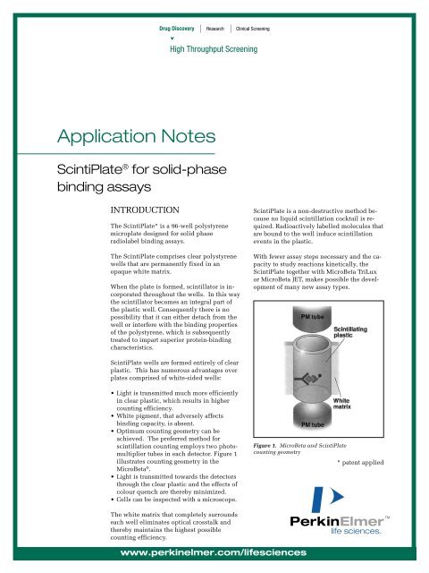
ScintiPlate for solid-phase binding assays - PerkinElmer
ScintiPlate for solid-phase binding assays - PerkinElmer
ScintiPlate for solid-phase binding assays - PerkinElmer

You also want an ePaper? Increase the reach of your titles
YUMPU automatically turns print PDFs into web optimized ePapers that Google loves.
Drug Discovery | Research |<br />
Application Notes<br />
Clinical Screening<br />
High Throughput Screening<br />
<strong>ScintiPlate</strong> ® <strong>for</strong> <strong>solid</strong>-<strong>phase</strong><br />
<strong>binding</strong> <strong>assays</strong><br />
INTRODUCTION<br />
The <strong>ScintiPlate</strong>* is a 96-well polystyrene<br />
microplate designed <strong>for</strong> <strong>solid</strong> <strong>phase</strong><br />
radiolabel <strong>binding</strong> <strong>assays</strong>.<br />
The <strong>ScintiPlate</strong> comprises clear polystyrene<br />
wells that are permanently fixed in an<br />
opaque white matrix.<br />
When the plate is <strong>for</strong>med, scintillator is incorporated<br />
throughout the wells. In this way<br />
the scintillator becomes an integral part of<br />
the plastic well. Consequently there is no<br />
possibility that it can either detach from the<br />
well or interfere with the <strong>binding</strong> properties<br />
of the polystyrene, which is subsequently<br />
treated to impart superior protein-<strong>binding</strong><br />
characteristics.<br />
<strong>ScintiPlate</strong> wells are <strong>for</strong>med entirely of clear<br />
plastic. This has numerous advantages over<br />
plates comprised of white-sided wells:<br />
• Light is transmitted much more efficiently<br />
in clear plastic, which results in higher<br />
counting efficiency.<br />
• White pigment, that adversely affects<br />
<strong>binding</strong> capacity, is absent.<br />
• Optimum counting geometry can be<br />
achieved. The preferred method <strong>for</strong><br />
scintillation counting employs two photomultiplier<br />
tubes in each detector. Figure 1<br />
illustrates counting geometry in the<br />
MicroBeta ® .<br />
• Light is transmitted towards the detectors<br />
through the clear plastic and the effects of<br />
colour quench are thereby minimized.<br />
• Cells can be inspected with a microscope.<br />
The white matrix that completely surrounds<br />
each well eliminates optical crosstalk and<br />
thereby maintains the highest possible<br />
counting efficiency.<br />
www.perkinelmer.com/lifesciences<br />
<strong>ScintiPlate</strong> is a non-destructive method because<br />
no liquid scintillation cocktail is required.<br />
Radioactively labelled molecules that<br />
are bound to the well induce scintillation<br />
events in the plastic.<br />
With fewer assay steps necessary and the capacity<br />
to study reactions kinetically, the<br />
<strong>ScintiPlate</strong> together with MicroBeta TriLux<br />
or MicroBeta JET, makes possible the development<br />
of many new assay types.<br />
Figure 1. MicroBeta and <strong>ScintiPlate</strong><br />
counting geometry<br />
* patent applied
2<br />
SCINTIPLATE PRODUCTS<br />
There are three <strong>ScintiPlate</strong> products.<br />
1450-501 <strong>ScintiPlate</strong> (25)<br />
1450-502 <strong>ScintiPlate</strong> (100)<br />
1450-551 <strong>ScintiPlate</strong> SA (10)<br />
Streptavidin coated<br />
The streptavidin <strong>binding</strong> capacity (6.0 x 10 6<br />
moles biotin) is maximized by attaching the<br />
protein to the plate using a covalent <strong>binding</strong><br />
technique.<br />
Other <strong>ScintiPlate</strong> coatings are available on<br />
request. Examples include anti-Mouse IgG,<br />
anti-rabbit IgG and poly-lysine.<br />
SCINTIPLATE APPLICATIONS<br />
BY MICROBETA<br />
The <strong>ScintiPlate</strong> can be used in all <strong>solid</strong><br />
<strong>phase</strong> <strong>binding</strong> <strong>assays</strong> where the label is a radioactive<br />
isotope. Examples of some typical<br />
<strong>binding</strong> <strong>assays</strong> are listed below.<br />
IRMA sandwich <strong>assays</strong> - e.g. T4 competitive<br />
immunoassay<br />
RIA competitive <strong>assays</strong> - e.g. estradiol<br />
Coated receptor <strong>assays</strong> - e.g. cloned G-protein<br />
or fast ion-receptor <strong>assays</strong><br />
Hybridization <strong>assays</strong> - e.g. DNA/RNA hybridization<br />
or detection of point mutations<br />
Enzyme activity <strong>assays</strong> - e.g. kinase with<br />
biotinylated substrate<br />
Cell <strong>binding</strong> studies - e.g. 2-site antibody to<br />
cell-surface receptors<br />
Binding experiments involving a labelled<br />
biotinylated compound - e.g. biotinylated<br />
substrate<br />
Kinetic <strong>assays</strong> involving a tritiated<br />
compound - e.g. an antibody-antigen or<br />
receptor-<strong>binding</strong> reaction.<br />
In the following section three applications of<br />
the <strong>ScintiPlate</strong> are illustrated in detail:<br />
estradiol radioimmunoassay, T4 RIA and<br />
<strong>binding</strong> of a biotinylated oligonucleotide.<br />
Estradiol RIA:<br />
Estradiol measurement was per<strong>for</strong>med as a<br />
<strong>solid</strong>-<strong>phase</strong> RIA using 3 H-labelled estradiol.<br />
The reference DELFIA ® Estradiol kit<br />
(Wallac, 1244-056) was made according to<br />
the kit instructions.<br />
Procedure: A <strong>ScintiPlate</strong> was coated with<br />
anti-rabbit IgG (1,5 µg/well). For the sample<br />
preparation, danazoli was diluted to a concentration<br />
of 5 µg/mL in assay buffer. 25 µL<br />
of standards, samples and controls and 100<br />
µL estradiol antiserum (concentration of 0.06<br />
µg/mL, dilution in assay buffer) were<br />
pipetted in triplicate into the pre-washed<br />
<strong>ScintiPlate</strong>. The plate was incubated <strong>for</strong> 30<br />
minutes and 100 µL of 3 H-estradiol dilution<br />
was added to the wells (1-10 pmol/L). The<br />
<strong>ScintiPlate</strong> was incubated <strong>for</strong> a further 2<br />
hours and the wells were washed four times<br />
with an automatic DELFIA PlateWash using<br />
Wash Solution (Wallac Oy). The <strong>ScintiPlate</strong><br />
was dried <strong>for</strong> an hour at RT, be<strong>for</strong>e measurement<br />
with MicroBeta (counting time<br />
3 minutes).<br />
Results: The best signal-to-noise ratio was<br />
found to be at 1 pmol/L of 3 H-estradiol. The<br />
coefficient of variation (CV%) varied from<br />
0.4% to 11.7% (figure 2).<br />
Figure 2. RIA Estradiol measurement with dilution<br />
series of 3 H-labelled estradiol, and CV % <strong>for</strong><br />
the standards with 1 pmol/L of labelled estradiol.
The correlation between the RIA and DELFIA<br />
methods is shown in figure 3.<br />
RIA Estradiol (counts)<br />
Figure 3. RIA Estradiol vs. Estradiol DELFIA kit.<br />
T4 RIA:<br />
T4 measurement was per<strong>for</strong>med as a <strong>solid</strong><strong>phase</strong><br />
competitive RIA. 125 I-labelled<br />
streptavidin was used as a label. The reference<br />
T4 DELFIA kit (Wallac) was done according<br />
to the kit instructions.<br />
DELFIA Estradiol (counts)<br />
Procedure: Biotinylated T4 was diluted to<br />
achieve a concentration of 600 nmol/L. The<br />
solution, containing 60 µL biotinylated<br />
T4 -dilution, 60 µL antibody stock solution<br />
(Wallac, concentration 12 µg/mL) and 6 mL<br />
T4 buffer was prepared. 25 µL of standards<br />
were pipetted as duplicates to the prewashed<br />
wells and 200 µL of previously prepared<br />
solution was added to the wells. The<br />
<strong>ScintiPlate</strong> was incubated <strong>for</strong> 90 minutes.<br />
The plate was then washed four times with<br />
DELFIA PlateWash and Wash Solution<br />
(Wallac) and a dilution of 125 I-Streptavidin<br />
was made to achieve a concentration of<br />
100,000 DPM. 200 µL of the 125 I-Streptavidin<br />
-dilution was added to the wells and the<br />
plate was incubated <strong>for</strong> an hour. Finally the<br />
plate was washed six times. The plate was<br />
dried <strong>for</strong> an hour at RT be<strong>for</strong>e measurement<br />
with MicroBeta (counting time 3 minutes).<br />
Results: The coefficient of variation (CV%)<br />
varied from 1.1 to 13.6. The linear area of the<br />
standard curve was between 20-300 nmol/L<br />
(figure 4).<br />
CPM<br />
Figure 4. RIA T4 and CV %.<br />
The correlation between T4 RIA and<br />
DELFIA, T4 analyses is seen in figure 5.<br />
RIA T4 (counts)<br />
Figure 5. Solid <strong>phase</strong> RIA T4 vs DELFIA T4 kit.<br />
The enzyme activity assay:<br />
An enzyme activity assay was per<strong>for</strong>med using<br />
a kinase and its biotinylated substrates.<br />
The effects of two concentrations of<br />
substrate, a) 3 and b) 6 µM were tested.<br />
CV %<br />
DELFIA T4 (counts)<br />
Procedure: The enzymatic reaction took<br />
place in Eppendorf tubes. The reactions contained<br />
0.25 µg enzyme, 3 or 6 µM substrate, 1<br />
mM DTT, 10 mM MgCl 2 , 50 mM HEPES pH<br />
6.8, 0.015 % Brij35, 2.5% DMSO and 50 mM<br />
ATP + 92 nM [γ- 33 P] -ATP in a total volume of<br />
25 µL. The reactions were incubated at<br />
+30 °C <strong>for</strong> two hours. After the incubation<br />
the reactions were diluted a) 1:100 and b)<br />
1:200 to obtain a substrate concentration of<br />
30 nM. Diluted reactions were pipetted in<br />
triplicate into pre-washed Streptavidin<br />
3
coated <strong>ScintiPlate</strong>. The plate was incubated<br />
with slow shaking <strong>for</strong> half an hour.<br />
After incubation the <strong>ScintiPlate</strong> was measured<br />
with MicroBeta using a default 33 P<br />
counting protocol.<br />
Results:<br />
Table 1 shows the signal and background values<br />
<strong>for</strong> reactions containing 3 or 6 µM<br />
substrate. In this case, an adequate signal-tonoise<br />
ratio is obtained with the lower<br />
substrate concentration.<br />
[S] signal noise S:N<br />
3 µm 511 12 43<br />
6 µm 763 15 51<br />
Table 1. The signal-to-noise ratio with two<br />
concentrations of substrate.<br />
OPTIMIZATION OF COUNTING<br />
<strong>ScintiPlate</strong> <strong>assays</strong>, like all <strong>assays</strong>, must be<br />
optimized to get the best results. Optimization<br />
concerns the bio-reaction part of coating<br />
as well as counting in the MicroBeta.<br />
Coating optimization<br />
Coating of the <strong>ScintiPlate</strong> is the same as all<br />
other plate coatings. First, the protein to be<br />
coated is diluted to a concentration of 0.5-10<br />
µg/mL in, <strong>for</strong> example, phosphate buffer. The<br />
plates are incubated overnight and then<br />
washed. All the remaining <strong>binding</strong> sites can<br />
be blocked (saturated) by dispensing, <strong>for</strong> example,<br />
0,5 % bovine serum albumin into the<br />
wells and by incubating the wells <strong>for</strong> a few<br />
hours at RT. The exact coating procedure<br />
must be fully optimized <strong>for</strong> each coating.<br />
The following factors should be considered.<br />
• amount of protein per well<br />
• pH of the coating buffer<br />
• ionic strength of the buffer<br />
• coating and saturation times<br />
• incubation temperature<br />
• number of washing cycles<br />
after coating and saturation<br />
The stability and reproducibility of the coating<br />
will also depend on the protein structure.<br />
Usually, antibody coatings are quite stable<br />
whereas other smaller recombinant proteins<br />
may need special stabilizing treatments.<br />
Once the optimum coating procedure <strong>for</strong> the<br />
<strong>ScintiPlate</strong> has been determined, the<br />
immuno-reaction parameters that are listed<br />
below should be considered.<br />
• time<br />
• temperature<br />
• buffer<br />
• assay volume<br />
Typical buffers that are used include TRIS-<br />
HCl and PBS. Also, DELFIA Assay Buffer,<br />
which contains appropriate additives and<br />
preservatives, has been optimized <strong>for</strong> a broad<br />
range of immuno<strong>assays</strong>.<br />
The use of extreme conditions, such as high /<br />
low pH or high detergent concentrations<br />
should be avoided.<br />
Optimization of counting per<strong>for</strong>mance<br />
The best counting per<strong>for</strong>mance is obtained if<br />
the <strong>ScintiPlate</strong> is washed and dried after the<br />
incubation <strong>phase</strong>.<br />
The wash step will reduce non-specific <strong>binding</strong><br />
and also eliminate counting efficiency<br />
variations caused by different coloured compounds.<br />
Tritium and other weak beta emitters can be<br />
partially absorbed or deflected by any residual<br />
water or buffer solution that remains<br />
on the well surface after washing. To avoid<br />
this the plates can be dried in an oven at<br />
37°C or left <strong>for</strong> 1 to 3 hours at RT.<br />
4
5<br />
COUNTING GEOMETRY OF<br />
SOLID PHASE BINDING<br />
ASSAYS<br />
Radioactive labels bound to a microplate or a<br />
bead exhibit “2π“ counting geometry regardless<br />
of whether liquid or <strong>solid</strong> scintillation<br />
counting is per<strong>for</strong>med. In other words, as the<br />
isotope decays, the emitted beta particle can<br />
move in any direction in the “4π“ <strong>solid</strong> angle<br />
that surrounds the labelled compound. If the<br />
labelled compound is attached to a surface,<br />
only a half of the total events can be detected.<br />
In the case of the <strong>ScintiPlate</strong> or a scintillating<br />
bead, only beta particles that move into the<br />
wall or the bead produce scintillation events.<br />
(Conversely, in the case of a coated non-scintillating<br />
plate assay, only beta particles that<br />
move into the cocktail will produce scintillation<br />
events.)<br />
Thus the counting efficiency of <strong>solid</strong>-<strong>phase</strong><br />
<strong>binding</strong> <strong>assays</strong> have systematically lower<br />
counting efficiencies compared to <strong>assays</strong> that<br />
have the labelled compounds fully dissolved<br />
in liquid cocktail.<br />
Figure 6. Counting geometry <strong>for</strong> <strong>solid</strong>- <strong>phase</strong><br />
counting compared to liquid samples.<br />
Isotope Counting efficiency, %<br />
3 H 30<br />
125 I 61<br />
33 P 60<br />
Table 2. Typical <strong>ScintiPlate</strong> counting efficiencies.<br />
REFERENCES<br />
1. Nakayama, G., Nova, M., Parandoosh, Z.<br />
(1998) A Scintillation Microplate Assay<br />
<strong>for</strong> the Assessment of Protein Kinase<br />
Activity. J. Biomol. Screen 3(1)<br />
2. Siitari, H., Oikari, T. (1992) Scintillating<br />
Microtitration Plates in Immunoassay.<br />
Liquid Scintillation Spectrometry,<br />
edited by Noakes, J<br />
3. Häggblad, J., Carlsson, B., Kivelä, P.,<br />
Siitari, H. (1995) Scintillating<br />
Microtitration Plates as Plat<strong>for</strong>m <strong>for</strong><br />
Determination of [3H] Estradiol Binding<br />
Constants <strong>for</strong> hER-HBD<br />
BioTechniques 18 (1)<br />
4. Häggblad, J., Carlsson, B., Raynaud, J-P.<br />
(1994) Evaluation of Con<strong>for</strong>mational<br />
Changes in hER-HBD by Pharmacological<br />
Dissection of Hormone Dissociation Rates<br />
in a Homogeneous Hormone Binding<br />
Assay. Hormonal Cancer Proceedings<br />
5. Ihalainen, J., Siitari, H., Laine, S.,<br />
Syvänen, A.-C., Palotie, A. (1994) Towards<br />
Automatic Detection of Point Mutations:<br />
Use of Scintillating MicroPlates in<br />
Solid-Phase Minisequencing.<br />
BioTechniques 16 (5)<br />
6. Braunwalder, A., Wennogle, L., Gay, B.,<br />
Lipson, K., Sills, M. (1996) Application of<br />
Scintillating Microtiter Plates to Measure<br />
Phosphopeptide Interactions with the<br />
GRB2-SH2 Binding Domain J. Biomol.<br />
Screen. 1 (1)
World Headquarters: <strong>PerkinElmer</strong> Life Sciences, 549 Albany Street, Boston, MA 02118-2512 USA (800) 551-2121<br />
In Europe: <strong>PerkinElmer</strong> Life Sciences, B-1930 Zaventem, Belgium, +32 2 717 7924 • P.O.Box 10, FIN-20101 Turku, Finland, +358 2 2678 111<br />
MicroBeta, <strong>ScintiPlate</strong> and DELFIA are registered trademarks of <strong>PerkinElmer</strong>, Inc.<br />
www.perkinelmer.com/lifesciences<br />
AAABH-0001-02, 3 March 2000 Printed in Finland by Offset House Oy Naantali 2000