Ankle Arthrodesis Using Ring External Fixation - Orthofix.com
Ankle Arthrodesis Using Ring External Fixation - Orthofix.com
Ankle Arthrodesis Using Ring External Fixation - Orthofix.com

Create successful ePaper yourself
Turn your PDF publications into a flip-book with our unique Google optimized e-Paper software.
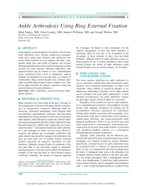
Techniques in Foot and <strong>Ankle</strong> Surgery 5(3):150–163, 2006 Ó 2006 Lippincott Williams & Wilkins, Philadelphia<br />
. S P E C I A L F O C U S .<br />
<strong>Ankle</strong> <strong>Arthrodesis</strong> <strong>Using</strong> <strong>Ring</strong> <strong>External</strong> <strong>Fixation</strong><br />
Mark Easley, MD, Colin Looney, MD, Samuel Wellman, MD, and Joseph Wilson, MD<br />
Division of Orthopaedic Surgery<br />
Duke University Medical Center<br />
Durham, NC<br />
| ABSTRACT<br />
Contemporary re<strong>com</strong>mendations for primary and revision<br />
ankle arthrodesis favor internal <strong>com</strong>pression techniques<br />
using screw and/or plate fixation, with satisfactory out<strong>com</strong>es<br />
being reported for most patients. Recently, <strong>com</strong>parable<br />
union rates and results of primary and revision<br />
tibiotalar arthrodeses have been reported using ring external<br />
fixation. In select patients, tibiotalar arthrodesis with<br />
internal fixation may be limited or even contraindicated<br />
given insufficient bone stock to adequately support<br />
implants, an abundance of avascular bone, or a history of<br />
osteomyelitis. <strong>Ring</strong> external fixation may facilitate clinically<br />
acceptable limb salvage in these <strong>com</strong>plex cases. This<br />
review paper describes the authors’ experience using ring<br />
external fixation for ankle arthrodesis.<br />
Keywords: ankle, arthrodesis, external fixation, multiplanar,<br />
Ilizarov<br />
| HISTORICAL PERSPECTIVE<br />
Many advances have been made in the past 2 decades in<br />
the management of advanced tibiotalar arthritis unresponsive<br />
to nonoperative treatment. Although ankle arthroscopy,<br />
distraction arthroplasty, fresh allograft shell<br />
reconstruction, supramalleolar osteotomy, and total ankle<br />
arthroplasty have shown promise in select cases of advanced<br />
ankle arthritis, ankle arthrodesis remains the criterion standard<br />
surgical management. Results of ankle arthrodesis using<br />
internal fixation for stability and <strong>com</strong>pression are consistently<br />
favorable, 1,2 and internal fixation for ankle arthrodesis<br />
is the technique of choice in most cases. However,<br />
surgeons and patients are occasionally confronted with<br />
ankle arthritis that should be managed with arthrodesis but<br />
does not lend itself well to internal fixation. <strong>External</strong><br />
fixation represents an attractive alternative in such<br />
situations. Although external fixation for problems of the<br />
foot and ankle is not universally taught in orthopedic<br />
training programs, it has been used routinely for numerous<br />
disorders of the foot and ankle in select medical centers<br />
worldwide. Gradually, the number of surgeons applying<br />
Address correspondence and reprint requests to Mark Easley, MD,<br />
Duke Health Center, 3116 North Duke Street, Rm 243, Durham,<br />
NC 27704. E-mail: easle004@mc.duke.edu.<br />
150<br />
Techniques in Foot and <strong>Ankle</strong> Surgery<br />
the techniques developed in these institutions for the<br />
surgical management of foot and ankle disorders is<br />
increasing, either by necessity or by recognition of the<br />
advantages of these methods in select foot and ankle<br />
problems. Although results of ankle arthrodesis using external<br />
fixation are not as widely published as those using<br />
internal fixation, the results of ankle arthrodesis using<br />
external fixation, even in revision surgery, are favorable.<br />
| INDICATIONS AND<br />
CONTRAINDICATIONS<br />
The most <strong>com</strong>mon indication for ankle arthrodesis is<br />
severe, destructive arthritis that fails nonoperative management,<br />
most <strong>com</strong>monly posttraumatic in origin. Less<br />
frequently, ankle arthritis is caused by primary or inflammatory<br />
arthropathy. Currently, severe ankle arthritis<br />
can be managed with total ankle replacement 3 or fresh<br />
allograft reconstruction, 4 but ankle arthrodesis remains<br />
the procedure against which all others are measured.<br />
Regardless of the method of fixation, ankle arthrodesis<br />
is contraindicated in patients with peripheral vascular<br />
disease until adequate circulation for healing can be<br />
reestablished. Absolute indications for arthrodesis<br />
include ankle arthritis associated with neuropathy, avascular<br />
necrosis of the talus or distal tibia, severe coronal or<br />
sagittal plane deformity, and nonreconstructable ankle<br />
ligament insufficiency. That is to say, in such cases, total<br />
ankle replacement is contraindicated. Joint-sparing procedures<br />
may be favored when ankle arthritis is associated<br />
with ipsilateral hindfoot arthrosis or a contralateral<br />
tibiotalar/tibiotalocalcaneal arthrodesis.<br />
Typically, ankle arthrodesis can be effectively performed<br />
with internal fixation. <strong>External</strong> fixation may confer<br />
advantages over internal fixation in several situations.<br />
Specifically, external fixation for ankle arthrodesis may<br />
be considered with 1) a history of sepsis/osteomyelitis<br />
(Figs. 1A, B); 2) a <strong>com</strong>promised soft tissue envelope about<br />
the ankle; 3) inadequate bone stock at the arthrodesis site<br />
to support internal fixation limited to the tibia and talus;<br />
4) a leg length discrepancy that is inadequately treated<br />
with a shoe modification; 5) failed prior ankle arthrodesis<br />
using internal fixation; and 6) anticipated non<strong>com</strong>pliance<br />
with a nonYweight-bearing/touchdownYweight-bearing<br />
Copyright © Lippincott Williams & Wilkins. Unauthorized reproduction of this article is prohibited.
Special Focus: <strong>Ankle</strong> <strong>Arthrodesis</strong> <strong>Using</strong> <strong>Ring</strong> <strong>External</strong> <strong>Fixation</strong><br />
FIGURE 1. A and B, <strong>Ankle</strong> arthrodesis using external fixation should be considered in septic ankle arthritis.<br />
status. Relative contraindications to external fixation for<br />
ankle arthrodesis are patients with total joint arthroplasties<br />
(pin tract infections may lead to infection of the joint<br />
replacement) and neuropathy of the contralateral leg<br />
(external fixator may inflict wounds in the contralateral<br />
lower extremity without the patient recognizing it).<br />
| PREOPERATIVE PLANNING<br />
History, clinical evaluation, and routine ankle radiographs<br />
establish the diagnosis of symptomatic tibiotalar arthritis.<br />
Weight-bearing radiographs of the ankle usually confirm<br />
the diagnosis. The soft tissue envelope about the ankle and<br />
the vascular status must be evaluated. Poor soft tissues<br />
about the ankle (affected by trauma and/or prior surgery)<br />
typically necessitate a less invasive exposure for joint<br />
preparation. Compromised perfusion of the extremity<br />
should prompt vascular consultation to optimize the<br />
chance for union and proper soft tissue healing.<br />
Routine weight-bearing ankle radiographs generally<br />
suffice, but if there is any suspicion on clinical evaluation<br />
for limb malalignment or foot pathology, then<br />
mechanical axis, hip and knee, and foot radiographs<br />
should also be evaluated. Computed tomography (CT)<br />
and magnetic resonance imaging (MRI) are not routinely<br />
required. In select cases, CT may help identify<br />
hindfoot arthritis/deformity if plain radiographs of the<br />
foot and ankle fail to adequately reveal these clinical<br />
findings. MRI should be considered if avascular<br />
necrosis of either the tibial plafond or talus is suggested<br />
by the radiographs; this may dictate how much bone<br />
resection is required at the arthrodesis site to access<br />
vascular surfaces for fusion.<br />
Simply placing a tibiotalar arthrodesis in neutral<br />
plantarflexion/dorsiflexion, slight hindfoot valgus, and<br />
slight external rotation will typically create a functional,<br />
plantigrade foot. However, disregard for whole limb and<br />
foot alignment may lead to a poor functional out<strong>com</strong>e,<br />
despite successful ankle fusion. Limb malalignment may<br />
need to be addressed before or simultaneous to ankle<br />
arthrodesis. An advantage with external fixation is that<br />
simultaneous <strong>com</strong>bined procedures can be performed,<br />
such as proximal tibial corticotomy with lengthening/<br />
realignment and ankle arthrodesis or con<strong>com</strong>itant ankle<br />
and foot procedures.<br />
When arthrodesis is planned in the face of infection,<br />
preoperative evaluation includes <strong>com</strong>plete blood cell<br />
FIGURE 2. A support under the calf facilitates positioning<br />
of the external fixator. The surgeon must ensure that<br />
when the support is removed, the proximal ring does not<br />
impinge on the calf.<br />
Volume 5, Issue 3 151<br />
Copyright © Lippincott Williams & Wilkins. Unauthorized reproduction of this article is prohibited.
FIGURE 3. <strong>External</strong> fixation alone does not create<br />
fusion; proper joint preparation is necessary.<br />
count with differential and blood cultures. Consideration<br />
may be given to a MRI or a <strong>com</strong>bination technetium bone<br />
scan/indium-labeled white blood cells scan to confirm/<br />
evaluate the extent of infection, keeping in mind that<br />
peripheral vascular disease and antibiotics will interfere<br />
with the effectiveness of these studies. To obtain useful<br />
intraoperative cultures, antibiotics should be stopped at<br />
least 3 days before surgery. Planning should include the<br />
potential for a staged procedure. Irrigation and debridement,<br />
hardware removal, and possible antibiotic bead<br />
placement should be planned as the first stage. If the ankle<br />
is determined to be unstable at that time, the external<br />
fixator should be placed for support. In a second stage, the<br />
antibiotic beads can be removed, the ankle again debrided,<br />
and <strong>com</strong>pression may be applied through the ankle if there<br />
is no evidence of persistent infection. Alternatively, the<br />
FIGURE 4. Ideal position for arthrodesis: neutral sagittal<br />
plane (plantigrade foot), slight hindfoot valgus, tibial shaft<br />
axis aligned with the second metatarsal, and no anterior<br />
talar translation within the ankle mortise. This is particularly<br />
important if a prebuilt frame is applied.<br />
152<br />
Special Focus: Easley et al<br />
plan can be for a single-stage procedure if the surgeon is<br />
satisfied that all of the infected tissue has been removed.<br />
| TECHNIQUE<br />
Techniques in Foot and <strong>Ankle</strong> Surgery<br />
A proximal regional anesthetic typically suffices for<br />
ankle arthrodesis using external fixation. Tourniquet use<br />
is generally limited to the approach for tibiotalar joint<br />
preparation. <strong>External</strong> fixation in itself cannot create a<br />
fusion; proper joint preparation is essential. Therefore,<br />
if there is any concern for the perfusion of either the<br />
tibial plafond or (more likely) the talar dome, then the<br />
tourniquet must be released before wound closure to confirm<br />
bleeding at the arthrodesis site. There is no need for<br />
tourniquet use during application of the external fixator.<br />
The patient is positioned supine, often with a support<br />
under the hip of the operated leg. The goal is to orient the<br />
lower leg directly perpendicular to the operating room<br />
table/floor to facilitate proper placement of the external<br />
fixator. A sterile proximal calf support (Bbump^) (Fig.2)<br />
should be created to suspend the lower leg during<br />
external fixator placement.<br />
FIGURE 5. A and B, Intraoperative fluoroscopy in 2<br />
planes confirms proper alignment. In this case, provisional<br />
pin fixation was used.<br />
Copyright © Lippincott Williams & Wilkins. Unauthorized reproduction of this article is prohibited.
FIGURE 6. The proximal ring block for tibial fixation.<br />
Special Focus: <strong>Ankle</strong> <strong>Arthrodesis</strong> <strong>Using</strong> <strong>Ring</strong> <strong>External</strong> <strong>Fixation</strong><br />
The lower leg is prepared and draped in usual sterile<br />
fashion, and the leg is exsanguinated using a tension<br />
bandage (unless persistent infection is suspected). The<br />
tibiotalar joint is approached and prepared using the<br />
surgeon’s preferred technique (Fig. 3). If there is any<br />
concern that the arthrodesis site may not maintain the ideal<br />
position for fusion (Fig. 4), then provisional fixation<br />
should be used. Although violation of the subtalar joint in<br />
a patient undergoing isolated tibiotalar joint arthrodesis<br />
in not ideal, one simple method of maintaining provisional<br />
fixation is a longitudinal Steinmann pin from the<br />
calcaneus into the talus. This pin is placed similar to the<br />
guide pin used for retrograde intramedullary nail procedures<br />
(Figs. 5A, B). Provisional fixation should maintain<br />
adequate reduction of the prepared talus within the<br />
prepared ankle mortise. Optimal function and potential<br />
for fusion is established with maximum contact between<br />
the arthrodesis surfaces, avoiding anterior translation of<br />
the talus within the ankle mortise. Valgus is generally<br />
established in the hindfoot; varus malalignment at the<br />
tibiotalar joint must be avoided. Furthermore, calcaneus<br />
or equinus positioning must be corrected to a neutral<br />
sagittal plane alignment. Finally, the second metatarsal<br />
should be in line with the tibial crest to ensure proper<br />
rotational positioning. After proper joint preparation and<br />
positioning, the wound can be closed in routine fashion.<br />
We re<strong>com</strong>mend closing the wound before applying the<br />
external fixator because access to the wound be<strong>com</strong>es<br />
challenging with the external fixator in place. A drain<br />
may be used, but it must be positioned so that it does not<br />
interfere with ensuing pin placement.<br />
Most external fixators have several consistent <strong>com</strong>ponents:<br />
1) a proximal ring or proximal ring block (Fig. 6),<br />
2) an intermediate ring or connector, and 3) a foot ring or<br />
frame. The proximal ring/ring block usually consists of 1<br />
or 2 rings attached to the distal tibia. The intermediate<br />
ring serves to support thin wires secured to the talus. The<br />
foot frame connects directly to the hindfoot, and in most<br />
constructs, to the midfoot and forefoot as well. The individual<br />
<strong>com</strong>ponents are secured to their respective bones<br />
using either thin wires (Bskinny wires^) orhalfpins.The<br />
<strong>com</strong>ponents of the external fixator are attached to one<br />
another by threaded rods, adjustable struts, or sockets.<br />
The connectors between the <strong>com</strong>ponents that span the<br />
tibiotalar joint must be able to <strong>com</strong>press.<br />
The external fixator can be constructed and applied<br />
in 2 basic ways: 1) preassembled and applied en bloc<br />
or 2) step-by-step assembly as it is applied on the<br />
patient. Theoretically, prebuilding the external fixator<br />
may save some time and guarantees a symmetrical<br />
frame construct, with limited modifications to connect<br />
FIGURE 7. A, Prebuilt Sheffield<br />
frame (<strong>Orthofix</strong>) with proximal<br />
ring/Sheffield clamp, intermediate<br />
two-thirds rings for talar<br />
fixation, and foot frame. B, Traditional<br />
Ilizarov frame with proximal<br />
extension to allow for<br />
simultaneous limb lengthening.<br />
Volume 5, Issue 3 153<br />
Copyright © Lippincott Williams & Wilkins. Unauthorized reproduction of this article is prohibited.
FIGURE 8. One to 2 cm must be maintained between the<br />
posterior heel and the foot frame.<br />
the various rings (Figs. 7A, B). Moreover, if the prebuilt<br />
external fixator has been symmetrically assembled, it<br />
serves as a template for proper lower leg and foot<br />
alignment. One challenge with a prebuilt frame is that it<br />
may be difficult to position both the proximal rings<br />
orthogonally with the tibia and the foot frame<br />
(Bhorseshoe ring^) parallel with the foot.<br />
Alternatively, if the external fixator is assembled in<br />
a stepwise manner, the proximal ring block is traditionally<br />
attached first. The foot frame is then applied, along<br />
with an intermediate ring or connection for the talar<br />
wires. Lastly, the proximal ring block is connected to the<br />
distal (foot and talar) construct. In assembling the frame<br />
in this graduated fashion, it may be useful to place<br />
threaded rods longitudinally from the proximal to distal<br />
construct to facilitate proper positioning of 3 or 4 threaded<br />
FIGURE 9. The foot should be plantar to the foot frame.<br />
154<br />
Special Focus: Easley et al<br />
Techniques in Foot and <strong>Ankle</strong> Surgery<br />
FIGURE 10. Foot positioned symmetrically within the<br />
foot frame.<br />
rods that will ultimately be used for <strong>com</strong>pression across<br />
the arthrodesis site.<br />
There are several technique tips that facilitate appropriate<br />
ring application and typically avoid intraoperative<br />
fixator revisions:<br />
1. Allow adequate space for the calf so that the proximal<br />
ring does not impinge on the soft tissues. One <strong>com</strong>mon<br />
FIGURE 11. To facilitate proper foot positioning within<br />
the foot frame, the forefoot wire can be placed first to<br />
suspend the foot. This helps ensure that the heel does<br />
not contact the foot frame, and subtle adjustments to the<br />
foot position are still possible.<br />
Copyright © Lippincott Williams & Wilkins. Unauthorized reproduction of this article is prohibited.
mistake is to attach the proximal ring with the bump<br />
behind the calf to suspend the leg and no regard for<br />
where the calf will be once the bump is removed. If not<br />
assessed proactively, the ring will impinge on the calf,<br />
necessitating removal of the ring and potential repositioning<br />
of thin wires/half pins (Fig. 2).<br />
2. Allow adequate room between the posterior heel and<br />
the foot frame. This space does not need to be more<br />
than 1.5 cm, as virtually no swelling occurs directly<br />
posteriorly at the calcaneus, irrespective of how<br />
extensive the surgery is. However, with direct contact<br />
of the foot frame to the calcaneus, the external fixator<br />
potentially may need to be disassembled and<br />
remounted in its entirety (Fig. 8).<br />
3. Be sure the foot is plantar to the foot frame. The<br />
plantar foot pad is thicker than it appears, and if the<br />
foot frame is positioned too low, it will not be<br />
possible to pass the thin wires through the bones of<br />
the foot, only the plantar soft tissues. Poor foot frame<br />
positioning may again necessitate disassembly of a<br />
major portion of the entire construct (Fig. 9).<br />
FIGURE 13. The thin wires in the foot are tensioned.<br />
Special Focus: <strong>Ankle</strong> <strong>Arthrodesis</strong> <strong>Using</strong> <strong>Ring</strong> <strong>External</strong> <strong>Fixation</strong><br />
FIGURE 12. The calcaneus is stabilized with 2 oblique<br />
thin wires.<br />
Applying a Prebuilt <strong>External</strong> Fixator<br />
We do not suggest that a prebuilt external fixator is the<br />
favored method for ankle arthrodesis using external<br />
fixation, but particularly for the surgeon inexperienced<br />
with traditional techniques of external fixation, this method<br />
may facilitate the procedure. After satisfactory joint<br />
preparation, this sequence of steps could be considered.<br />
1. Provisional pin fixation of the arthrodesis site.<br />
2. Intraoperative fluoroscopy to confirm satisfactory<br />
position of the tibiotalar arthrodesis site (Figs. 5A, B).<br />
3. Support under the calf (Fig. 2).<br />
4. Positioning of the prebuilt external fixator (Fig. 2).<br />
5. Position the foot frame portion symmetrically about<br />
the foot, avoiding contact between the posterior heel<br />
and the foot frame (Fig. 10).<br />
6. Next, thin wires and half pins are added to the<br />
construct. For ankle arthrodesis, thin wires are smooth,<br />
and there is no need to use olive wires. Both thin wires<br />
and half pins should be inserted without tensioning the<br />
skin. Cold water or saline irrigation or a moist sponge<br />
to gently hold the thin wire should be used to cool the<br />
wire during insertion. Once the thin wire has exited the<br />
opposite cortex, a mallet should be used to <strong>com</strong>plete<br />
the insertion. Half pin insertion requires a small stab<br />
incision; thin wires can be placed directly through the<br />
skin without an incision.<br />
7. Suspend the foot frame from a single wire or 2 forefoot<br />
wires (Fig. 11). Traditional positioning of forefoot<br />
wires is 1 wire through the first and second<br />
metatarsals and the second wire through the fifth<br />
through the third metatarsals. This orientation is safe<br />
and stable, but necessitates that the wires extend<br />
above and below the frame, which may make weightbearing<br />
more difficult. Alternatively, a single forefoot<br />
wire can be placed from the fifth metatarsal to the<br />
first. This wire is usually placed obliquely to the foot<br />
FIGURE 14. Proximal ring fixation, in this case, using<br />
half pins.<br />
Volume 5, Issue 3 155<br />
Copyright © Lippincott Williams & Wilkins. Unauthorized reproduction of this article is prohibited.
FIGURE 15. A and B, A supplemental half pin in the calcaneus is optional.<br />
frame and often needs to be connected off the medial<br />
side using a post (extended connector) to avoid<br />
excessive plantar flexion of the first ray.<br />
8. Two oblique calcaneal wires are placed (Fig. 12).<br />
9. A fourth thin wire is placed in the midfoot/forefoot.<br />
10. The foot wires are tensioned. If there is an open foot<br />
frame construct (open distally), then the forefoot<br />
wires need to be tensioned before the calcaneal wires<br />
to ensure that tension is maintained in all foot wires<br />
(if the calcaneal wires are tensioned before the<br />
forefoot wires are tensioned, the calcaneal wires<br />
may lose tension, depending on the inherent stiffness<br />
of the foot frame) (Fig. 13).<br />
11. With the foot frame securely applied to the foot, the<br />
proximal ring block is secured to the tibia. Although<br />
provisional fixation remains across the tibiotalar<br />
joint, subtle adjustments are still possible (ie,<br />
slightly greater dorsiflexion or a shift in rotation<br />
can be ac<strong>com</strong>plished). The proper proximal ring<br />
position can be maintained with a transverse thin<br />
wire that is tensioned. Proximal ring fixation can be<br />
performed solely with the use of thin wires, as<br />
originally described, but half pins are also an option<br />
(Fig. 14). Many different <strong>com</strong>binations of pins are<br />
possible. Generally, when a single tibial ring is<br />
used, a clamp that supports 2 half pins in the medial<br />
face of the tibia is attached to the superior portion of<br />
the the ring, and a clamp that supports 1 additional<br />
pin in the anterior tibia is suspended from the<br />
inferior portion. When using 2 proximal rings, at<br />
least 1 half pin is attached to each ring into the<br />
medial face of the tibia, and at least 1 additional pin<br />
is clamped to either ring in another plane.<br />
12. Consideration may be given to a supplemental half<br />
pin placed posterior to anterior in the calcaneus and<br />
attached to the foot frame (Figs. 15A, B).<br />
13. With the thin wires properly tensioned and the half<br />
pins adequately secured, <strong>com</strong>pression can now be<br />
applied across the tibiotalar arthrodesis site. In a<br />
patient with a prior subtalar fusion, <strong>com</strong>pression<br />
FIGURE 16. A and B, Talar wires are placed from an intermediate ring not only to provide greater support but to protect<br />
the subtalar joint during tibiotalar <strong>com</strong>pression.<br />
156<br />
Special Focus: Easley et al<br />
Techniques in Foot and <strong>Ankle</strong> Surgery<br />
Copyright © Lippincott Williams & Wilkins. Unauthorized reproduction of this article is prohibited.
Special Focus: <strong>Ankle</strong> <strong>Arthrodesis</strong> <strong>Using</strong> <strong>Ring</strong> <strong>External</strong> <strong>Fixation</strong><br />
FIGURE 17. Compression is applied through the tibiotalar<br />
joint.<br />
between the calcaneus and distal tibia is sufficient.<br />
However, in patients with intact subtalar joints,<br />
<strong>com</strong>pression should only be applied across the<br />
tibiotalar arthrodesis site. To ac<strong>com</strong>plish this, a<br />
fixed Bdistal ring block^ is applied to isolate the<br />
talus from the calcaneus and protect the subtalar<br />
joint. Through either extended connectors from the<br />
foot frame or an additional intermediate ring, 1 or<br />
preferably 2 thin wires are placed in the talus and<br />
appropriately tensioned. Care must be taken to<br />
ensure that the pins are indeed in the talus and do<br />
not engage the malleoli (Figs. 16A, B).<br />
14. Compression may be applied after all <strong>com</strong>ponents<br />
of the external fixator have been tightened, and all<br />
thin wires have been tensioned. If the Steinmann pin<br />
placed longitudinally for provisional fixation is<br />
directly perpendicular to the arthrodesis site, then<br />
it can be used as a Brail^ during <strong>com</strong>pression. The<br />
provisional fixation is typically removed at the end<br />
of the case (Fig. 17).<br />
FIGURE 18. Intraoperative fluoroscopy after <strong>com</strong>pression.<br />
Note the bending of the tensioned thin wires in the talus.<br />
15. Intraoperative fluoroscopy is used to confirm<br />
adequate bony apposition at the arthrodesis site<br />
and proper pin position (Fig. 18).<br />
16. If there is concern for inadequate <strong>com</strong>pression or a<br />
tendency toward varus malalignment at the tibiotalar<br />
joint, consideration should be given to a fibular osteotomy,<br />
if it was not already cut or resected during the<br />
approach. We favor an oblique osteotomy, proximal<br />
lateral to distal medial through a 2- to 3-cm incision.<br />
17. The wires are appropriately cut and bent. Alternatively,<br />
the pins can be cut flush with the external fixator.<br />
However, if a wire needs to be tensioned later, leaving<br />
a carefully bent residual wire allows for this.<br />
18. Sutures are used to close any extension of the stab<br />
incisions created for pin placement.<br />
FIGURE 19. A and B, Example of Taylor Spatial Frame being<br />
used for revision ankle arthrodesis that required posterior<br />
translation of the talus in addition to tibiotalar <strong>com</strong>pression.<br />
Volume 5, Issue 3 157<br />
Copyright © Lippincott Williams & Wilkins. Unauthorized reproduction of this article is prohibited.
FIGURE 20. Patient weight-bearing with external fixator.<br />
A foot plate with tread has been applied to the foot frame.<br />
19. Sterile dressings are applied and wrapped relatively<br />
tightly over the pins to provide stability at the<br />
skin/wire interface to decrease the likelihood of pin<br />
tract infections.<br />
Traditional Step-by-Step <strong>External</strong><br />
<strong>Fixation</strong> Method<br />
1. The proximal ring/ring block is usually attached first<br />
(Fig. 6). A reference (thin) wire can be placed perpendicular<br />
to the anatomical axis of the tibia. The<br />
proximal ring is attached to this reference wire. A<br />
second thin wire is placed at a different angle and<br />
attached to the proximal ring that is being held in an<br />
orthogonal position. Ideally, this second wire should<br />
be placed as close to 90 degrees from the first to create<br />
FIGURE 22. Radiograph demonstrating loose proximal<br />
pins suspicious for pin tract infection/osteomyelitis.<br />
the greatest stability. In placing 2 thin wires on the<br />
same ring, one should be above and the other below the<br />
ring. If the pins are placed on the same side of the ring,<br />
they will collide and displace. These thin wires are<br />
tensioned. A second ring can be placed and secured in a<br />
similar manner. The 2 rings are connected by sockets or<br />
short threaded rods. The spacing between these 2 rings<br />
is variable and should allow for adequate stability (ie,<br />
adequate space should be left to permit half pin<br />
placement between the rings. To ensure that the 2 rings<br />
are symmetrically oriented, it may be easier to first<br />
connect the 2 rings, slide them onto the lower leg, have<br />
FIGURE 21. A, <strong>Ankle</strong> arthrodesis site obscured by external fixator, making assessment of fusion difficult. B, CT scan to<br />
facilitate assessment of fusion site.<br />
158<br />
Special Focus: Easley et al<br />
Techniques in Foot and <strong>Ankle</strong> Surgery<br />
Copyright © Lippincott Williams & Wilkins. Unauthorized reproduction of this article is prohibited.
Special Focus: <strong>Ankle</strong> <strong>Arthrodesis</strong> <strong>Using</strong> <strong>Ring</strong> <strong>External</strong> <strong>Fixation</strong><br />
FIGURE 23. A, Poor pin care and unstable skin around pins led to pin tract infection. B, Same patient after 10 days of<br />
proper pin care, and dressings to stabilize the skin-pin interface oral antibiotics.<br />
the assistant hold the rings in an orthogonal position,<br />
and place thin wires in both rings to maintain proper<br />
ring position. Half pins may then be added to both<br />
proximal rings if desired. Half pins stiffen the construct<br />
but are not essential. In fact, traditional Ilizarov<br />
teaching does not include half pins. We typically add<br />
2 half pins from the most proximal ring attached to a<br />
clamp and at least 1 additional half pin from the lower<br />
ring. Spacing between the proximal ring block and the<br />
distal construct should allow for <strong>com</strong>pression.<br />
2. The foot frame is attached to the foot in the manner<br />
described for the prebuilt frame above (Fig. 7B).<br />
FIGURE 24. Patient with persistent postoperative infection.<br />
<strong>External</strong> fixator was not removed and maintained<br />
tibiolar <strong>com</strong>pression during multiple debriments and even<br />
soft-tissue transfer.<br />
3. An intermediate partial ring or connectors from the<br />
foot frame are incorporated to attach to 1 or<br />
preferably 2 talar wires. The talar wires are tensioned<br />
after the intermediate partial ring or connectors have<br />
been secured to the foot frame. Next, threaded rods<br />
are added to connect the proximal ring block and<br />
foot frame construct. It is not problematic if the<br />
threaded rods do not line up exactly congruently/<br />
symmetrically with both portions of the external<br />
fixator. Various connectors can easily be added to<br />
the proximal ring or foot frame to allow for<br />
longitudinal threaded rods that can be used for<br />
<strong>com</strong>pression across the tibiotalar joint. To facilitate<br />
simplicity, the external fixator can be assembled in a<br />
graduated manner but, as mentioned above, the<br />
proximal and distal <strong>com</strong>ponents are symmetrically<br />
oriented by loosely placing longitudinal threaded<br />
rods through the proximal and distal external<br />
fixator <strong>com</strong>ponents.<br />
4. Adjustable struts to provide software-directed dynamic<br />
multiplanar correction are available (Taylor Spatial<br />
Frame, Smith & Nephew Richards, Memphis, TN) if<br />
alignment changes are necessary postoperatively.<br />
Whereas traditional frame constructs allow only immediate<br />
and subsequent <strong>com</strong>pression at the arthrodesis<br />
site, the Taylor Spatial Frame readily affords angular,<br />
rotational, and translational correction to fine tune the<br />
relative positions of the tibia and talus for fusion. The<br />
details of this technique are beyond the scope of this<br />
paper (Figs. 19A, B).<br />
| POSTOPERATIVE MANAGEMENT<br />
The surgical incision must be protected adequately until it<br />
is sealed. Pin care should be initiated within the first week<br />
after application of the external fixator, but without<br />
Volume 5, Issue 3 159<br />
Copyright © Lippincott Williams & Wilkins. Unauthorized reproduction of this article is prohibited.
contaminating the surgical wound. Many techniques have<br />
been suggested for pin care. Our preferred technique is to<br />
perform a mechanical debridement of the pin/skin interface<br />
using a 4 8-inch sponge moistened with a 50:50<br />
mixture of sterile saline and hydrogen peroxide. The<br />
moistened sponge is used to Bshoeshine^ the pins at the<br />
pin/skin interface and to debride the crusting that<br />
routinely develops. Skin <strong>com</strong>promise can be kept to a<br />
minimum by maintaining mild pressure on the skin at<br />
the pin site. Problems arise if the pin/skin interface is<br />
unstable. This constant irritation leads to pin site problems.<br />
Foam pads, vial caps, and specialized wraps have<br />
been devised; we prefer to use surgical sponges wrapped<br />
around adjacent pins in a figure 8 pattern and stacked to<br />
the level of the respective ring/frame <strong>com</strong>ponent to<br />
which the pin is fastened. Irrespective of method, it is<br />
important to maintain this skin stabilization at pin sites<br />
that are problematic.<br />
We generally delay weight-bearing until the wound<br />
is healed. Then, we apply a foot plate with a tread for the<br />
patient to advance weight-bearing as tolerated (Figs.<br />
20A, B). Frequenly, by 4 to 6 weeks after ankle<br />
arthrodesis, full weight-bearing is tolerated by the patient<br />
in the external fixator. We routinely <strong>com</strong>press an additional<br />
1 to 2 mm at 2 to 3 follow-up visits, particularly if<br />
there is a suggestion of in<strong>com</strong>plete bony apposition at the<br />
tibiotalar arthrodesis site.<br />
Removal of the external fixator is determined based<br />
on follow-up radiographs and the suggestion of bridging<br />
trabeculation in at least 2 planes at the arthrodesis site. If<br />
the arthrodesis site is obscured by the external fixator on<br />
follow-up radiographs, a limited CT scan in the coronal<br />
and sagittal planes aids in assessing fusion (Fig. 21)<br />
Approximately 50% of external fixators are removed in<br />
the clinic setting. We re<strong>com</strong>mend that external fixators<br />
that use hydroxyapatite-coated half pins be removed in<br />
the operating room. We routinely re<strong>com</strong>mend that<br />
patients wear a walking cast for 3 to 4 weeks immediately<br />
after external fixator removal and graduate to<br />
an ankle foot orthosis, with a rockerbottom shoe<br />
modification, to be worn an additional 2 to 3 months<br />
as the pin sites fully heal, and the patient adapts to the<br />
ankle fusion.<br />
| COMPLICATIONS<br />
Intraoperative<br />
Pin placement, particularly thin wire application<br />
cannot be haphazard. Safe zones have been established<br />
to avoid neurovascular injury. Pins should be placed<br />
with the skin in a relaxed, tensionless position. If<br />
excessive skin tension occurs, the pin should be replaced.<br />
Alternatively, a small relaxing incision can be<br />
made. We re<strong>com</strong>mend cold water/saline irrigation or<br />
holding pins with moistened sponges during pin placement<br />
to decrease the risk of bone and/or skin necrosis<br />
that may lead to pin tract infections, pin loosening,<br />
and osteomyelitis.<br />
Short-Term Postoperative<br />
Poor pin care will invariably lead to pin tract infections<br />
and, potentially, osteomyelitis (Fig. 22). Pin sites should<br />
be cleaned regularly, and the skin needs to be stabilized<br />
around the pin sites (Figs. 23A, B). If a pin tract should<br />
be<strong>com</strong>e infected and fails to respond to proper pin care<br />
and antibiotic management, the involved pin will need<br />
to be removed and replaced with a different pin in a minor<br />
operative procedure. Wound dehiscence is relatively rare<br />
because the external fixator maintains excellent stability<br />
FIGURE 25. A, Failed ankle arthrodesis using internal fixation. B, Same patient after revision ankle arthrodesis using<br />
external fixation. C, Follow-up radiographs after external fixator removal, demonstrating successful fusion.<br />
160<br />
Special Focus: Easley et al<br />
Techniques in Foot and <strong>Ankle</strong> Surgery<br />
Copyright © Lippincott Williams & Wilkins. Unauthorized reproduction of this article is prohibited.
Special Focus: <strong>Ankle</strong> <strong>Arthrodesis</strong> <strong>Using</strong> <strong>Ring</strong> <strong>External</strong> <strong>Fixation</strong><br />
of the ankle arthrodesis site, but just like for any surgical<br />
procedure, infections occasionally occur. The advantage is<br />
that no implant is located at the arthrodesis site; instead, all<br />
wires are far removed from the arthrodesis site. Another<br />
advantage is that the infection can often be managed<br />
without removing the external fixator, thereby maintaining<br />
<strong>com</strong>pression at the arthrodesis site (Fig. 24).<br />
<strong>Ankle</strong> arthrodesis, regardless of technique, may go on<br />
to delayed union, nonunion, and malunion. Generally,<br />
delayed union is effectively managed with further<br />
<strong>com</strong>pression applied to the arthrodesis site in the office<br />
setting. The risk of malunion (equinus, varus, and<br />
excessive valgus), as for arthrodesis using internal<br />
fixation, is typically avoided by proper intraoperative<br />
positioning of the tibiotalar joint. The advantage of<br />
external fixation is that subtle adjustments are typically<br />
still possible postoperatively.<br />
After external fixator removal, the patient should be<br />
protected in a walking cast, boot, or brace until the tibial<br />
pin sites have healed. With the stiffness at the ankle<br />
arthrodesis site, immediate full weight-bearing after<br />
external fixator removal may lead to stress fracture<br />
through a tibial pin site.<br />
Intermediate/Long-Term Postoperative<br />
Adjacent joint arthritis invariably develops over time<br />
but is not consistently symptomatic. Simultaneous<br />
subtalar <strong>com</strong>pression may accelerate the development<br />
of subtalar arthritis and should be avoided with the<br />
proper frame construct.<br />
| RESULTS<br />
Results for ankle arthrodesis using ring external fixation<br />
have not been widely published. Hawkins et al 5 reported<br />
an 80% (16 of 20) good results in cases of <strong>com</strong>plex<br />
distal tibial pathology or failed ankle arthrodesis treated<br />
with an Ilizarov external fixator. The authors pointed<br />
out that the Ilizarov fixator represents a reasonable tool<br />
for limb salvage and an alternative to amputation in<br />
difficult cases of ankle pathology (Hawkins et al). Calif<br />
et al 6 noted solid fusion in 14 of 14 ankle arthrodeses<br />
FIGURE 26. A, Failed total ankle arthroplasty. B, Implant removed, demonstrating bony defect at ankle. C, Proximal<br />
corticotomy with external fixator applied. D, Patient weight-bearing during simultaneous ankle <strong>com</strong>pression and proximal<br />
distraction osteogenesis. E, Lateral radiograph of ankle arthrodesis site after removal of failed total ankle implants.<br />
F, Lateral radiograph of proximal tibia/fibula, demonstrating distraction osteogenesis.<br />
Volume 5, Issue 3 161<br />
Copyright © Lippincott Williams & Wilkins. Unauthorized reproduction of this article is prohibited.
performed with Ilizarov external fixation for ankle<br />
pathology associated with extensive periarticular bone<br />
loss and severe deformity. All joints were fused in<br />
anatomical alignment, and patients had greatly<br />
improved their functional status. Sakurakichi et al 7<br />
noted that <strong>com</strong>plex ankle arthrodesis could be effectively<br />
be performed with simultaneous proximal tibial<br />
lengthening using the Ilizarov external fixator. Although<br />
the series included only 6 patients, all patients healed<br />
their arthrodesis sites and gained 2 to 3 mm with<br />
simultaneous lengthening.<br />
Probably the most <strong>com</strong>pelling article is Katsenis<br />
et al’s 8,9 report of 21 revision ankle arthrodeses<br />
performed using the Ilizarov external fixator to manage<br />
nonunions/malunions about the ankle. Despite the<br />
patients having <strong>com</strong>plex hindfoot pathology, all 21<br />
healed and with a plantigrade foot. At an average<br />
follow-up of 12 years, 18 of 21 had good to excellent<br />
results. The authors pointed out that external fixation<br />
requires considerable attention and reported 20 major<br />
<strong>com</strong>plications intraoperatively and 7 postoperatively.<br />
We conducted a similar study at our institution (on<br />
a program for American Orthopaedic Foot and <strong>Ankle</strong><br />
Society Summer Meeting, San Diego, CA, July 2006).<br />
We analyzed 22 consecutive revision ankle arthrodesis<br />
performed for nonunions using ring external fixation<br />
(Figs. 25AYC). All patients had at least 1 prior attempt<br />
at arthrodesis using internal fixation. The average<br />
number of surgeries before revision arthrodesis was 2<br />
(range, 1Y8). <strong>External</strong> fixation was maintained for an<br />
average of 15 weeks (range, 12Y44 weeks). Union (time<br />
to removal of external fixation) was suggested by<br />
evidence of bridging trabeculation at the arthrodesis<br />
site in 3 standard radiographic views of the ankle. In<br />
cases where union could not be adequately determined<br />
on radiographic views or the arthrodesis site was<br />
obscured by the external fixator, a limited CT scan<br />
was obtained to assess union. All 22 patients were<br />
available for follow-up at an average of 51 months<br />
(range, 15Y62 months). The average American Orthopaedic<br />
Foot and <strong>Ankle</strong> Society ankle-hindfoot score<br />
improved from 26 points (range, 0Y45 points) preoperatively<br />
to 64 points (range, 0Y87 points) at final follow-up.<br />
Tibiotalar fusion was achieved in 19 (86%) of 22<br />
patients. In the 3 patients with persistent nonunions, one<br />
had avascular necrosis of the talus and two had persistent<br />
osteomyelitis. Two of these patients underwent rerevision<br />
arthrodesis and one opted for amputation. Over the<br />
course of treatment with external fixation, 34 minor<br />
<strong>com</strong>plications (pin tract infections [n = 24], broken pins<br />
[n = 3], and cellulitis [n = 7]) were managed effectively<br />
with local wound care, oral antibiotics, and/or pin<br />
removal in the clinic setting. Four major <strong>com</strong>plications<br />
(deep infection [n = 2] and wound dehiscence [n = 2])<br />
162<br />
Special Focus: Easley et al<br />
were surgically addressed, while maintaining <strong>com</strong>pression<br />
at the arthrodesis site by external fixation. Three<br />
patients had symptomatic malunions: varus (n = 2),<br />
excessive valgus (n = 1), and equinus (n = 1). Hindfoot<br />
motion was less than physiological in all patients<br />
(<strong>com</strong>pared with the contralateral extremity), despite<br />
the external fixator being constructed to protect the<br />
subtalar joint from con<strong>com</strong>itant <strong>com</strong>pression.<br />
Possible Concerns and Future<br />
of This Technique<br />
<strong>External</strong> fixation techniques are not practical for all<br />
patients or surgeons. However, it is our opinion that<br />
surgeons managing <strong>com</strong>plex foot and ankle disorders<br />
should have external fixation in their armamentarium<br />
because there are situations when internal fixation<br />
techniques are limited or contraindicated. Specifically,<br />
revision arthrodesis and cases of septic arthritis/osteomyelitis<br />
frequently lend themselves poorly to internal<br />
fixation. With some focused training in learning<br />
centers and residency training programs, most surgeons<br />
can acquire skills to apply external fixation to ankle<br />
arthrodesis. The future of this technique depends on<br />
adequate teaching being made available and userfriendly<br />
external fixation systems being developed. A<br />
limiting factor remains that many surgeons without<br />
formal external fixation training are intimidated by the<br />
<strong>com</strong>plexity of the traditional Ilizarov system (Figs.<br />
26AYF). Newer systems have been introduced that tend<br />
to be more forgiving, although they may forfeit some of<br />
the versatility of the seemingly endless <strong>com</strong>binations of<br />
external fixation constructs afforded by the original<br />
systems. Although these newer systems may appear<br />
simpler, surgeons must be<strong>com</strong>e familiar at least with the<br />
fundamentals of the original Ilizarov method to consistently<br />
and successfully use external fixation in<br />
treating their patients with <strong>com</strong>plex foot and ankle<br />
disorders. We foresee that basic external fixation<br />
techniques will be<strong>com</strong>e part of most residency training<br />
programs, more courses will be offered through learning<br />
centers as interest in external fixation grows, and<br />
surgeons will be<strong>com</strong>e more facile with techniques of<br />
external fixation. With emphasis on minimally invasive<br />
approaches and the evolution of <strong>com</strong>puter-assisted<br />
surgery, external fixation will continue its development<br />
as an integral part of precise surgical management of<br />
<strong>com</strong>plex disorders of the foot and ankle.<br />
| REFERENCES<br />
Techniques in Foot and <strong>Ankle</strong> Surgery<br />
1. Monroe MT, Beals TC, Manol A 2nd. Clinical out<strong>com</strong>e of<br />
arthrodesis of the ankle using rigid internal fixation with<br />
cancellous screws. Foot <strong>Ankle</strong> Int. 1999;20:227Y231.<br />
[526Y535].<br />
Copyright © Lippincott Williams & Wilkins. Unauthorized reproduction of this article is prohibited.
Special Focus: <strong>Ankle</strong> <strong>Arthrodesis</strong> <strong>Using</strong> <strong>Ring</strong> <strong>External</strong> <strong>Fixation</strong><br />
2. Thomas R, Daniels TR, Parker K. Gait analysis and functional<br />
out<strong>com</strong>es following ankle arthrodesis for isolated ankle<br />
arthritis. J Bone Joint Surg Am. 2006;88:526Y535.<br />
3. Knecht SI, Estin M, Callahan JJ, et al. The agility total<br />
ankle arthroplasty. Seven to sixteen-year follow-up. J Bone<br />
Joint Surg Am. 2004;86A:1161Y1171.<br />
4. Meehan R, McFarlin S, Bugbee W, et al. Fresh ankle<br />
osteochondral allograft transplantation for tibiotalar joint<br />
arthritis. Foot <strong>Ankle</strong> Int. 2005;26:793Y802.<br />
5. Hawkins BJ, Langermann RJ, Anger DM, et al. The<br />
Ilizarov technique in ankle fusion. Clin Orthop Relat Res.<br />
Jun 1994;217Y225.<br />
6. Calif E, Stein H, Lerner A. The Ilizarov external fixation<br />
frame in <strong>com</strong>pression arthrodesis of large, weight bearing<br />
joints. Acta Orthop Belg. 2004;70:51Y56.<br />
7. Sakurakichi K, Tsuchiya H, Uehara K, et al. <strong>Ankle</strong><br />
arthrodesis <strong>com</strong>bined with tibial lengthening using the<br />
Ilizarov apparatus. J Orthop Sci. 2003;8:20Y25.<br />
8. Katsenis D, Bhave A, Paley D, et al. Treatment of malunion<br />
and nonunion at the site of an ankle fusion with<br />
Ilizarov apparatus. J Bone Joint Surg Am. 2005;87:<br />
186Y191.<br />
9. Paley D, Lamm BM, Katsenis D, et al. Treatment of malunion<br />
and nonunion at the site of an ankle fusion with the<br />
Ilizarov apparatus. Surgical technique. J Bone Joint Surg<br />
Am. 2006;88:S119Y134. [Review].<br />
Volume 5, Issue 3 163<br />
Copyright © Lippincott Williams & Wilkins. Unauthorized reproduction of this article is prohibited.