Ankle Arthrodesis Using Ring External Fixation - Orthofix.com
Ankle Arthrodesis Using Ring External Fixation - Orthofix.com
Ankle Arthrodesis Using Ring External Fixation - Orthofix.com

Create successful ePaper yourself
Turn your PDF publications into a flip-book with our unique Google optimized e-Paper software.
Special Focus: <strong>Ankle</strong> <strong>Arthrodesis</strong> <strong>Using</strong> <strong>Ring</strong> <strong>External</strong> <strong>Fixation</strong><br />
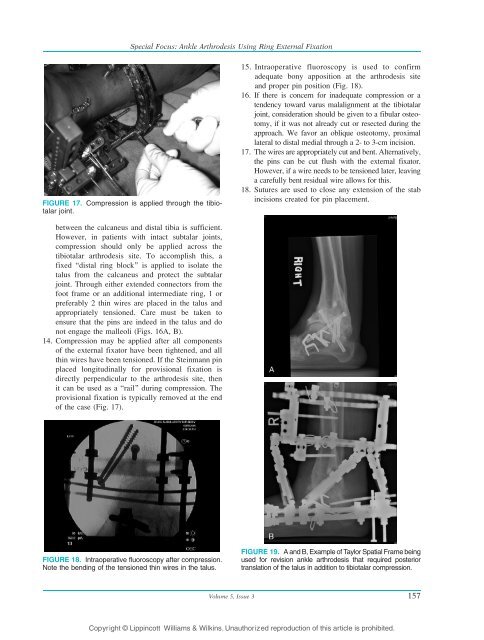
FIGURE 17. Compression is applied through the tibiotalar<br />
joint.<br />
between the calcaneus and distal tibia is sufficient.<br />
However, in patients with intact subtalar joints,<br />
<strong>com</strong>pression should only be applied across the<br />
tibiotalar arthrodesis site. To ac<strong>com</strong>plish this, a<br />
fixed Bdistal ring block^ is applied to isolate the<br />
talus from the calcaneus and protect the subtalar<br />
joint. Through either extended connectors from the<br />
foot frame or an additional intermediate ring, 1 or<br />
preferably 2 thin wires are placed in the talus and<br />
appropriately tensioned. Care must be taken to<br />
ensure that the pins are indeed in the talus and do<br />
not engage the malleoli (Figs. 16A, B).<br />
14. Compression may be applied after all <strong>com</strong>ponents<br />
of the external fixator have been tightened, and all<br />
thin wires have been tensioned. If the Steinmann pin<br />
placed longitudinally for provisional fixation is<br />
directly perpendicular to the arthrodesis site, then<br />
it can be used as a Brail^ during <strong>com</strong>pression. The<br />
provisional fixation is typically removed at the end<br />
of the case (Fig. 17).<br />
FIGURE 18. Intraoperative fluoroscopy after <strong>com</strong>pression.<br />
Note the bending of the tensioned thin wires in the talus.<br />
15. Intraoperative fluoroscopy is used to confirm<br />
adequate bony apposition at the arthrodesis site<br />
and proper pin position (Fig. 18).<br />
16. If there is concern for inadequate <strong>com</strong>pression or a<br />
tendency toward varus malalignment at the tibiotalar<br />
joint, consideration should be given to a fibular osteotomy,<br />
if it was not already cut or resected during the<br />
approach. We favor an oblique osteotomy, proximal<br />
lateral to distal medial through a 2- to 3-cm incision.<br />
17. The wires are appropriately cut and bent. Alternatively,<br />
the pins can be cut flush with the external fixator.<br />
However, if a wire needs to be tensioned later, leaving<br />
a carefully bent residual wire allows for this.<br />
18. Sutures are used to close any extension of the stab<br />
incisions created for pin placement.<br />
FIGURE 19. A and B, Example of Taylor Spatial Frame being<br />
used for revision ankle arthrodesis that required posterior<br />
translation of the talus in addition to tibiotalar <strong>com</strong>pression.<br />
Volume 5, Issue 3 157<br />
Copyright © Lippincott Williams & Wilkins. Unauthorized reproduction of this article is prohibited.