Ankle Arthrodesis Using Ring External Fixation - Orthofix.com
Ankle Arthrodesis Using Ring External Fixation - Orthofix.com
Ankle Arthrodesis Using Ring External Fixation - Orthofix.com

Create successful ePaper yourself
Turn your PDF publications into a flip-book with our unique Google optimized e-Paper software.
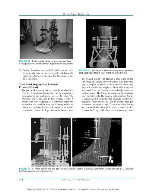
FIGURE 20. Patient weight-bearing with external fixator.<br />
A foot plate with tread has been applied to the foot frame.<br />
19. Sterile dressings are applied and wrapped relatively<br />
tightly over the pins to provide stability at the<br />
skin/wire interface to decrease the likelihood of pin<br />
tract infections.<br />
Traditional Step-by-Step <strong>External</strong><br />
<strong>Fixation</strong> Method<br />
1. The proximal ring/ring block is usually attached first<br />
(Fig. 6). A reference (thin) wire can be placed perpendicular<br />
to the anatomical axis of the tibia. The<br />
proximal ring is attached to this reference wire. A<br />
second thin wire is placed at a different angle and<br />
attached to the proximal ring that is being held in an<br />
orthogonal position. Ideally, this second wire should<br />
be placed as close to 90 degrees from the first to create<br />
FIGURE 22. Radiograph demonstrating loose proximal<br />
pins suspicious for pin tract infection/osteomyelitis.<br />
the greatest stability. In placing 2 thin wires on the<br />
same ring, one should be above and the other below the<br />
ring. If the pins are placed on the same side of the ring,<br />
they will collide and displace. These thin wires are<br />
tensioned. A second ring can be placed and secured in a<br />
similar manner. The 2 rings are connected by sockets or<br />
short threaded rods. The spacing between these 2 rings<br />
is variable and should allow for adequate stability (ie,<br />
adequate space should be left to permit half pin<br />
placement between the rings. To ensure that the 2 rings<br />
are symmetrically oriented, it may be easier to first<br />
connect the 2 rings, slide them onto the lower leg, have<br />
FIGURE 21. A, <strong>Ankle</strong> arthrodesis site obscured by external fixator, making assessment of fusion difficult. B, CT scan to<br />
facilitate assessment of fusion site.<br />
158<br />
Special Focus: Easley et al<br />
Techniques in Foot and <strong>Ankle</strong> Surgery<br />
Copyright © Lippincott Williams & Wilkins. Unauthorized reproduction of this article is prohibited.