

lateral pain syndromes of the foot and ankle - The Podiatry Institute
lateral pain syndromes of the foot and ankle - The Podiatry Institute
lateral pain syndromes of the foot and ankle - The Podiatry Institute

Create successful ePaper yourself
Turn your PDF publications into a flip-book with our unique Google optimized e-Paper software.
14<br />
CHAPTER 3<br />
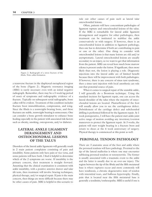
Figure 2. Radiograph <strong>of</strong> a stress fracture <strong>of</strong> <strong>the</strong><br />
fibula. Note callus formation.<br />
a transverse fracture in <strong>the</strong> diaphyseal-metaphyseal region<br />
<strong>of</strong> <strong>the</strong> bone (Figure 2). Magnetic resonance imaging<br />
(MRI) is rarely necessary even with an initial negative<br />
radiograph. Remember, <strong>the</strong>re may be a 3-week lag period<br />
<strong>of</strong> onset <strong>of</strong> symptoms <strong>and</strong> radiographic evidence <strong>of</strong> a<br />
fracture. Typically on subsequent serial radiographs, bone<br />
callus will be evident. Treatment <strong>of</strong> this condition includes<br />
fracture boot immobilization, compression, <strong>and</strong> icing.<br />
Since <strong>the</strong> fibula is a nonweight-bearing bone, <strong>and</strong> <strong>the</strong>se<br />
fractures are stable, nonweight bearing is unnecessary. One<br />
can consider a bone growth stimulator to enhance bone<br />
healing especially in <strong>the</strong> patient with associated risk factors<br />
such as obesity, smoking, osteoporosis, <strong>and</strong>/or diabetes.<br />
LATERAL ANKLE LIGAMENT<br />
DERANGEMENT AND<br />
OSTEOCHONDRAL LESIONS<br />
OF THE TALUS<br />
Disorders <strong>of</strong> <strong>the</strong> <strong>lateral</strong> <strong>ankle</strong> ligaments will generally result<br />
in 2 main patient complaints consisting <strong>of</strong> <strong>pain</strong> <strong>and</strong><br />
instability. Some patients only have <strong>pain</strong> or vice versa, <strong>and</strong><br />
some patients will have both. I find it helpful to determine<br />
which <strong>of</strong> <strong>the</strong> 2 symptoms are worse. If instability is <strong>the</strong><br />
primary concern, <strong>the</strong>n treatment is straight forward.<br />
Assuming that <strong>the</strong> clinical examination is consistent with<br />
<strong>ankle</strong> instability, with a positive anterior drawer <strong>and</strong> talar<br />
tilt tests, <strong>the</strong>n treatment will involve bracing techniques,<br />
physical <strong>the</strong>rapy, <strong>and</strong>/or surgical repair. If <strong>pain</strong> is <strong>the</strong> main<br />
concern, <strong>the</strong>n things are more difficult because <strong>the</strong>re may<br />
be o<strong>the</strong>r causes <strong>of</strong> <strong>pain</strong>. MRI is helpful in this scenario to<br />
rule out o<strong>the</strong>r causes <strong>of</strong> <strong>pain</strong> such as <strong>lateral</strong> talar<br />
osteochondral lesions.<br />
Often, patients will have concomitant pathologies <strong>of</strong><br />
ligament rupture <strong>and</strong> osteochondral lesions <strong>of</strong> <strong>the</strong> talus.<br />
If <strong>the</strong> MRI is remarkable for <strong>lateral</strong> <strong>ankle</strong> ligament<br />
derangement <strong>and</strong> negative for o<strong>the</strong>r pathologies, <strong>the</strong>n<br />
treatment can be instituted to stabilize <strong>the</strong> <strong>ankle</strong><br />
conservatively or with surgery. If however, <strong>the</strong>re is an<br />
osteochondral lesion in addition to ligament pathology,<br />
<strong>the</strong>n one has to determine if both are contributing to <strong>pain</strong><br />
or one or <strong>the</strong> o<strong>the</strong>r. One thing to consider with<br />
osteochondral lesions is that many lesions are old <strong>and</strong>/or<br />
asymptomatic. Lateral osteochondral lesions are usually<br />
secondary to an injury, so we want to get that information<br />
from <strong>the</strong> patient. MRI can reveal how much bone marrow<br />
edema is present under <strong>the</strong> lesion. If significant, <strong>the</strong>n more<br />
likely than not, <strong>the</strong> lesion is playing a role. Diagnostic<br />
injections into <strong>the</strong> <strong>lateral</strong> <strong>ankle</strong> are <strong>of</strong> limited benefit<br />
because <strong>the</strong>re will be improvement with both pathologies.<br />
If however, <strong>the</strong>re is any concern <strong>of</strong> sinus tarsi syndrome,<br />
<strong>the</strong>n a sinus tarsi injection with local anes<strong>the</strong>sia can rule<br />
out that potential source <strong>of</strong> <strong>pain</strong>.<br />
When it comes to surgical repair <strong>of</strong> <strong>the</strong> unstable <strong>ankle</strong>,<br />
I will generally do a Brostrom technique. Using <strong>the</strong><br />
st<strong>and</strong>ard incision for ligament repair, one can access <strong>the</strong><br />
<strong>lateral</strong> shoulder <strong>of</strong> <strong>the</strong> talus where <strong>the</strong> majority <strong>of</strong> osteochondral<br />
lesions are located. Plantarflexion <strong>of</strong> <strong>the</strong> <strong>foot</strong><br />
will usually allow you to see <strong>the</strong> cartilaginous defect.<br />
Debridement <strong>of</strong> <strong>the</strong> cartilage defect <strong>and</strong> subchondral<br />
drilling is performed followed by <strong>the</strong> ligament repair. At 1<br />
week postoperative, I will have <strong>the</strong> patient start <strong>ankle</strong> joint<br />
active range <strong>of</strong> motion avoiding any inversion/eversion<br />
maneuvers to protect <strong>the</strong> ligament repair. At 3 weeks, <strong>the</strong><br />
patient will start weight bearing in a fracture boot <strong>and</strong><br />
return to shoes at <strong>the</strong> 6 week anniversary <strong>of</strong> surgery.<br />
Physical <strong>the</strong>rapy is commenced at this point as well.<br />
PERONEAL TENDON PATHOLOGY<br />
<strong>The</strong>re are 3 anatomic areas <strong>of</strong> <strong>the</strong> <strong>foot</strong> <strong>and</strong> <strong>ankle</strong> where<br />
<strong>the</strong> peroneal tendons will have pathology. Proximal to <strong>the</strong><br />
tip <strong>of</strong> <strong>the</strong> <strong>lateral</strong> malleolus is where one may encounter<br />
subluxing peroneal tendons or tenosynovitis. <strong>The</strong> former<br />
is usually associated with a traumatic event to <strong>the</strong> <strong>ankle</strong><br />
<strong>and</strong> <strong>the</strong> latter is usually due to an over-use injury. <strong>The</strong><br />
region between <strong>the</strong> tip <strong>of</strong> <strong>the</strong> fibula <strong>and</strong> <strong>the</strong> fifth metatarsal<br />
base is where ruptures occur <strong>and</strong> more commonly will<br />
have tendinosis, a chronic degenerative state <strong>of</strong> tendon<br />
with interstitial tears, <strong>and</strong> bulbous hypertrophy. Finally,<br />
<strong>pain</strong> that is located near <strong>the</strong> fifth metatarsal base is<br />
generally a tendinitis/en<strong>the</strong>sopathy. MRI is useful if one is


