

lateral pain syndromes of the foot and ankle - The Podiatry Institute
lateral pain syndromes of the foot and ankle - The Podiatry Institute
lateral pain syndromes of the foot and ankle - The Podiatry Institute

You also want an ePaper? Increase the reach of your titles
YUMPU automatically turns print PDFs into web optimized ePapers that Google loves.
C H A P T E R 3<br />
LATERAL PAIN SYNDROMES OF THE FOOT<br />
AND ANKLE<br />
William D. Fishco, DPM<br />
<strong>The</strong> majority <strong>of</strong> patient encounters with <strong>the</strong> podiatrist are<br />
secondary to <strong>pain</strong> in <strong>the</strong> <strong>foot</strong> <strong>and</strong>/or <strong>ankle</strong>. If we draw an<br />
imaginary line bisecting <strong>the</strong> lower leg <strong>and</strong> extending<br />
distally to <strong>the</strong> third toe, <strong>pain</strong> in <strong>the</strong> medial aspect <strong>of</strong> <strong>the</strong><br />
<strong>foot</strong> <strong>and</strong> <strong>ankle</strong> is typically straight forward. <strong>The</strong>re is a<br />
predominance <strong>of</strong> medial heel <strong>pain</strong>, which is usually<br />
plantar fasciitis. Medial arch symptoms are seen frequently<br />
in <strong>the</strong> pes valgus <strong>foot</strong> type such has posterior tibial<br />
tendinitis. Less frequently one may see tarsal tunnel<br />
syndrome. First ray <strong>pain</strong> <strong>syndromes</strong> are typically associated<br />
with hallux valgus or hallux limitus; <strong>and</strong> finally,<br />
degenerative joint disease is frequently seen in <strong>the</strong> medial<br />
column <strong>and</strong> <strong>the</strong> second tarsometatarsal joint. Evaluation<br />
<strong>and</strong> diagnosis <strong>of</strong> <strong>the</strong>se disorders can typically be done<br />
without much ambiguity.<br />
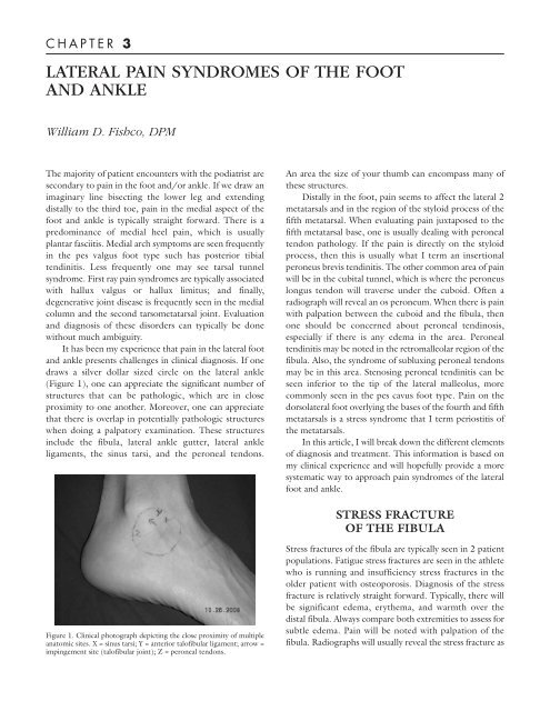
It has been my experience that <strong>pain</strong> in <strong>the</strong> <strong>lateral</strong> <strong>foot</strong><br />
<strong>and</strong> <strong>ankle</strong> presents challenges in clinical diagnosis. If one<br />
draws a silver dollar sized circle on <strong>the</strong> <strong>lateral</strong> <strong>ankle</strong><br />
(Figure 1), one can appreciate <strong>the</strong> significant number <strong>of</strong><br />
structures that can be pathologic, which are in close<br />
proximity to one ano<strong>the</strong>r. Moreover, one can appreciate<br />
that <strong>the</strong>re is overlap in potentially pathologic structures<br />
when doing a palpatory examination. <strong>The</strong>se structures<br />
include <strong>the</strong> fibula, <strong>lateral</strong> <strong>ankle</strong> gutter, <strong>lateral</strong> <strong>ankle</strong><br />
ligaments, <strong>the</strong> sinus tarsi, <strong>and</strong> <strong>the</strong> peroneal tendons.<br />
Figure 1. Clinical photograph depicting <strong>the</strong> close proximity <strong>of</strong> multiple<br />
anatomic sites. X = sinus tarsi; Y = anterior tal<strong>of</strong>ibular ligament; arrow =<br />
impingement site (tal<strong>of</strong>ibular joint); Z = peroneal tendons.<br />
An area <strong>the</strong> size <strong>of</strong> your thumb can encompass many <strong>of</strong><br />
<strong>the</strong>se structures.<br />
Distally in <strong>the</strong> <strong>foot</strong>, <strong>pain</strong> seems to affect <strong>the</strong> <strong>lateral</strong> 2<br />
metatarsals <strong>and</strong> in <strong>the</strong> region <strong>of</strong> <strong>the</strong> styloid process <strong>of</strong> <strong>the</strong><br />
fifth metatarsal. When evaluating <strong>pain</strong> juxtaposed to <strong>the</strong><br />
fifth metatarsal base, one is usually dealing with peroneal<br />
tendon pathology. If <strong>the</strong> <strong>pain</strong> is directly on <strong>the</strong> styloid<br />
process, <strong>the</strong>n this is usually what I term an insertional<br />
peroneus brevis tendinitis. <strong>The</strong> o<strong>the</strong>r common area <strong>of</strong> <strong>pain</strong><br />
will be in <strong>the</strong> cubital tunnel, which is where <strong>the</strong> peroneus<br />
longus tendon will traverse under <strong>the</strong> cuboid. Often a<br />
radiograph will reveal an os peroneum. When <strong>the</strong>re is <strong>pain</strong><br />
with palpation between <strong>the</strong> cuboid <strong>and</strong> <strong>the</strong> fibula, <strong>the</strong>n<br />
one should be concerned about peroneal tendinosis,<br />
especially if <strong>the</strong>re is any edema in <strong>the</strong> area. Peroneal<br />
tendinitis may be noted in <strong>the</strong> retromalleolar region <strong>of</strong> <strong>the</strong><br />
fibula. Also, <strong>the</strong> syndrome <strong>of</strong> subluxing peroneal tendons<br />
may be in this area. Stenosing peroneal tendinitis can be<br />
seen inferior to <strong>the</strong> tip <strong>of</strong> <strong>the</strong> <strong>lateral</strong> malleolus, more<br />
commonly seen in <strong>the</strong> pes cavus <strong>foot</strong> type. Pain on <strong>the</strong><br />
dorso<strong>lateral</strong> <strong>foot</strong> overlying <strong>the</strong> bases <strong>of</strong> <strong>the</strong> fourth <strong>and</strong> fifth<br />
metatarsals is a stress syndrome that I term periostitis <strong>of</strong><br />
<strong>the</strong> metatarsals.<br />
In this article, I will break down <strong>the</strong> different elements<br />
<strong>of</strong> diagnosis <strong>and</strong> treatment. This information is based on<br />
my clinical experience <strong>and</strong> will hopefully provide a more<br />
systematic way to approach <strong>pain</strong> <strong>syndromes</strong> <strong>of</strong> <strong>the</strong> <strong>lateral</strong><br />
<strong>foot</strong> <strong>and</strong> <strong>ankle</strong>.<br />
STRESS FRACTURE<br />
OF THE FIBULA<br />
Stress fractures <strong>of</strong> <strong>the</strong> fibula are typically seen in 2 patient<br />
populations. Fatigue stress fractures are seen in <strong>the</strong> athlete<br />
who is running <strong>and</strong> insufficiency stress fractures in <strong>the</strong><br />
older patient with osteoporosis. Diagnosis <strong>of</strong> <strong>the</strong> stress<br />
fracture is relatively straight forward. Typically, <strong>the</strong>re will<br />
be significant edema, ery<strong>the</strong>ma, <strong>and</strong> warmth over <strong>the</strong><br />
distal fibula. Always compare both extremities to assess for<br />
subtle edema. Pain will be noted with palpation <strong>of</strong> <strong>the</strong><br />
fibula. Radiographs will usually reveal <strong>the</strong> stress fracture as
14<br />
CHAPTER 3<br />
Figure 2. Radiograph <strong>of</strong> a stress fracture <strong>of</strong> <strong>the</strong><br />
fibula. Note callus formation.<br />
a transverse fracture in <strong>the</strong> diaphyseal-metaphyseal region<br />
<strong>of</strong> <strong>the</strong> bone (Figure 2). Magnetic resonance imaging<br />
(MRI) is rarely necessary even with an initial negative<br />
radiograph. Remember, <strong>the</strong>re may be a 3-week lag period<br />
<strong>of</strong> onset <strong>of</strong> symptoms <strong>and</strong> radiographic evidence <strong>of</strong> a<br />
fracture. Typically on subsequent serial radiographs, bone<br />
callus will be evident. Treatment <strong>of</strong> this condition includes<br />
fracture boot immobilization, compression, <strong>and</strong> icing.<br />
Since <strong>the</strong> fibula is a nonweight-bearing bone, <strong>and</strong> <strong>the</strong>se<br />
fractures are stable, nonweight bearing is unnecessary. One<br />
can consider a bone growth stimulator to enhance bone<br />
healing especially in <strong>the</strong> patient with associated risk factors<br />
such as obesity, smoking, osteoporosis, <strong>and</strong>/or diabetes.<br />
LATERAL ANKLE LIGAMENT<br />
DERANGEMENT AND<br />
OSTEOCHONDRAL LESIONS<br />
OF THE TALUS<br />
Disorders <strong>of</strong> <strong>the</strong> <strong>lateral</strong> <strong>ankle</strong> ligaments will generally result<br />
in 2 main patient complaints consisting <strong>of</strong> <strong>pain</strong> <strong>and</strong><br />
instability. Some patients only have <strong>pain</strong> or vice versa, <strong>and</strong><br />
some patients will have both. I find it helpful to determine<br />
which <strong>of</strong> <strong>the</strong> 2 symptoms are worse. If instability is <strong>the</strong><br />
primary concern, <strong>the</strong>n treatment is straight forward.<br />
Assuming that <strong>the</strong> clinical examination is consistent with<br />
<strong>ankle</strong> instability, with a positive anterior drawer <strong>and</strong> talar<br />
tilt tests, <strong>the</strong>n treatment will involve bracing techniques,<br />
physical <strong>the</strong>rapy, <strong>and</strong>/or surgical repair. If <strong>pain</strong> is <strong>the</strong> main<br />
concern, <strong>the</strong>n things are more difficult because <strong>the</strong>re may<br />
be o<strong>the</strong>r causes <strong>of</strong> <strong>pain</strong>. MRI is helpful in this scenario to<br />
rule out o<strong>the</strong>r causes <strong>of</strong> <strong>pain</strong> such as <strong>lateral</strong> talar<br />
osteochondral lesions.<br />
Often, patients will have concomitant pathologies <strong>of</strong><br />
ligament rupture <strong>and</strong> osteochondral lesions <strong>of</strong> <strong>the</strong> talus.<br />
If <strong>the</strong> MRI is remarkable for <strong>lateral</strong> <strong>ankle</strong> ligament<br />
derangement <strong>and</strong> negative for o<strong>the</strong>r pathologies, <strong>the</strong>n<br />
treatment can be instituted to stabilize <strong>the</strong> <strong>ankle</strong><br />
conservatively or with surgery. If however, <strong>the</strong>re is an<br />
osteochondral lesion in addition to ligament pathology,<br />
<strong>the</strong>n one has to determine if both are contributing to <strong>pain</strong><br />
or one or <strong>the</strong> o<strong>the</strong>r. One thing to consider with<br />
osteochondral lesions is that many lesions are old <strong>and</strong>/or<br />
asymptomatic. Lateral osteochondral lesions are usually<br />
secondary to an injury, so we want to get that information<br />
from <strong>the</strong> patient. MRI can reveal how much bone marrow<br />
edema is present under <strong>the</strong> lesion. If significant, <strong>the</strong>n more<br />
likely than not, <strong>the</strong> lesion is playing a role. Diagnostic<br />
injections into <strong>the</strong> <strong>lateral</strong> <strong>ankle</strong> are <strong>of</strong> limited benefit<br />
because <strong>the</strong>re will be improvement with both pathologies.<br />
If however, <strong>the</strong>re is any concern <strong>of</strong> sinus tarsi syndrome,<br />
<strong>the</strong>n a sinus tarsi injection with local anes<strong>the</strong>sia can rule<br />
out that potential source <strong>of</strong> <strong>pain</strong>.<br />
When it comes to surgical repair <strong>of</strong> <strong>the</strong> unstable <strong>ankle</strong>,<br />
I will generally do a Brostrom technique. Using <strong>the</strong><br />
st<strong>and</strong>ard incision for ligament repair, one can access <strong>the</strong><br />
<strong>lateral</strong> shoulder <strong>of</strong> <strong>the</strong> talus where <strong>the</strong> majority <strong>of</strong> osteochondral<br />
lesions are located. Plantarflexion <strong>of</strong> <strong>the</strong> <strong>foot</strong><br />
will usually allow you to see <strong>the</strong> cartilaginous defect.<br />
Debridement <strong>of</strong> <strong>the</strong> cartilage defect <strong>and</strong> subchondral<br />
drilling is performed followed by <strong>the</strong> ligament repair. At 1<br />
week postoperative, I will have <strong>the</strong> patient start <strong>ankle</strong> joint<br />
active range <strong>of</strong> motion avoiding any inversion/eversion<br />
maneuvers to protect <strong>the</strong> ligament repair. At 3 weeks, <strong>the</strong><br />
patient will start weight bearing in a fracture boot <strong>and</strong><br />
return to shoes at <strong>the</strong> 6 week anniversary <strong>of</strong> surgery.<br />
Physical <strong>the</strong>rapy is commenced at this point as well.<br />
PERONEAL TENDON PATHOLOGY<br />
<strong>The</strong>re are 3 anatomic areas <strong>of</strong> <strong>the</strong> <strong>foot</strong> <strong>and</strong> <strong>ankle</strong> where<br />
<strong>the</strong> peroneal tendons will have pathology. Proximal to <strong>the</strong><br />
tip <strong>of</strong> <strong>the</strong> <strong>lateral</strong> malleolus is where one may encounter<br />
subluxing peroneal tendons or tenosynovitis. <strong>The</strong> former<br />
is usually associated with a traumatic event to <strong>the</strong> <strong>ankle</strong><br />
<strong>and</strong> <strong>the</strong> latter is usually due to an over-use injury. <strong>The</strong><br />
region between <strong>the</strong> tip <strong>of</strong> <strong>the</strong> fibula <strong>and</strong> <strong>the</strong> fifth metatarsal<br />
base is where ruptures occur <strong>and</strong> more commonly will<br />
have tendinosis, a chronic degenerative state <strong>of</strong> tendon<br />
with interstitial tears, <strong>and</strong> bulbous hypertrophy. Finally,<br />
<strong>pain</strong> that is located near <strong>the</strong> fifth metatarsal base is<br />
generally a tendinitis/en<strong>the</strong>sopathy. MRI is useful if one is
concerned about tendinosis. Conservative treatments are<br />
rarely helpful for tendinosis <strong>and</strong> will generally require a<br />
surgical intervention.<br />
<strong>The</strong>re is a favorable prognosis with conservative<br />
treatment for peroneal tendinitis that generally will include<br />
some combination <strong>of</strong> anti-inflammatory medication,<br />
immobilization, physical <strong>the</strong>rapy, <strong>and</strong> orthoses. A word <strong>of</strong><br />
caution with cortisone injections is prudent in peroneal<br />
pathology. One may consider a cortisone injection into <strong>the</strong><br />
cubital tunnel for localized <strong>pain</strong> that does not respond to<br />
o<strong>the</strong>r treatments. <strong>The</strong> injection into <strong>the</strong> cubital tunnel is<br />
inferior to <strong>the</strong> peroneus brevis tendon. For o<strong>the</strong>r areas<br />
along <strong>the</strong> course <strong>of</strong> <strong>the</strong> tendons, unless <strong>the</strong> patient will be<br />
immobilized, I will not consider a cortisone injection.<br />
I have seen too many tendon ruptures in this area after<br />
cortisone injections (Figures 3 <strong>and</strong> 4).<br />
SINUS TARSI SYNDROME<br />
This is one <strong>of</strong> those disorders that nobody really knows exactly<br />
what it is, but it can be successfully treated.<br />
Diagnosis <strong>of</strong> sinus tarsi syndrome is straight forward. When<br />
<strong>the</strong>re is <strong>pain</strong> in <strong>the</strong> sinus tarsi on palpation <strong>and</strong> symptoms<br />
resolve after injection <strong>of</strong> local anes<strong>the</strong>tic, one can be<br />
confident with <strong>the</strong> diagnosis even without fur<strong>the</strong>r testing.<br />
Certainly radiographic evaluation is necessary to assess for<br />
advanced arthritis <strong>of</strong> <strong>the</strong> subtalar joint. MRI can be done<br />
to exclude o<strong>the</strong>r pathologies. My personal opinion is that<br />
sinus tarsi syndrome is a synovitis whe<strong>the</strong>r it is a chronic<br />
condition caused by mechanical influences <strong>of</strong> <strong>the</strong> pes valgus<br />
<strong>foot</strong> type or some kind <strong>of</strong> acute inversion injury.<br />
Instability <strong>of</strong> <strong>the</strong> subtalar joint has been postulated as<br />
Figure 3. Clinical view <strong>of</strong> <strong>the</strong> area <strong>of</strong> <strong>the</strong> <strong>foot</strong> that cortisone should be<br />
avoided (danger zone) <strong>and</strong> a safe area for cortisone in <strong>the</strong> cubital tunnel<br />
(OK). <strong>The</strong> dashed line represents <strong>the</strong> peroneal tendon course.<br />
CHAPTER 3 15<br />
a source <strong>of</strong> apparent <strong>ankle</strong> instability. If <strong>the</strong>re is disruption<br />
<strong>of</strong> <strong>the</strong> talocalcaneal ligaments <strong>and</strong> <strong>the</strong> cervical ligament,<br />
<strong>the</strong>n it seems likely that like <strong>the</strong> <strong>ankle</strong>, this can contribute<br />
to instability. It is difficult to make a clinical call on<br />
subtalar joint instability. Stress testing <strong>of</strong> <strong>the</strong> subtalar joint<br />
is difficult to do without influence <strong>of</strong> <strong>the</strong> <strong>ankle</strong> joint. My<br />
personal opinion is that if <strong>the</strong> talar tilt test is equivocal <strong>and</strong><br />
<strong>the</strong> anterior drawer test is positive for laxity, <strong>the</strong>n it is<br />
generally <strong>ankle</strong> ligament instability. Conversely, if <strong>the</strong><br />
anterior drawer test seems normal <strong>and</strong> <strong>the</strong> talar tilt test is<br />
not, <strong>the</strong>n subtalar joint instability may be playing a role.<br />
We know that in <strong>ankle</strong> sprains, it would be unlikely that<br />
<strong>the</strong> calcane<strong>of</strong>ibular ligament ruptures leaving an intact<br />
anterior tal<strong>of</strong>ibular ligament.<br />
If instability <strong>of</strong> subtalar joint plays a clinical role, <strong>the</strong>n<br />
why aren’t <strong>the</strong>re reports <strong>of</strong> instability following sinus tarsi<br />
decompressions that involve excision <strong>of</strong> <strong>the</strong> entire contents<br />
including <strong>the</strong> ligaments? It would seem likely that<br />
everybody who had a sinus tarsi decompression would<br />
have symptoms <strong>of</strong> instability postoperatively, however that<br />
does not seem to be <strong>the</strong> case.<br />
Treatment <strong>of</strong> sinus tarsi syndrome will typically<br />
involve oral anti-inflammatory medications, cortisone<br />
injections, physical <strong>the</strong>rapy, <strong>and</strong>/or biomechanical control<br />
with orthotic devices. In cases <strong>of</strong> arthritis, subtalar<br />
arthrodesis may be necessary. Sinus tarsi decompression<br />
may be <strong>of</strong> benefit in recalcitrant cases. In confirmed<br />
instability <strong>of</strong> <strong>the</strong> subtalar joint, a split peroneus longus<br />
tenodesis <strong>ankle</strong> stabilization procedure may be a good<br />
approach as <strong>the</strong> 2-ligament repair will also cross <strong>the</strong><br />
subtalar joint. <strong>The</strong>refore, both <strong>the</strong> <strong>ankle</strong> <strong>and</strong> subtalar joints<br />
are stabilized.<br />
Figure 4. Intraoperative view that depicts a partial rupture <strong>of</strong> <strong>the</strong><br />
peroneus brevis tendon. <strong>The</strong>re is calcification within <strong>the</strong> tendon. This<br />
patient had prior cortisone injections in this area.
16<br />
CHAPTER 3<br />
IMPINGEMENT OF THE<br />
TALOFIBULAR JOINT<br />
Ano<strong>the</strong>r source <strong>of</strong> <strong>lateral</strong> <strong>ankle</strong> <strong>pain</strong> may be an impingement<br />
between <strong>the</strong> fibula <strong>and</strong> talus in <strong>the</strong> <strong>lateral</strong> <strong>ankle</strong> gutter<br />
(Figure 5). In this syndrome, <strong>pain</strong> is usually well defined at<br />
<strong>the</strong> tip <strong>of</strong> <strong>the</strong> fibula. A lesion marker at <strong>the</strong> site <strong>of</strong> <strong>pain</strong> may<br />
help with radiographic interpretation. If <strong>the</strong> palpatory<br />
examination reveals <strong>pain</strong> not only on <strong>the</strong> tip <strong>of</strong> <strong>the</strong><br />
malleolus, but also in <strong>the</strong> gutter <strong>and</strong> antero<strong>lateral</strong> <strong>ankle</strong> joint,<br />
<strong>the</strong>n consider an MRI for assessment <strong>of</strong> an osteochondral<br />
lesion (Figure 6). A diagnostic injection <strong>of</strong> local anes<strong>the</strong>sia<br />
may aid in <strong>the</strong> diagnosis especially with ruling out o<strong>the</strong>r<br />
sources <strong>of</strong> <strong>pain</strong>. Treatment will usually require surgical<br />
intervention, which can be done arthroscopically or through<br />
an open approach. Conservative treatments will typically<br />
involve biomechanical control with orthotics.<br />
PERIOSITIS OF THE<br />
METATARSALS<br />
Pain in <strong>the</strong> dorso<strong>lateral</strong> <strong>foot</strong> overlying <strong>the</strong> bases <strong>of</strong> <strong>the</strong><br />
fourth <strong>and</strong> fifth metatarsals is usually a stress syndrome<br />
(Figure 7). Patients will complain <strong>of</strong> a sharp stabbing <strong>pain</strong><br />
or a dull ache upon activity. Aggravating symptoms include<br />
bare<strong>foot</strong> walking or wearing flimsy shoes. In my practice,<br />
I see more <strong>of</strong> this problem in <strong>the</strong> summer when people are<br />
wearing flip flops <strong>and</strong> <strong>and</strong>/or going bare<strong>foot</strong> around <strong>the</strong><br />
Figure 5. Radiograph <strong>of</strong> an <strong>ankle</strong> illustrating an<br />
osseous impingement <strong>of</strong> <strong>the</strong> tal<strong>of</strong>ibular joint as<br />
well as a suspicious looking <strong>lateral</strong> shoulder <strong>of</strong> <strong>the</strong><br />
talus. This patient had chronic <strong>lateral</strong> <strong>ankle</strong> <strong>pain</strong><br />
that upon examination revealed <strong>pain</strong> in <strong>the</strong><br />
antero<strong>lateral</strong> gutter <strong>of</strong> <strong>the</strong> <strong>ankle</strong> <strong>and</strong> on <strong>the</strong><br />
inferior aspect <strong>of</strong> <strong>the</strong> fibula.<br />
house. Treatment involves anti-inflammatory medication,<br />
wearing supportive stiff soled shoes, <strong>and</strong> orthotics.<br />
Surgery is usually unnecessary as this problem generally<br />
gets better with nonoperative <strong>the</strong>rapies. <strong>The</strong> notable<br />
exception would be <strong>the</strong> patient that presents with <strong>the</strong>se<br />
symptoms <strong>and</strong> <strong>the</strong>re is an underlying compound deformity<br />
<strong>of</strong> <strong>the</strong> <strong>foot</strong>, such as <strong>the</strong> equinovarus <strong>foot</strong>, contributing to<br />
excessive stress.<br />
COMMON DENOMINATOR IN<br />
LATERAL PAIN SYNDROMES<br />
When we carefully look at <strong>the</strong> common link with most<br />
<strong>of</strong> <strong>the</strong>se <strong>lateral</strong> <strong>pain</strong> <strong>syndromes</strong>, <strong>the</strong> cavus <strong>foot</strong> <strong>and</strong><br />
metatarsus adductus <strong>foot</strong> type are typically seen. <strong>The</strong><br />
notable exception is sinus tarsi syndrome, which is seen<br />
more <strong>of</strong>ten in <strong>the</strong> pronated <strong>foot</strong> type. <strong>The</strong> high arched<br />
<strong>foot</strong> is vulnerable to <strong>lateral</strong> <strong>ankle</strong> injuries especially if <strong>the</strong>re<br />
is a fore<strong>foot</strong> valgus. <strong>The</strong> metatarsus adductus <strong>foot</strong> type will<br />
be a <strong>lateral</strong> over loader in gait, which will contribute to<br />
periostitis <strong>of</strong> <strong>the</strong> metatarsals <strong>and</strong> peroneal tendon<br />
pathology. Lateral <strong>ankle</strong> bony impingement is most likely<br />
a due to repetitive strain <strong>and</strong> stresses on <strong>the</strong> <strong>lateral</strong> <strong>ankle</strong><br />
joint <strong>and</strong>/or acute injuries resulting in avulsions that heal<br />
causing exostoses. When reviewing cases <strong>of</strong> Jones fractures<br />
<strong>and</strong> avulsion fractures <strong>of</strong> <strong>the</strong> fifth metatarsal, more <strong>of</strong>ten<br />
than not, <strong>the</strong>se patients have a component <strong>of</strong> metatarsus<br />
adductus (Figures 8 <strong>and</strong> 9).<br />
Figure 6. MRI <strong>of</strong> <strong>the</strong> same patient in Figure 5. Note <strong>the</strong> tal<strong>of</strong>ibular<br />
impingement <strong>and</strong> osteochondral lesion <strong>of</strong> <strong>the</strong> <strong>lateral</strong> shoulder <strong>of</strong> <strong>the</strong> talus.
Figure 7. Radiographs <strong>of</strong> a patient who underwent<br />
cavus <strong>foot</strong> reconstructive surgery. Note that <strong>the</strong>re<br />
is cortical hypertrophy <strong>of</strong> <strong>the</strong> fourth <strong>and</strong> fifth<br />
metatarsals. This is consistent with <strong>lateral</strong> overloading<br />
<strong>of</strong> <strong>the</strong> <strong>foot</strong> that can cause dorso<strong>lateral</strong> <strong>foot</strong><br />
<strong>pain</strong>.<br />
Figure 9. Radiograph <strong>of</strong> a Jones fracture. Note <strong>the</strong><br />
underlying metatarsus adductus.<br />
When a patient presents to <strong>the</strong> <strong>of</strong>fice with <strong>lateral</strong> <strong>ankle</strong><br />
<strong>and</strong>/or hind <strong>foot</strong> complaints, diagnosis is more difficult<br />
than medial complaints due to <strong>the</strong> proximity <strong>of</strong> anatomic<br />
structures <strong>of</strong> joints, ligaments, <strong>and</strong> tendons. Careful interpretation<br />
<strong>of</strong> symptoms including <strong>pain</strong> <strong>and</strong> instability in<br />
conjunction with a thorough clinical examination is<br />
paramount. <strong>The</strong> examination should include careful<br />
palpatory <strong>and</strong> range <strong>of</strong> motion maneuvers, laxity testing<br />
<strong>of</strong> <strong>the</strong> <strong>ankle</strong>, manual muscle testing <strong>of</strong> <strong>the</strong> peroneals,<br />
CHAPTER 3 17<br />
Figure 8. Radiograph <strong>of</strong> an avulsion fracture <strong>of</strong> <strong>the</strong><br />
fifth metatarsal. Note <strong>the</strong> underlying metatarsus<br />
adductus.<br />
Figure 10. MRI illustrating a stress fracture <strong>of</strong> <strong>the</strong> cuboid. This is a less<br />
common source <strong>lateral</strong> <strong>foot</strong> <strong>pain</strong>.<br />
diagnostic anes<strong>the</strong>tic injections, radiographs <strong>and</strong> MRI if<br />
necessary. MRI is generally used as a confirmatory study as<br />
we typically know what is wrong <strong>and</strong> want to rule out<br />
o<strong>the</strong>r less common pathologies (Figure 10).<br />
For <strong>the</strong>se clinically challenging cases, I find it<br />
particularly helpful to examine patients multiple times.<br />
Subsequent examinations on a patient with a complicated<br />
case make <strong>the</strong> clinical diagnosis clearer. I always reevaluate<br />
a patient after an MRI so that I can focus on MRI
18<br />
CHAPTER 3<br />
pathology <strong>and</strong> confirm clinical correlation. Remember, we<br />
do not treat radiographs/MRIs. I have found that<br />
peroneal pathology such as split tears are very common in<br />
older patients, but many times do not clinically correlate<br />
with <strong>the</strong> patient’s symptoms. Once <strong>the</strong> clinical diagnosis is<br />
made, effective treatment can be instituted.<br />
Surgery is typically considered for <strong>lateral</strong> <strong>ankle</strong><br />
instability with <strong>pain</strong>, osteochondral lesions <strong>of</strong> <strong>the</strong> talus,<br />
impingement <strong>syndromes</strong>, <strong>and</strong> tendinosis <strong>of</strong> <strong>the</strong> peroneal<br />
tendons. Conservative care is usually successful in <strong>the</strong><br />
treatment <strong>of</strong> periostitis, stress fractures, acute tendinitis,<br />
sinus tarsi syndrome, <strong>and</strong> acute <strong>ankle</strong> sprains. For orthotic<br />
management in <strong>the</strong> cavus <strong>foot</strong> type, I will typically try to<br />
address <strong>the</strong> fore<strong>foot</strong> valgus with a first ray cutout <strong>and</strong> a<br />
dancer’s pad. A valgus wedge on <strong>the</strong> orthotic device may<br />
also be <strong>of</strong> benefit to reduce <strong>lateral</strong> <strong>foot</strong> strain.<br />
I have some final thoughts on <strong>the</strong> subject <strong>of</strong><br />
reconstructive surgery. When conservative care is not<br />
satisfactory for <strong>the</strong> patient, <strong>the</strong>n one needs to consider<br />
surgical intervention. I think it is important to determine<br />
<strong>the</strong> biomechanic reason for <strong>the</strong> underlying pathology. For<br />
example, in <strong>the</strong> case <strong>of</strong> a rigid pes cavus deformity with<br />
<strong>the</strong> chief complaint/diagnosis <strong>of</strong> <strong>lateral</strong> <strong>ankle</strong> instability<br />
<strong>and</strong> peroneal tendinosis, <strong>the</strong> repair <strong>of</strong> <strong>the</strong> tendon <strong>and</strong><br />
ligaments may not be prudent in <strong>the</strong> long run. This is one<br />
<strong>of</strong> those scenarios when a long discussion with your<br />
patient is necessary to explain why <strong>the</strong> problem started <strong>and</strong><br />
why merely repairing <strong>the</strong> “defects” may not prevent<br />
recurrence. It may seem aggressive or overkill to <strong>the</strong><br />
patient to suggest a Dwyer calcaneal osteotomy, DFWO <strong>of</strong><br />
<strong>the</strong> first metatarsal, along with <strong>lateral</strong> <strong>ankle</strong> ligament<br />
repair, for a “weak <strong>ankle</strong>.” In a nutshell, this is what makes<br />
<strong>the</strong> podiatrist special, not only fixing a problem (i.e., a<br />
torn tendon), but assessing <strong>the</strong> biomechanics <strong>of</strong> <strong>the</strong><br />
lower extremity <strong>and</strong> incorporating that into <strong>the</strong> overall<br />
treatment plan.


