Insect Dissection Lab (III)
Insect Dissection Lab (III)
Insect Dissection Lab (III)

Create successful ePaper yourself
Turn your PDF publications into a flip-book with our unique Google optimized e-Paper software.
Name:______________________<br />
<strong>Insect</strong> <strong>Dissection</strong> <strong>Lab</strong> (Part 3)<br />
<strong>Insect</strong> Physiology (Ento 306)<br />
Introduction: Like the two previous labs, this handout provides you with the basic instructions<br />
for dissection and an outline of the tissues, organs, and structures that you will need to be<br />
familiar with and draw. All questions you need to ask or things you need to draw are<br />
underlined. Attached is a sheet of circles in which you will make all your drawings which must<br />
include a short description and magnification used. Draw the structures as accurately as<br />
possible but don’t worry; you’re being graded on accuracy and participation, not artistic ability.<br />
Make use of the appendix which is a collection of diagrams from the textbook and literature<br />
that will be helpful in finding the correct structures and organs.<br />
Supplies:<br />
-‐ Saline Solution<br />
-‐ <strong>Dissection</strong> probes<br />
-‐ Forceps<br />
-‐ Scissors<br />
-‐ Razor blade<br />
-‐ <strong>Insect</strong>/dissection pins<br />
-‐ <strong>Dissection</strong> pan<br />
-‐ Stereoscopic dissecting microscope<br />
-‐ Compound microscope<br />
-‐ Microscope slide and coverslip<br />
-‐ Freshly sacrificed organism:<br />
-‐ South American cockroach (Blaberus sp.)<br />
-‐ Tobacco/Tomato hornworm (Manduca sexta)<br />
-‐ Tobacco budworm (Heliothis virescens)
<strong>Dissection</strong> of cockroach nervous system<br />
Ventral nerve cord and ganglia<br />
<strong>Insect</strong> Physiology (Ento 306)<br />
Remove the wings by cutting their attachments with a pair of scissors. The dissection is<br />
facilitated if the legs are removed by cutting across their trochanters. Insert the blade of a pair<br />
of fine scissors in the lateral margin of the body between the tergite and sternite. Insert the<br />
blade only as deep as is necessary to penetrate the exoskeleton and be careful you do not<br />
damage internal organs with deep cuts. Cut anteriorly along the right side of the tergites all the<br />
way to the anterior end of the pronotum. Cut transversely across the anterior margin of the<br />
pronotum, just posterior to the head, and upon reaching the left side, change directions and cut<br />
posteriorly along the left side all the way back to the last tergite. Make a transverse cut through<br />
the exoskeleton across the posterior border of the last tergite. You have now cut all of the way<br />
around the dorsum.<br />
Remove the dorsal diaphragm along with the tergal muscles, tracheae, and heart from the<br />
abdomen. Accomplish this by cutting with fine scissors around the periphery of the abdomen.<br />
Remove the gut, fat body, reproductive organs from the thorax and abdomen. Remove the<br />
longitudinal body wall muscles (sternal muscles) and connective tissue as necessary from the<br />
floor of the abdominal cavity to reveal the ventral nerve cord.<br />
Identify and draw one of the three thoracic ganglia. From each extend several pairs of nerves to<br />
the abundant muscles of these segments.<br />
Cockroach brain<br />
Using your scissors and forceps or a razor blade carefully remove the epicranium (top of the<br />
head) from the region between the compound eyes and antennae. Remove muscles as<br />
necessary to reveal the bright white, dorsal brain between the compound eyes.<br />
Draw the cockroach brain in as much detail as possible. <strong>Lab</strong>el at least two of the following<br />
features:<br />
-‐ Optic lobe<br />
-‐ Protocerebrum<br />
-‐ Deutocerebrum<br />
-‐ Tritocerebrum<br />
-‐ Corpora cardiac<br />
-‐ Corpora allata<br />
-‐ Circumesophogeal connective
<strong>Dissection</strong> of Tobacco hornworm caterpillar<br />
External features<br />
<strong>Insect</strong> Physiology (Ento 306)<br />
Manipulate the caterpillar so you are able to draw it in an en face position. Draw what you see<br />
and label the following features using the stereo dissecting microscope:<br />
-‐ <strong>Lab</strong>rum<br />
-‐ Mandibles<br />
-‐ Maxilla/maxillary palp<br />
-‐ Spinneret (for producing silk)<br />
-‐ Antennae<br />
-‐ Ommatidia<br />
-‐ Legs<br />
Identify the following structures involved in walking on the caterpillar<br />
-‐ Proleg<br />
-‐ Anal proleg/Anal clasper<br />
Identify and draw a spiracle<br />
Caterpillar hemocoel dissection<br />
Cut off the caudal horn at its base. Insert scissors into the hole from the cut-‐off horn and cut<br />
through skin and body muscle along a line down the center of the caterpillar’s dorsal side. Cut<br />
from caudal to rostral (head) end. Pull open the exoskeleton and use dissection pins to hold the<br />
exoskeleton and body muscles open by pinning through the body wall at an outward angle.<br />
Bathe the caterpillar in saline solution.<br />
Identify the gastrointestinal tract, fat body, and trachea.<br />
Gastro-‐intestinal tract<br />
Use forceps to tear the esophagus and lift out the digestive track carefully.<br />
Identify the following features<br />
-‐ Crop/midgut<br />
-‐ Malphigian tubules<br />
-‐ Proctodeal valve<br />
-‐ Intestine<br />
-‐ Rectum
<strong>Insect</strong> Physiology (Ento 306)<br />
Note that the malphigian tubule tips are attached to the rectum. This is termed the crypto-‐<br />
nephridial arrangement.<br />
Estimate what % of the caterpillar body cavity is made up of the caterpillar gut<br />
________% of body cavity<br />
Nervous system<br />
Locate the caterpillar’s ventral nerve cord. Identify the abdominal and thoracic ganglia.<br />
Draw the entire dissected caterpillar under low magnification highlighting the nerve cord and<br />
label the ganglia. Do not include the gastrointestinal tract, trachea, or fatbody.<br />
Tease or cut open the caterpillar’s head capsule and locate the brain.<br />
<strong>Dissection</strong> of the Tobacco budworm moth<br />
External features<br />
Draw the moth’s head and label the following features:<br />
-‐ <strong>Lab</strong>ial palps<br />
-‐ Proboscis<br />
-‐ Compound eyes<br />
-‐ Antennae<br />
Moth hemocoel cross section<br />
Cut off the moth’s wings and lay it ventral side up in the dissection pan. Using a razor blade cut<br />
through the moth sagitally. Lay the two halves on their side and use a probe or forceps to<br />
spread open the abdomen. Identify the flight muscles, trachea reproductive organs (include the<br />
male ‘hair pencil’ or female ovarioles), and if possible the gastrointestinal tract. Draw the<br />
moth’s internal anatomy in cross section labeling the flight muscle and reproductive organs.<br />
Estimate what percentage of the body cavity these two sets of tissues occupy.<br />
__________%<br />
Think about the generalized anatomy of a grasshopper or cockroach compared to that of<br />
the caterpillar and moth. What does this say about the behavioral ‘priorities’ of these<br />
different organisms?
<strong>Dissection</strong> illustration sheets<br />
Description:<br />
Magnification:<br />
Description:<br />
Magnification:<br />
Description:<br />
Magnification:<br />
Description:<br />
Magnification:<br />
<strong>Insect</strong> Physiology (Ento 306)
Description:<br />
Magnification:<br />
Description:<br />
Magnification:<br />
Description:<br />
Magnification:<br />
Description:<br />
Magnification:<br />
<strong>Insect</strong> Physiology (Ento 306)
Description:<br />
Magnification:<br />
Description:<br />
Magnification:<br />
Description:<br />
Magnification:<br />
Description:<br />
Magnification:<br />
<strong>Insect</strong> Physiology (Ento 306)
APPENDIX<br />
Anterior view of the head of Periplaneta fuliginosa<br />
Illustrations by Richard Fox http://webs.lander.edu/rsfox/invertebrates/periplaneta.html<br />
<strong>Insect</strong> Physiology (Ento 306)<br />
Dorsal view of the perivisceral coelom of a male P. americana. The dorsal diaphragm,<br />
heart, and tergal muscles have been removed. Abdominal segments are numbered.<br />
Malpighian tubules have been shortened and reduced in number for clarity.<br />
Illustrations by Richard Fox http://webs.lander.edu/rsfox/invertebrates/periplaneta.html
<strong>Insect</strong> Physiology (Ento 306)<br />
<strong>Dissection</strong> of P. americana showing nervous system. T = thoracic ganglion, A =<br />
abdominal ganglion. The abdominal segments are numbered.<br />
Generalized insect brain in lateral view.
<strong>Insect</strong> Physiology (Ento 306)<br />
Diagrammatic cross section of a generalized insect abdominal segment. Redrawn from<br />
Snodgrass (1935)<br />
Illustrations by Richard Fox (http://webs.lander.edu/rsfox/invertebrates/romalea.html)<br />
External anatomy of a generalized caterpillar. Redrawn and modified from Snodgrass<br />
(1935).<br />
Illustrations by Richard Fox (http://webs.lander.edu/rsfox/invertebrates/papilio.html)
<strong>Insect</strong> Physiology (Ento 306)<br />
Dorsal view of the head capsule of the larvae of Papilio polyxenes. Anterior is at the top.<br />
Internal anatomy of a generalized caterpillar viewed from the left. Two of the three left<br />
Malpighian tubules have been truncated for clarity. Redrawn and modified from<br />
Snodgrass (1935).
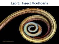
Lepidopteran mouthparts<br />
Internal anatomy of an adult and larval lepidopteran.<br />
<strong>Insect</strong> Physiology (Ento 306)