munson necropsy - UC Davis School of Veterinary Medicine ...
munson necropsy - UC Davis School of Veterinary Medicine ...
munson necropsy - UC Davis School of Veterinary Medicine ...

Create successful ePaper yourself
Turn your PDF publications into a flip-book with our unique Google optimized e-Paper software.
Compiled by:<br />
Linda Munson, DVM, PhD, D-ACVP<br />
Wildlife Health Center<br />
<strong>School</strong> <strong>of</strong> <strong>Veterinary</strong> <strong>Medicine</strong><br />
University <strong>of</strong> California, <strong>Davis</strong><br />
Send correspondence to:<br />
Dr. Linda Munson<br />
VM: PMI, University <strong>of</strong> California<br />
<strong>Davis</strong>, CA 95616 USA<br />
e-mail: l<strong>munson</strong>@ucdavis.edu<br />
Necropsy <strong>of</strong> Wild Animals<br />
The author thanks the following veterinarians and veterinary pathologists for their valuable input: W.B. Karesh, M.F. McEntee, L.J. Lowenstine,<br />
M.E. Roelke-Parker, E. Williams, M.H. Woodford and D. Haines for her illustrations.<br />
The “Necropsy Procedures for Wild Animals” is also available in Portugese, French, and Spanish from the Wildlife Conservation Society website<br />
(www.wcs.org/science/wildlifehealthsci/fieldvet/fvp-techpages.html).
Introduction: Why <strong>necropsy</strong> wild animals?<br />
Preparing for the <strong>necropsy</strong><br />
Equipment<br />
Safety precautions<br />
Preparation <strong>of</strong> sample containers<br />
Performing the Necropsy<br />
General considerations<br />
Checking for anthrax<br />
Handling decomposed carcasses<br />
Examining the carcass and its environment<br />
TABLE OF CONTENTS<br />
Tissue sampling procedures<br />
For histology, microbiology, serology, toxicology, parasitology, and cytology<br />
General steps to performing the <strong>necropsy</strong><br />
Carcass dissection using the carnivore as a model<br />
Carnivores<br />
Ungulates (including ruminants and equids)<br />
Birds<br />
Reptiles (snake)<br />
Post-Necropsy<br />
Disinfecting the <strong>necropsy</strong> site<br />
Storage or submission <strong>of</strong> samples<br />
Dealing with an epidemic<br />
APPENDIX I<br />
Formulations for sample preservatives<br />
APPENDIX II<br />
a. Tissue checklist for microbiology and toxicology<br />
b. Fixed tissue checklist for histology<br />
c. Specimen submission form<br />
d. Necropsy protocol<br />
e. Gross examination worksheet<br />
f. Field report <strong>of</strong> wildlife death
I. INTROD<strong>UC</strong>TION. Why <strong>necropsy</strong> wild animals?<br />
Disease is one <strong>of</strong> many factors affecting the viability <strong>of</strong> wild populations. In a balanced ecosystem, most populations<br />
survive with low levels <strong>of</strong> disease or with periodic epidemics. However, as wildlife populations become more dense<br />
from habitat restriction, the risks <strong>of</strong> a catastrophic epidemic within wildlife populations increase. Transmission <strong>of</strong><br />
diseases between wild and domestic animals also becomes more likely.<br />
To determine the disease risks to a population, the causes <strong>of</strong> morbidity and mortalities in that population must<br />
be identified. Risk assessment also includes an understanding <strong>of</strong> the natural history <strong>of</strong> infectious diseases in that<br />
environment, including the history <strong>of</strong> previous epidemics. Many wildlife disease epidemics affecting valuable wildlife<br />
resources or livestock have gone undetected because appropriate samples were not collected for diagnostic testing<br />
from animals that died during the epidemic. When appropriate samples and accurate written and photographic<br />
records are taken, the cause <strong>of</strong> an epidemic can be determined in most cases.<br />
While it is ideal to transport sick or recently dead animals to a pathology laboratory for <strong>necropsy</strong> by trained<br />
personnel, in most circumstances transport is not possible. However, appropriate tissue samples can be obtained by<br />
field personnel if trained in <strong>necropsy</strong> procedures and sample collection. The purpose <strong>of</strong> this manual is to provide<br />
practical guidelines for performing field necropsies on wild animals and for collecting, storing and shipping samples<br />
in the field for diagnostic testing.<br />
We strongly recommend that complete tissue and blood samples be obtained from carcasses . If only selected samples<br />
are taken because a particular disease is suspected and the animal does not have that disease, these samples may be<br />
inadequate to test for other diseases that might be causing the epidemic. Furthermore, selective sampling limits the<br />
information that could be procured from a wild animal <strong>necropsy</strong> that could aid in future population or ecosystem<br />
management.<br />
Before performing a <strong>necropsy</strong> on an animal two important points need to be considered:<br />
1. ZOONOTIC DISEASES : Could this species have a disease that is transmissible to humans? Diseases such as rabies<br />
or echinococcosis (Hydatid disease) in carnivores, anthrax or rabies in ungulates, or psitticosis in birds can cause<br />
serious and fatal diseases in humans. Many primate diseases also can cause human illness. FOR THIS REASON, THE<br />
PERSON PERFORMING THE NECROPSY SHOULD WEAR A MASK AND PROTECTIVE CLOTHING. Wearing a<br />
mask is particularly important when preforming a <strong>necropsy</strong> on a primate, bird, or a carnivore suspected <strong>of</strong> rabies or<br />
hydatid disease. Also, all samples should be handled with care and unfixed samples should be placed in LEAKPROOF<br />
containers so that dangerous infectious materials do not leak during transport.<br />
2. REPORTABLE AND INFECTIOUS DISEASES : Could this animal have a disease that is infectious to livestock or<br />
other wild animals? Diseases such as anthrax, foot and mouth disease, or tuberculosis can spread to other animals<br />
through contamination <strong>of</strong> the environment during the <strong>necropsy</strong> procedure. Anyone <strong>necropsy</strong>ing wild animals should<br />
be aware <strong>of</strong> the typical lesions <strong>of</strong> these diseases and take extra precautions when decontaminating a <strong>necropsy</strong> site.<br />
Suspicious carcases should be deeply buried to prevent scavenger access if anthrax is suspected.<br />
IMPORTANT DISEASES TO RECOGNIZE<br />
The following important diseases have lesions that can be recognized at <strong>necropsy</strong> and should be handled<br />
appropriately. Note that animals with rabies do not have any typical gross lesions.<br />
ANTHRAX<br />
Ruminants have a large, dark “tarry” spleen; Carnivores have a swollen head or neck due to edema in the s<strong>of</strong>t tissues.<br />
Blood smears contain large bacterial rods (3-10 µm long by 1 µm wide) with blunt ends surrounded by a capsule.<br />
Bacteria occur singly or in short chains. Carcasses with anthrax should not be necropsied.<br />
ECHINOCOCCUS (“Hydatid disease”)<br />
Clear cysts in the liver <strong>of</strong> carnivores or rodents
TUBERCULOSIS<br />
Nodules in the lungs, enlarged lymph nodes or thickened intestinal wall.<br />
Equipment<br />
A basic <strong>necropsy</strong> kit can be assembled in preparation for transport to a field <strong>necropsy</strong> site on short notice. The kit<br />
should contain the following items:<br />
Protective clothing :<br />
Rubber Gloves<br />
Rubber Boots or Plastic Foot Protectors<br />
Rubber Apron<br />
Coveralls<br />
Mask (to cover mouth and nose) and Eye Goggles or Face Shield<br />
Necropsy documentation<br />
Camera and film<br />
Field notebook<br />
Necropsy equipment<br />
Sharp knife (including sharpening stone or steel)<br />
Scissors (small and large)<br />
Forceps<br />
String<br />
Ax or hatchet<br />
Hack saw or bone saw<br />
Small and large shears<br />
Chisel and mallet<br />
“Come-along” or block and tackle<br />
Scalpels and razor blades<br />
Alcohol lamp or gas burner for sterilizing instruments<br />
Plastic ruler or measuring tape<br />
Specimen containers and sampling equipment<br />
Rigid plastic containers with tight fitting lids (approximately 1 liter)<br />
Small vials, tissue cassettes, or tags to identify specific samples<br />
Sterile vials or blood tubes<br />
Plastic bags with closure tops (zip-lock or whirl-pack)<br />
Parafilm or sealing tape<br />
Aluminum foil<br />
Sterile syringes and needles (20 g)<br />
Sterile swabs in transport tubes<br />
Labeling tape or tags, waterpro<strong>of</strong> labeling pens, and pencil<br />
Microscope slides and slide boxes for transport<br />
WHO rabies kit (or drinking straw in a small jar <strong>of</strong> glycerin)<br />
Transport materials<br />
Ice coolers<br />
Leak-pro<strong>of</strong>, break-pro<strong>of</strong> containers<br />
Absorptive packing materials<br />
Sealing tape<br />
Sterile buffered glycerin (50%) (see Appendix I for formulation)
“Easy blood” (see Appendix I for formulation)<br />
Fixatives<br />
10% buffered formalin (see Appendix I for formulation)<br />
100% acetone for cytologies<br />
70% ethyl alcohol for parasites<br />
Disinfecting materials<br />
Pail and brush<br />
Disinfectant<br />
Borax<br />
Sodium hypoclorite (0.5%) (10% Chlorox)<br />
70% ethyl alcohol (for disinfecting instruments)<br />
Equipment<br />
Microscope: A microscope with a mirror for a light source or adapted for car batteries (A field scope will permit<br />
assessment for anthrax before opening a carcass).<br />
Centrifuge: A portable centrifuge for spinning blood is optimal. (Eg. Mobilespin centrifuge from Vulcon Technologies,<br />
718 Main, Grandview, MO 640330 USA; 816-966-1212 or FAX 816-966-8879)<br />
Safety Precautions<br />
Personal safety<br />
Because some diseases <strong>of</strong> wildlife can cause serious illness or death in humans, all carcasses should be handled as if<br />
they were harboring potentially dangerous diseases and precautions for personal safety should be exercised. Minimal<br />
protective clothing includes coveralls, gloves and a mask that covers the nose and mouth, rubber boots. When<br />
<strong>necropsy</strong>ing a primate, a full face shield, coveralls, and double gloves should be worn. A washable rubber apron also<br />
is recommended.<br />
Carcass handling and disposal<br />
Diseased wildlife also should be handled to minimize exposure <strong>of</strong> other wild and domestic animals. If ANTHRAX<br />
is suspected, a blood smear should made by nicking an ear vein or other available vein and checking for Bacillus<br />
anthracis by microscopy before the carcass is opened. Carcasses with anthrax or other infectious diseases should be<br />
buried (preferably covered with a disinfectant and buried at least 2 m deep to prevent scavenging).<br />
Shipping samples<br />
Fresh and frozen samples should be packaged so that no leakage occurs.<br />
Preparing Sample Containers<br />
All containers, tubes, slides, and bags should be labeled using a waterpro<strong>of</strong> marker. Placing a second label in a plastic<br />
bag that is then attached to the container adds further security. For formalin-fixed tissues, a paper label with the<br />
animal identification written in pencil can be submerged in formalin with the tissues.<br />
The following information should be included on the labels:<br />
Date<br />
Geographic location (Park name or nearest town, country)<br />
Species<br />
Sex and approximate age<br />
Tissue Identification (this is not necessary for formalin fixed tissue samples)<br />
Person taking sample<br />
Animal ID (if available)
Performing for the Necropsy: General Considerations<br />
Checking for Anthrax<br />
Determine if ANTHRAX is present<br />
Blood Smear Procedure<br />
Before opening a carcass, blood smears should be obtained from peripheral areas (in ungulates) or from areas <strong>of</strong><br />
swelling (in carnivores).<br />
IF ANTHRAX BACTERIA ARE SEEN ON BLOOD SMEARS, THE CARCASS SHOULD NOT BE OPENED.<br />
Opening a carcass with anthrax will cause the bacteria to sporulate and disperse the spores throughout the<br />
environment.<br />
To screen for anthrax before a carcass is opened, a small amount <strong>of</strong> blood should be obtained from an ear vein or<br />
coronary band. In carnivores, blood should be obtained from areas on the face and neck that are swollen.<br />
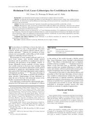
Take a single small drop <strong>of</strong> whole blood and place it near one end <strong>of</strong> the microscope slide (A). Bring the end <strong>of</strong> a<br />
second slide (held at a 45 degree angle) up to the drop until the drop disperses along the edge <strong>of</strong> the second slide(B).<br />
Then with the first slide placed on a flat surface, push the second slide quickly and evenly toward the opposite end <strong>of</strong><br />
the first slide (C). The results should appear with an irregular thin edge, as illustrated (D). Several thick blood smears<br />
should also be made by spreading a drop <strong>of</strong> blood in a small circle on the slides. When the smears are dry, label one<br />
end <strong>of</strong> the slide (with a solvent resistant pen or pencil) with the date, species, animal ID, and location. Slides can then<br />
be transported to a laboratory or stained in the field with New Methylene Blue or a Wright’s stain kit.
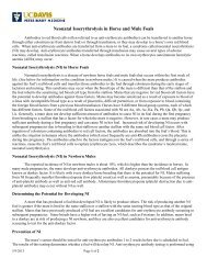
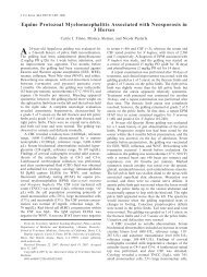
Anthrax Bacteria Diagram<br />
A. Bacillus anthracis, X2000.<br />
Note 2 main characteristics:<br />
1. sharp, squared ends<br />
2. pale capsule around bacteria<br />
B. Clostridium septicum, X2250.<br />
Note 3 characteristics:<br />
1. smaller size<br />
2. no capsule<br />
3. rounded ends<br />
Anthrax bacteria are commonly confused with Clostridium bacteria (see fig. above). Anthrax bacteria appear as large<br />
(up to 10 µm long) rectangular rods, alone or in chains and surrounded by a capsule. Anthrax bacilli obtained from a<br />
recently dead carcass usually do not form spores.<br />
Handling Decomposed Carcasses<br />
Examining the Carcass and its Environment<br />
Assess the Condition <strong>of</strong> the Environment<br />
Note recent weather conditions that could have caused animal deaths (drought, floods, electrical storm, etc)<br />
Note the ambient temperature<br />
Note signs <strong>of</strong> struggle<br />
Assess the Condition <strong>of</strong> the Animal
Note any bite WOUNDS or other signs <strong>of</strong> predation. If wounds are present, look for bruising and bleeding in the<br />
tissues near the wounds which would indicate that they occurred before the animal died. Otherwise these wound<br />
most likely were caused from the carcass being scavanged.<br />
Look for broken bones, missing hair, broken or missing teeth or other signs <strong>of</strong> trauma.<br />
Look for and preserve any external parasites.<br />
Determine Nutritional Status <strong>of</strong> the Animal<br />
Take weight (if possible) and/or body length and girth. Assess fat stores under the skin and in body cavities.<br />
Note the amount <strong>of</strong> fat around the heart and kidneys<br />
Note the muscle mass <strong>of</strong> the animal<br />
Note the amount <strong>of</strong> food in the digestive tract<br />
Note the condition <strong>of</strong> the teeth<br />
Most carcasses will have some AUTOLYSIS, but diagnostic tests can still be performed if tissues are properly handled.<br />
Handle autolyzed tissues for histopathology very gently<br />
Hold tissues at the edges only<br />
Cut with a sharp knife or scapel<br />
Quickly place in formalin<br />
Freeze or refrigerate samples as soon as possible for infectious disease or toxicology testing.<br />
Autolysis can cause many artifacts in tissues that can be confused with a disease process. However, it is always best<br />
to take a sample from an area that looks abnormal rather than assume that the change was caused by autolysis.<br />
Histopathology will be able to distinguish between true lesions and post-mortem changes.<br />
Tissue Sampling Procedures<br />
Histology<br />
(see Tissue Check List in Appendix II)<br />
Samples should be taken from all major organs and ANY ABNORMAL AREAS<br />
All samples should be placed in a common container <strong>of</strong> 10% buffered formalin<br />
SUBMERGE TISSUES IN 10 TIMES THE VOLUME OF FORMALIN AS THE VOLUME OF TISSUE<br />
Samples should be no thicker than 1 cm so that they can fix, but long and wide enough to represent the different areas<br />
<strong>of</strong> a tissue as well as any abnormalities. Samples that include abnormal areas and surrounding normal areas are best.<br />
Samples should be handled carefully by grasping at the edges. Do not scrape surfaces <strong>of</strong> tissues or compress them with<br />
forceps.<br />
Most tissues do not need individual labeling. If a tissue needs special labeling (eg. a specific lymph node), place it in<br />
a different container (or tissue cassette) or attach a piece <strong>of</strong> paper to the tissue with string or a pin and label the paper<br />
or container with pencil or waterpro<strong>of</strong> marking pen.<br />
Microbiology (Bacteriology and Virology)
To take samples without contaminating them, the samples need to be taken BEFORE tissues are touched and the<br />
instruments need to be sterilized. These samples also should be placed in sterile containers. To sterilize instruments:<br />
dip the tips in alcohol and then flame them or flame the tips until they are red and then let them cool. Samples also<br />
can be taken with a sterile swab, sterile syringe, or by placing a large (3 cm2) section <strong>of</strong> tissue in a sterile container<br />
(the center <strong>of</strong> the tissue will be uncontaminated).<br />
Take samples that contain abnormal areas . Appropriate samples include: whole blood, pus, areas with abscesses or<br />
nodules, or intestinal contents (within a loop <strong>of</strong> intestines).When taking samples from infected tissues, select an area<br />
near the edge <strong>of</strong> the affected tissue where live organisms are most likely to be found. If no abnormal areas are present,<br />
take standard tissue samples <strong>of</strong> lung, liver, kidney, spleen, tonsil, and intestines (see Check List in Appendix II)<br />
Keep samples moist with sterile transport media, sealed in a sterile container and cold. If refrigeration is not available,<br />
samples can be placed buffered glycerin.<br />
Smears <strong>of</strong> pus or infected tissues also are useful and can be air-dried and shipped with cultures.<br />
Serology<br />
Serum should be placed in sterile tubes then stored and shipped frozen.<br />
OBTAINING SERUM FROM A CARCASS<br />
In recently dead animals, the right heart usually contains plasma clots (yellow/tan material), unclotted blood, or<br />
clotted blood. Plasma or blood should be removed and left undisturbed for approximately 30 min to encourage<br />
clot formation, then centrifuged at approximately 2000 X G for 20 min. The Mobilespin centrifuge from Vulcon<br />
Technologies, 718 Main, Grandview, MO 640330 USA; 816-966-1212 or FAX 816-966-8879 is portable and easily<br />
adapted for field use. When a centrifuge is not available, serum can still be obtained by letting the clot or blood cells<br />
settle. The serum (clear/yellow fluid or red-tinged fluid if the animal is autolyzed) or plasma should be separated for<br />
the blood cells, divided into at least two aliquots, transferred to vials, and then refrigerated or frozen (-20° or -70°)<br />
until transported to a laboratory. Serum vials should be labeled with the species, animal ID, date, and owner (e.g.,<br />
country and park) using a waterpro<strong>of</strong> marker.<br />
If a centrifuge is not available and blood is obtained from a live animal or a dead animal whose blood has not yet<br />
clotted, remove whole blood into a blood tube, let the blood clot with the tube inverted (rubber stopper down), then<br />
turn the tube right side up and very carefully remove the stopper with the blot clot attached, leaving the serum in the<br />
tube.<br />
Toxicology<br />
Take samples and place half <strong>of</strong> each sample in aluminum foil and half in plastic bags or containers (aluminum or<br />
plastic interfere with the testing <strong>of</strong> some toxins). Samples should be stored frozen (if possible) until shipped to a<br />
laboratory.<br />
(see check list in Appendix II)<br />
Parasitology<br />
Make at least 3 blood smears on clean glass slides (see procedure under “ Checking for Anthrax .”<br />
Fix approximately 2 g feces in 70% ethyl alcohol or formalin.<br />
Making Slides for Cytology<br />
Make a clean cut with a scalpel blade across the surface <strong>of</strong> the abnormal area <strong>of</strong> the tissue you wish to examine.<br />
Grasp the sample firmly with forceps, placing the cut surface down.<br />
Blot the cut surface <strong>of</strong> the sample across a paper towel or other absorbent surface until no blood or fluids are evident.<br />
Then gently touch the blotted surface in several locations on clean slides.<br />
Air dry slides.<br />
General Steps to Performing the Necropsy<br />
Carcass Dissection Using the Carnivore as an Example
Introduction<br />
The <strong>necropsy</strong> method outlined below provides a simple consistent method to examine a carcass and its body organs.<br />
A CARNIVORE has been used to demonstrate carcass dissection and tissue sampling procedures. Procedures for<br />
<strong>necropsy</strong> <strong>of</strong> UNGULATES, BIRDS, and REPTILES also are demonstrated. Very small animals (less than 100 g) can be<br />
fixed whole by opening the body cavity and submerging the entire animal in formalin.<br />
All carnivores and ungulates are placed ON THE LEFT SIDE so that the right side <strong>of</strong> the carcass is opened. All birds,<br />
reptiles, and primates are placed ON THEIR BACK.<br />
After the body cavities are opened, the general nutritional condition <strong>of</strong> the animal and location <strong>of</strong> all organs should<br />
be assessed (to determine if any organs are displaced) before organs are removed. At this time, a sterile blood sample<br />
for culture can be removed from the heart (the right atrium is the best location), then additional blood can be taken<br />
to obtain serum for serological tests. Also sterile samples <strong>of</strong> other organs should be taken for culture before organs are<br />
handled.<br />
After the general condition <strong>of</strong> the animal has been recorded, individual organs can be removed, examined, and<br />
sampled in a systematic manner. Any abnormal findings (lesions) should be described. Photographs <strong>of</strong> abnormal<br />
findings provide the best documentation for records.<br />
Describing Abnormalities Found at Necropsy<br />
Any abnormality should be described by the following criteria:<br />
Location<br />
Number and distribution<br />
Color<br />
Size<br />
Shape<br />
Consistency and texture<br />
For example: “The liver contains multiple tan, firm nodules ranging from 1 to 3 cm in diameter that are distributed<br />
throughout all liver lobes. The nodules are gritty on cut surface.”<br />
Procedure for Dissecting Carnivores (See check lists in the Appendix IIb for tissues to sample)<br />
Figure 1. Opening the Carnivore Carcass<br />
Examine the carcass for wounds and note the general condition <strong>of</strong> the fur.<br />
Place the carcass on its left side.<br />
Cut the skin along the ventral midline from the chin to the tail (Step 1, Fig. 1 below). On females, examine the<br />
mammary glands. On males, examine the prepuce and penis. On neonates, examine the umbilicus.<br />
On the right side, reflect the skin to the level <strong>of</strong> the backbone.<br />
Reflect the right limbs by cutting through the muscles and the hip and shoulder joints.<br />
Open all three body cavities (abdomen, chest, heart):<br />
Remove the flap <strong>of</strong> muscle and other tissues that cover the right side <strong>of</strong> the abdominal cavity.<br />
Open the right side <strong>of</strong> the chest cavity by cutting the ribs along the sternum and backbone (Step 2, Fig.1 below).<br />
Open the sac surrounding the heart.<br />
Record any abnormal locations or size <strong>of</strong> organs.<br />
Record the quantity, color, and contents <strong>of</strong> any fluids in the body cavities. Look for abnormal attachments <strong>of</strong> organs to<br />
the body cavity and determine if these attachments are easy to break.
General Organ Sampling Procedures (Figures 2-4)<br />
Stomach and intestines (Remove stomach and intestines first but open them last to prevent contamination <strong>of</strong> the<br />
<strong>necropsy</strong> site)<br />
Find where the esophagus enters the stomach and cut across the esophagus while holding the entry to the stomach<br />
closed to keep any food inside. Remove the stomach and intestines as a unit by cutting the mesentery where they<br />
attach to the intestines. Sample several lymph nodes along the attachments <strong>of</strong> the intestines. Leave the pancreas<br />
attached to the intestines and the spleen attached to the stomach. Cut across the rectum while holding it closed<br />
to prevent feces from escaping. Open the intestines along their length (intestines are best opened at the end <strong>of</strong> the<br />
<strong>necropsy</strong> to prevent contamination <strong>of</strong> other organs with food and fecal material). Note the content <strong>of</strong> the intestines<br />
(amount <strong>of</strong> food, presence <strong>of</strong> abnormal materials such as poisonous plants). Take stomach contents for toxicology.<br />
Take tissue samples from all areas <strong>of</strong> the GI tract and the pancreas.
Spleen, liver, pancreas<br />
Remove the spleen from the stomach, examine it for abnormalities by slicing it across in multiple sites and then<br />
remove samples for histology. Remove the liver; open the gall bladder. Examine the liver by cutting it across in<br />
multiple sites. Take samples for histology and toxicology.<br />
Kidneys and adrenals<br />
Remove and examine the adrenal glands and take transverse samples for histology. Remove the kidneys, examine<br />
them for abnormalities and take samples for histology that include the cortex, medulla, and pelvis and take samples<br />
for toxicology. Remove the bladder and take a section for histology.
Reproductive tract<br />
Remove the reproductive tract (testis if male; uterus and ovaries if female) and make a cut across the gonads and into<br />
the uterine lumen before placing in fixative for histopathology.<br />
Heart and lungs<br />
Separate the bones <strong>of</strong> the larynx behind the tongue and dissect out the trachea with esophagus attached. Continue<br />
into the chest including the lungs, heart, and large blood vessels. Take a sample from the thymus if present.
Open, examine and sample the trachea and other airways and the esophagus. Take samples from lymph nodes<br />
surrounding the airways.<br />
Examine the lungs for lumps and areas <strong>of</strong> firmness. Take samples <strong>of</strong> any abnormal areas, as well as areas that look<br />
normal.<br />
Open the chambers <strong>of</strong> the heart and examine the heart valves between the chambers. Take samples <strong>of</strong> the heart<br />
including valve. Open the great vessels and take a sample .<br />
Head and oral cavity<br />
Examine the eyes, mouth and nostrils for ulcers and abnormal discharges. Remove an eye for histology by cutting the<br />
muscles around the eyeball. Cut between the lower jaw bone and tongue and remove the tongue from below. Examine<br />
the inside <strong>of</strong> the mouth, tonsils, and teeth. Remove the other tonsil and several lymph nodes under the skin at the<br />
angle <strong>of</strong> the jaw and above the larynx for histology.<br />
Brain<br />
To remove the entire brain: Separate the skull from the neck at the junction with the vertebra. Remove the skin from<br />
the top <strong>of</strong> the head then the top <strong>of</strong> the skull using the illustrated landmarks.<br />
Remove the brain and cut it in half. Preserve one half in formalin and split the other half into containers for virology<br />
and toxicology.<br />
Removing a brain if rabies is suspected<br />
If rabies is suspected, extra precaution should be used when removing the brain. The person removing the brain<br />
should wear a face shield or mask and eye goggles. The safest procedure is to remove the head and insert a drinking<br />
straw through the hole at the base <strong>of</strong> the skull where it attached to the neck. The straw should be inserted in the
direction <strong>of</strong> the eye. Pinch the base <strong>of</strong> the straw and remove the straw with the brain sample. Then cut the straw (with<br />
the brain sample still inside) into 1 cm lengths and drop the sample in either glycerin or formalin. Although this<br />
procedure is very safe, it only allows testing for rabies. So if the samples are negative for rabies, no other brain tissues<br />
are available to test for other diseases.<br />
The best procedure is to remove the entire brain, then cut it lengthwise down the middle. Send one half to a<br />
laboratory to test for rabies and fix one half for histology.<br />
Skeletal Muscles and Nerves<br />
Take samples <strong>of</strong> the diaphragm and several leg muscles.<br />
Take a sample <strong>of</strong> the large nerve between the muscle bundles <strong>of</strong> the back leg.<br />
Bone Marrow<br />
Remove a long bone from the leg and crack it open in the middle. Fix one half for histology and store half for<br />
microbiology<br />
Ungulates<br />
The procedures for ungulates are the same as for carnivores, except that all forestomach chambers should be opened.
Birds<br />
THE PROSECTOR SHOULD WEAR A MASK because humans can acquire psitticosis, tuberculosis, and fungal<br />
diseases from birds.<br />
Dip the carcass in water containing a disinfectant or spray it to wet the feathers.<br />
Examine the carcass for evidence <strong>of</strong> trauma and ectoparasites.<br />
Place the bird on its back and open the skin from the beak to the vent.<br />
Retract the skin to expose the keel and breast muscles, ribs, and muscles over the lower celomic cavity. Assess the<br />
amount <strong>of</strong> body fat under the skin and in the body cavity. Assess the amount <strong>of</strong> musculature over the keel.<br />
Open the celomic cavity by making a horizontal cut at the bottom edge <strong>of</strong> the keel extending on each side through the<br />
pectoral muscles and then lifting the sternum. Cut the ribs and clavicle near the attachment to the sternum. Open the<br />
caudal part <strong>of</strong> the sternum long the midline.<br />
Inspect the location and size <strong>of</strong> all organs. Examine the air sacs for transparency and note any plaques or opaque<br />
areas. Note any abnormal fluids<br />
Using sterile instruments, take sterile samples <strong>of</strong> any visible lesions as well as <strong>of</strong> spleen, lung, and liver.<br />
Remove the tongue, trachea, esophagus and heart as a unit. Take the thyroid glands for histology (thyroids are located<br />
at the thoracic inlet where the major blood vessels above the heart branch). Open the trachea and heart and take<br />
samples for histology.<br />
Gastrointestinal tract, liver, and spleen: Remove the liver and take samples for histology and toxicology. Remove the<br />
proventriculus and ventriculus (gizzard) and intestines, including the cloaca and bursa <strong>of</strong> Fabricius. Note the spleen<br />
at the junction <strong>of</strong> the proventriculus and ventriculus. Fix what is remaining <strong>of</strong> the spleen (after taking a sample for<br />
culture) for histology. Open the intestinal tract along its length, noting the content and taking samples for toxicology.<br />
Leave the pancreas attached to intestines and take samples<br />
Lungs: Dissect the lungs away from the body wall, examine them for firmness or lumps, and take samples for<br />
histology.<br />
Reproductive tract, adrenal glands, and kidneys: The gonads (testes in the male or left ovary in the female) are located<br />
in front <strong>of</strong> the kidneys along the backbone. The adrenal glands are located just in front <strong>of</strong> the gonads and are also<br />
attached to the body below the spine. The female also should have an oviduct visible. Bluntly dissect the kidneys from<br />
the body wall, leaving the gonads and adrenals attached. Fix for histology.<br />
Brain: Remove a section <strong>of</strong> skull covering the brain, remove a sterile sample (half <strong>of</strong> the brain, if possible) for<br />
microbiology and then fix the rest in the skull (in small birds) or remove the brain from skull before fixing (in larger<br />
birds).<br />
Bone marrow: Remove a tibiotarsal bone and crack it open before fixing.<br />
Nerves: Take samples from the large nerves between the wing or the leg and the body wall.
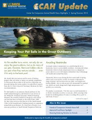
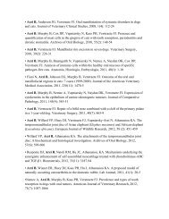
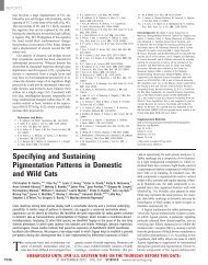
Avian Dissection<br />
Gastrointestinal Tract, Liver, and Spleen<br />
Remove the liver and take samples for histology and toxicology. Remove the proventriculus and ventriculus (gizzard)<br />
and intestines, including the cloaca and bursa <strong>of</strong> Fabricius. Note the spleen at the junction <strong>of</strong> the proventriculus and<br />
ventriculus. Fix what is remaining <strong>of</strong> the spleen (after taking a sample for culture) for histology. Open the intestinal<br />
tract along its length, noting the content and taking samples for toxicology. Leave the pancreas attached to intestines<br />
and take samples.
Gastrointestinal Tract<br />
Lungs<br />
Dissect the lungs away from the body wall,<br />
examine them for firmness or lumps, and take<br />
samples for histology.<br />
Lungs
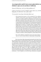
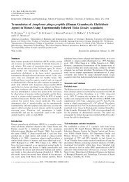
Reptiles<br />
The procedure for reptiles is similar to birds. A snake is used as an example. Other reptiles have similar organ<br />
anatomy, although the kidneys <strong>of</strong> lizards are further back in the pelvis.<br />
The animal is placed on its back and examined for evidence <strong>of</strong> trauma. Open the body along the midline. For turtles,<br />
the plastron is removed at the junction with the carapace with a saw. The amount <strong>of</strong> body fat and condition <strong>of</strong> the<br />
musculature is assessed and any abnormal fluids recorded. Take sterile samples <strong>of</strong> lungs, liver and spleen for culture<br />
before handling the tissues.<br />
Find the thyroid(s) anterior to the heart (single midline organ in some species and paired in other species) and<br />
remove and fix them for histology.<br />
Beginning at the mouth, remove the trachea, heart and lungs. Open the trachea and examine the lungs for firmness or<br />
lumps. Open the heart. Take samples <strong>of</strong> all organs for histology.<br />
Remove the intestinal tract as a unit,<br />
beginning in the oral cavity. Open the<br />
esophagus, stomach and intestines<br />
along their length and take samples for<br />
histology<br />
Remove the liver, spleen and pancreas.<br />
Examine and sample for histology<br />
Remove the gonads and adrenals (along<br />
the midline in front <strong>of</strong> the gonads). In<br />
females, remove the oviduct with the<br />
ovaries. Dissect the kidneys from the<br />
body wall. Take samples for histology.<br />
Reptile<br />
Dissection Shown on Snake
Post-<strong>necropsy</strong><br />
Disinfecting the <strong>necropsy</strong> site<br />
The carcass and all tissues from the carcass including blood soaked dirt should be buried or incinerated. All<br />
contaminated paper or plastic materials should be either thoroughly disinfected or incinerated. All blood and residual<br />
tissues should be removed from the instruments and tools with soap and water. Then the instruments should be<br />
disinfected. Necropsy boots and apron should be cleaned and any contaminated clothing thoroughly washed. The<br />
external surfaces <strong>of</strong> any containers with samples should also be washed.<br />
Storage or submission <strong>of</strong> samples<br />
TO SHIP SAMPLES:<br />
Formalin-fixed samples can be kept at a cool room temperature until shipped.<br />
Any samples for culture should be kept refrigerated (for parasitology or bacterial cultures) or frozen (for toxicology or<br />
virus cultures). Freezing at -70° C is preferable to 20° C (standard freezers).<br />
1. Contact the laboratory before shipping.<br />
2. Check regulations for shipping tissue samples. Get proper permits and use the correct containers.<br />
3. Frozen samples must be shipped in insulated containers and by express carrier. Pack specimens in dry ice or with<br />
ice blocks. Seal container to prevent leakage. Include proper permits and animal identification.<br />
4. Formalin-fixed tissues should be fixed for at least a week in 10 times as much formalin as tissue. Then most <strong>of</strong> the<br />
formalin can be removed and tissues can be wrapped in toweling soaked in formalin for shipping. The tissues soaked<br />
in formalin can then be placed in leakpro<strong>of</strong> containers for shipping.<br />
IT IS BEST TO SHIP FROZEN AND FIXED SAMPLES SEPARATELY. IF THEY MUST BE SHIPPED TOGETHER, then<br />
insulate the fixed tissues from freezing by wrapping in newpapers. Assure that there is no spillage <strong>of</strong> formalin, because<br />
fixation <strong>of</strong> frozen samples will make culturing for bacteria or viruses impossible and will alter cells on blood smears<br />
or cytology slides.<br />
Dealing with an Epidemic<br />
Complete the FIELD REPORT OF WILDLIFE DEATH form found in Appendix II.<br />
Contact appropriate local or national governmental personnel.<br />
Collect samples from as many dead or affected animals as possible.<br />
Collect the following information on the epidemic:<br />
Species and approximate number affected<br />
Signs the animals are showing<br />
Location <strong>of</strong> affected animals
Appendix I. Formulations for Sample Preservatives<br />
Sterile Buffered Glycerin (50%)<br />
For transporting tissues for culture when refrigeration is not available.<br />
To make sterile buffered glycerin, mix glycerin with an equal amount <strong>of</strong> buffer composed <strong>of</strong>:<br />
A. 21 g citric acid mixed in 1000 distilled water<br />
B. 28.4 g anhydrous sodium phosphate in 1000 distilled water<br />
Mix 9.15 ml <strong>of</strong> A and 90.85 ml <strong>of</strong> B<br />
Mix 100 ml <strong>of</strong> buffer with 100 ml <strong>of</strong> glycerin. Then sterilize in small tubes to take into the field<br />
“Easy Blood”<br />
For transporting DNA from blood cells for genetic studies when refrigeration is not available. Also can be used to<br />
preserve DNA for longer periods <strong>of</strong> time if refrigerated or frozen.<br />
1.2 g Tris HCl<br />
3.7 g Na2 EDTA<br />
2 g sodium dodecyl sulfate (SDS)<br />
Add water to 100 ml<br />
10% Buffered Formalin<br />
For fixation <strong>of</strong> tissues for histology<br />
To make one liter mix:<br />
100 ml formalin (38-40% formaldehyde)<br />
900 ml distilled water<br />
4 g sodium chloride (table salt) [OR 4.5 gm sodium phosphate (monobasic) and 3.6 gm sodium hydroxide]
Appendix IIa. Tissue Checklist for Microbiology and Toxicology<br />
Tissue Microbiology Toxicology<br />
Brain x x<br />
Fat x<br />
Kidney x x<br />
Stomach Contents x<br />
Hair x<br />
Liver x x<br />
Whole blood x x<br />
Lymph Nodes x x<br />
Tonsils x x<br />
Spleen x x<br />
Abscesses, granulomas x
Appendix IIb. Fixed Tissue Checklist for Histology<br />
Preserve the following tissues in 10 % buffered formalin at a ratio <strong>of</strong> 1 part tissue to 10 parts formalin. Tissues should<br />
be no thicker than 1 cm. INCLUDE SECTIONS OF ALL LESIONS AND SAMPLES OF ALL TISSUES ON THE<br />
TISSUE LIST.<br />
Recommended tissue sampling procedures:<br />
__ Salivary gland<br />
__ Oral/pharyngeal mucosa and tonsil -plus any areas with erosions, ulcerations or other lesions.<br />
__ Tongue - cross section near tip including both mucosal surfaces.<br />
__ Lung - sections from several lobes including a major bronchus<br />
__ Trachea<br />
__ Thyroid/parathyroids<br />
__ Lymph nodes - cervical, mediastinal, bronchial, mesenteric and lumbar. Cut transversely.<br />
__ Thymus<br />
__ Heart - Sections from both sides including valves<br />
__ Liver - sections from 3 different areas including gall bladder<br />
__ Spleen - Cross sections including capsule.<br />
__ GI Tract - 3 cm long sections <strong>of</strong>:<br />
Esophagus<br />
Stomach - multiple sections from all regions <strong>of</strong> the lining<br />
Intestines - multiple sections from different areas<br />
__ Omentum - ~3 cm square<br />
__ Pancreas - sections from two areas<br />
__ Adrenal - entire gland with transverse incision.<br />
__ Kidney -cortex and medulla from each kidney<br />
__ Urinary bladder, ureters, urethra - cross section <strong>of</strong> bladder and 2 cm sections <strong>of</strong> ureter and urethra.<br />
__ Reproductive tract - Entire uterus and ovaries with longitudinal cuts into lumens <strong>of</strong> uterine horns.<br />
Both testes (transversely cut) with epididymis. Entire prostate, transversely cut.<br />
__ Eye<br />
__ Brain - cut longitudinally along midline.<br />
__ Spinal cord (if neurologic disease) - sections from cervical, thoracic and lumbar cord.<br />
__ Diaphragm and Skeletal muscle - cross section <strong>of</strong> thigh muscles<br />
__ Opened rib or longitudinally sectioned _ femur - marrow must be exposed for proper fixation<br />
__ Skin - full thickness <strong>of</strong> abdominal skin, lip and ear pinna.<br />
__ Neonates: umbilical stump - include surrounding tissues
Appendix IIc. Specimen Submission Form<br />
Date<br />
Person submitting samples:<br />
Agency:<br />
Address:<br />
Phone #/Fax<br />
Species: Animal<br />
ID: Sex<br />
Died Killed (circle one)<br />
Number affected:<br />
Location where carcass found:<br />
Evidence <strong>of</strong> struggling?<br />
Environmental conditions:<br />
Other information/observations:
Appendix IId. Necropsy Protocol<br />
Name <strong>of</strong> prosector<br />
Address <strong>of</strong> prosector<br />
Location <strong>of</strong> carcass<br />
SPECIES &nb sp; ;<br />
ANY ID # WEIGHT SEX<br />
DATE OF DEATH DATE OF NECROPSY<br />
HISTORY: (briefly summarize clinical signs, circumstances <strong>of</strong> death):<br />
SHIPPING TISSUES: PLEASE OBTAIN PROPER CITES AND EXPORT PERMITS BEFORE SHIPPING TISSUES<br />
BETWEEN COUNTRIES. After 72 hrs in fixative, ship tissues in a leak-pro<strong>of</strong> container in adequate formalin to keep<br />
tissues moist.
Appendix IIe. Gross Examination Worksheet<br />
PROSECTOR:<br />
GENERAL CONDITION: (Nutritional condition, physical condition)<br />
Neonates: examine for malformations (cleft palate, deformed limbs etc).<br />
SKIN: (Including pinna, feet)<br />
MUSCULOSKELETAL SYSTEM: (Bones, joints, muscles)<br />
BODY CAVITIES: (Fat stores, abnormal fluids)<br />
Neonates : assess hydration (tissue moistness)<br />
HEMOLYMPHATIC: (Spleen, lymph nodes, thymus)<br />
RESPIRATORY SYSTEM: (Nasal cavity, larynx, trachea, lungs, regional lymph nodes)<br />
Neonates: determine if breathing occurred (do the lungs float in formalin?)<br />
CARDIOVASCULAR SYSTEM: (Heart, pericardium, great vessels)<br />
DIGESTIVE SYSTEM: (Mouth, teeth, esophagus, stomach, intestines, liver, pancreas, mesenteric lymph nodes).<br />
Neonates: is milk present in stomach?
URINARY SYSTEM: (Kidneys, ureters, urinary bladder, urethra)<br />
REPROD<strong>UC</strong>TIVE SYSTEM: (Testis/ovary, uterus, vagina, penis, prepuce, prostate, mammary glands, placenta)<br />
ENDOCRINE SYSTEM: (Adrenals, thyroid, parathyroids, pituitary)<br />
NERVOUS SYSTEM: (Brain, spinal cord, peripheral nerves)<br />
SENSORY ORGANS (Eyes, ears)<br />
PRELIMINARY DIAGNOSES:<br />
LABORATORY STUDIES:(List bacterial and viral cultures submitted and results, if available)
Appendix IIf. Field Report <strong>of</strong> Wildlife Death<br />
Date:<br />
Person reporting:<br />
Affiliation:<br />
Address<br />
Telephone #/Fax<br />
Species<br />
Clinical signs noted before death:<br />
Number affected:<br />
Other species in the region/Number affected: