Normal Anatomy of the California Sea Lion globe
Normal Anatomy of the California Sea Lion globe
Normal Anatomy of the California Sea Lion globe

You also want an ePaper? Increase the reach of your titles
YUMPU automatically turns print PDFs into web optimized ePapers that Google loves.
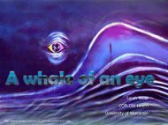
Through <strong>the</strong> eyes <strong>of</strong> a sea lion<br />
Sarah Miller<br />
Richard Dubielzig, DVM, DACVP
The Globe<br />
Large Diameter<br />
Cornea thicker peripherally<br />
flattened cornea<br />
Due to stroma<br />
Shorter internal axial length<br />
more accurately defines optical function
The Cornea<br />
Flattened region over a stenopaic pupil<br />
not seen histologically
Bowman’s membrane<br />
TEM<br />
Descemet’s thin<br />
Endo<strong>the</strong>lium thin<br />
The cornea
Extensive constriction <strong>of</strong> pupil<br />
protects from large changes in<br />
illumination<br />
Iridocorneal angle<br />
Thick pectinate ligament with<br />
intermittent open channels<br />
Intermittent corneoscleral<br />
trabecular meshwork<br />
Anterior Uvea
Dilator muscle<br />
absent at <strong>the</strong> pupil<br />
Robust near iris base where it<br />
abutts sphincter m.<br />
Near pectinate ligament<br />
Extends over widened base <strong>of</strong><br />
ciliary processes<br />
Sphincter muscle<br />
Thickest near iris base<br />
Near pectinate ligament<br />
Accomodation??<br />
Anterior Uvea
Ciliary Body<br />
Dilator muscle extending up <strong>the</strong> base <strong>of</strong> <strong>the</strong> ciliary processes<br />
Circumferential smooth muscle subtending zonular ligaments<br />
accomodation??<br />
Lens attachment: direct attachment <strong>of</strong> ciliary processes<br />
zonular fibers
The Lens<br />
Spherical lens<br />
Heterogeneous (multifocal)<br />
Lower R.I. in periphery<br />
Compensates for <strong>the</strong> inability <strong>of</strong> <strong>the</strong> cornea to refract<br />
underwater<br />
Spherical lens<br />
http://lifestyle.iloveindia.com/lounge/images/facts-about-pinniped-2.jpg
Tapetum lucidum<br />
• Tapetum lucidum covers 2/3 to entire fundus
Tapetum lucidum<br />
Extremely thick both dorsal & ventral to optic nerve
Monochromatic<br />
loss <strong>of</strong> S opsin<br />
Ganglion cells!!<br />
Very large but sparce<br />
Pinniped<br />
Ganglion cells!!<br />
Dog
Sclera<br />
• Thick<br />
– Near limbus<br />
– Near optic nerve<br />
• Thin<br />
– At equator<br />
Vascular Plexus<br />
Similar to <strong>the</strong> vascular plexus<br />
seen in cetaceans?
Vascular Plexus<br />
Similar to <strong>the</strong> vascular plexus seen in<br />
cetaceans?
References<br />
• Miller S., Coltiz C., Dubielzig R. (2010) <strong>Anatomy</strong> <strong>of</strong> <strong>the</strong> <strong>California</strong> sea lion<br />
<strong>globe</strong>. <strong>globe</strong>.<br />
Veterinary Ophthalmology supplement 1: 63‐71. 63 71.<br />
• Ya Supin, Supin,<br />
Alexander, et al. The Sensory Physiology <strong>of</strong> Aquatic Mammals.<br />
Boston, Kluwer Academic Publishers, 2001. (pp 239‐264) 239 264)<br />
• Mass A. & Ya Supin A. (2007) Adaptive Features <strong>of</strong> Aquatic Mammals’ Mammals<br />
Eye. The Anatomical Record 290:701‐715.<br />
290:701 715.<br />
• Peichl L. et al. (2001) For Whales and <strong>Sea</strong>ls <strong>the</strong> Ocean is Not Blue: A<br />
Visual Pigment Loss in Marine Mammals. European Journal <strong>of</strong><br />
Neuroscience 13: 1520‐1528. 1520 1528.