Marigold Cell Size and Polyploidy - Association for Biology ...
Marigold Cell Size and Polyploidy - Association for Biology ...
Marigold Cell Size and Polyploidy - Association for Biology ...
Create successful ePaper yourself
Turn your PDF publications into a flip-book with our unique Google optimized e-Paper software.
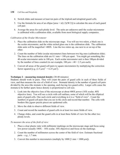
130 <strong>Cell</strong> <strong>Size</strong> <strong>and</strong> <strong>Polyploidy</strong><br />
4. Switch slides <strong>and</strong> measure at least ten pairs of the triploid <strong>and</strong> tetraploid guard cells.<br />
5. Use the <strong>for</strong>mula <strong>for</strong> area of an ellipse [area = π(L/2)(W/2)] to calculate the area of each guard<br />
cell pair.<br />
6. Average the areas <strong>for</strong> each ploidy level. The units are unknown until the ocular micrometer<br />
is calibrated with a calibration slide, available from most biological supply companies.<br />
Calibration of the Ocular Micrometer<br />
1. Place the calibration slide on the microscope stage. You will see two rulers, a black one is<br />
the ocular micrometer, <strong>and</strong> the white etched glass one is the calibration slide. The calibration<br />
slide units will be magnified ~400X. Line the two rulers up, one next to or on top of the<br />
other.<br />
2. Count the number of little (ocular micrometer) lines between two big ones (calibration slide).<br />
The lines on the calibration slide are 0.1 mm = 100 µm apart. You might get something like<br />
40 ocular micrometer units in 100 µm. Each ocular micrometer unit is then 100µm divided<br />
by the number of lines counted (in our example, 100 µm ÷ 40 = 2.5 µm each).<br />
3. Convert all areas of the guard cell pairs to square micrometers by multiplying the correction<br />
factor squared [e.g. (2.5 µm) 2 = 6.25 µm 2 ].<br />
Technique 2 – measuring stomatal density (30-40 minutes)<br />
Students should work in pairs. They will count the pairs of guard cells in each of three fields of<br />
view, <strong>and</strong> then measure the size of a field of view. Stomatal density is the number of guard cell pairs<br />
divided by the area (the stomate is the opening, each having two guard cells). Larger cells cause the<br />
stomata to be farther apart; hence density is proportional to cell size.<br />
1. Look into the objective lens of the microscope at about 400X power (10X ocular, 40X<br />
objective lens). You will see a circle with cell outlines, some of which will be the elliptical<br />
pairs of guard cells. One such circle <strong>and</strong> everything in it is a “field of view”. Count the<br />
numbers of guard cell pairs that you see in the circle <strong>and</strong> record that number. The cells with<br />
borders like jigsaw puzzle pieces are epidermal cells.<br />
2. Move the slide to observe different fields of view.<br />
3. Count <strong>and</strong> record the numbers of guard cells in at least two more fields of view.<br />
4. Change slides, <strong>and</strong> count the guard cells in at least three fields of view <strong>for</strong> the other two<br />
ploidy levels.<br />
To measure the area of the field of view:<br />
1. Place a clear plastic ruler with millimeter markings on the microscope stage <strong>and</strong> focus using<br />
low power (usually 100X – 10X ocular, 10X objective) <strong>and</strong> focus on the markings.<br />
2. Count the number of millimeters across the center of the field of view. Estimate fractional<br />
parts – e.g., 1.7 mm.<br />
3. Convert this number to micrometers (multiply by 1000 [1 mm = 1000 µm]).


