Organelle Isolation and Marker Enzyme Assay - Association for ...
Organelle Isolation and Marker Enzyme Assay - Association for ...
Organelle Isolation and Marker Enzyme Assay - Association for ...

Create successful ePaper yourself
Turn your PDF publications into a flip-book with our unique Google optimized e-Paper software.
Chapter 8<br />
<strong>Organelle</strong> <strong>Isolation</strong> <strong>and</strong> <strong>Marker</strong> <strong>Enzyme</strong> <strong>Assay</strong><br />
Harish Padh<br />
Department of Biochemistry <strong>and</strong> Molecular Biology<br />
The University of Chicago<br />
Chicago, Illinois 60637<br />
(312) 702-1326, FAX: (312) 702-0439<br />
Harish Padh received his B.S. in Microbiology from Gujarat University, M.S. in<br />
Biochemistry from the M. S. University of Baroda, <strong>and</strong> Ph.D. in Biochemistry<br />
from the University of Delhi, India. At present he is Research Assistant Professor<br />
at the University of Chicago. He has taught courses in biochemistry,<br />
microbiology, <strong>and</strong> cell biology at various levels. His research interest is studying<br />
aspects of cell biology in Dictyostelium discoideum.<br />
Reprinted from: Padh, H. 1992. <strong>Organelle</strong> isolation <strong>and</strong> marker enzyme assay. Pages 129–146, in Tested studies<br />
<strong>for</strong> laboratory teaching, Volume 13 (C. A. Goldman, Editor). Proceedings of the 13th Workshop/Conference of the<br />
<strong>Association</strong> <strong>for</strong> Biology Laboratory Education (ABLE), 191 pages.<br />
- Copyright policy: http://www.zoo.utoronto.ca/able/volumes/copyright.htm<br />
Although the laboratory exercises in ABLE proceedings volumes have been tested <strong>and</strong> due consideration has been<br />
given to safety, individuals per<strong>for</strong>ming these exercises must assume all responsibility <strong>for</strong> risk. The <strong>Association</strong> <strong>for</strong><br />
Biology Laboratory Education (ABLE) disclaims any liability with regards to safety in connection with the use of the<br />
exercises in its proceedings volumes.<br />
© 1992 Harish Padh<br />
<strong>Association</strong> <strong>for</strong> Biology Laboratory Education (ABLE) ~ http://www.zoo.utoronto.ca/able<br />
129
130 <strong>Organelle</strong> <strong>Isolation</strong><br />
Contents<br />
Student Outline ..............................................................................................................130<br />
Introduction....................................................................................................................130<br />
Cells/Tissue Homogenization........................................................................................130<br />
Differential Centrifugation ............................................................................................131<br />
Density-Equilibrium Centrifugation..............................................................................131<br />
<strong>Marker</strong> <strong>Enzyme</strong>s ............................................................................................................133<br />
The Organism, Dictyostelium discoideum.....................................................................133<br />
Synopsis of the Experiment ...........................................................................................134<br />
Laboratory Protocol .......................................................................................................135<br />
Preparation of Homogenate (steps 1–3).........................................................................135<br />
Centrifugation (steps 4–7) .............................................................................................135<br />
<strong>Assay</strong> <strong>for</strong> Succinate Dehydrogenase..............................................................................136<br />
<strong>Assay</strong> <strong>for</strong> Acid Phosphatase ..........................................................................................136<br />
<strong>Assay</strong> <strong>for</strong> Alkaline Phosphodiesterase...........................................................................137<br />
Tabulation of Results.....................................................................................................137<br />
Notes <strong>for</strong> the Instructor .................................................................................................138<br />
Acknowledgements........................................................................................................139<br />
Literature Cited..............................................................................................................139<br />
Appendix A: Expected Results <strong>and</strong> Examples of Assignment Questions.....................140<br />
Appendix B: Growing Cells of Dictyostelium discoideum............................................142<br />
Appendix C: Materials...................................................................................................144<br />
Student Outline<br />
Introduction<br />
Eukaryotic cells contain several types of intracellular membrane-bound structures known as<br />
organelles which per<strong>for</strong>m a variety of specific functions. For example, mitochondria synthesize<br />
ATP, chloroplasts convert light energy to chemical energy, while the endoplasmic reticulum is<br />
involved in protein synthesis. The types <strong>and</strong> number of organelles in cells range from no organelle<br />
(in mature mammalian red blood cells) to many <strong>and</strong> diverse organelles (<strong>for</strong> example, in cells active<br />
in the synthesis of proteins <strong>for</strong> secretion).<br />
Biochemical analysis of the structure <strong>and</strong> function of organelles requires a relatively pure<br />
sample. There are several methods <strong>for</strong> isolating subcellular organelles (Alberts et al., 1989; Darnell<br />
et al., 1990; Novikoff <strong>and</strong> Holtzman, 1976); in this laboratory you will employ one of the most<br />
widely-used methods, differential centrifugation.<br />
Cells/Tissue Homogenization<br />
An ubiquitous first step in the isolation of subcellular organelles is to homogenize the cells.<br />
This process involves breaking open the cell membrane (<strong>and</strong> the cell wall if present). Commonly<br />
used methods of homogenization include (1) dounce homogenization, where the cells are crushed<br />
between two revolving solid surfaces; (2) filtration, where cells are <strong>for</strong>ced through smaller pores in<br />
a filter; (3) grinding, where cells are ground by swirling with glass beads; (4) sonication, where<br />
cells are bombarded with ultrasonic vibrations; <strong>and</strong> (5) solubilization, in which cell membranes are<br />
dissolved in detergents such as Triton X-100. <strong>Enzyme</strong> digestion is also used to remove cell-wall
<strong>Organelle</strong> <strong>Isolation</strong> 131<br />
constituents. The method of choice depends on the type of tissue to be homogenized <strong>and</strong> the<br />
specific purpose of the experiment.<br />
Usually during the homogenization process isotonic sucrose is added to the homogenization<br />
buffer to prevent osmotic rupture of organelle membranes since it is usually important to purify<br />
intact organelles. Following homogenization, the homogenate is normally spun at low speed to<br />
remove any intact cells along with other large cellular debris. When this is done, the next step is<br />
subcellular fractionation.<br />
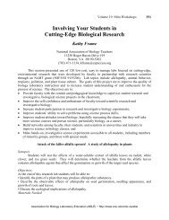
Differential Centrifugation<br />
Differential centrifugation is one of the widely-used techniques to separate cellular<br />
organelles. A slight modification of this technic known as rate zonal centrifugation is also used<br />
frequently in which organelles, after a single spin, b<strong>and</strong> in a tube according to their sedimentation<br />
rate. The technique of differential centrifugation is shown schematically in Figure 8.1 <strong>and</strong><br />
described below.<br />
Size, density, <strong>and</strong> shape influence the movement of a subcellular particle in a centrifugal<br />
field. This movement (sedimentation) results from the interaction between a particle's weight <strong>and</strong><br />
the resistance it encounters in moving through a suspension medium <strong>and</strong> the relative centrifugal<br />
<strong>for</strong>ce exerted on the particle. Under a given centrifugal <strong>for</strong>ce, particles that are relatively large or<br />
dense will sediment more rapidly than particles that are smaller <strong>and</strong> lighter. With respect to the<br />
major components found in cells, the order of sedimentation is typically (from most to least dense):<br />
nuclei, mitochondria, lysosomes, plasma membrane, endoplasmic reticulum, <strong>and</strong> contractile<br />
vacuoles. Depending on the specific cell type, however, this order can vary. Additionally,<br />
differences in the rate of sedimentation are sometimes not large enough to provide separation of<br />
one organelle from another.<br />
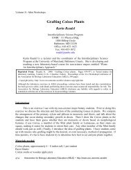
Density-Equilibrium Centrifugation<br />
A second widely-used procedure <strong>for</strong> separating organelles is known as density-equilibrium<br />
centrifugation (see Figure 8.2). In this procedure, subcellular particles are layered on a density<br />
gradient <strong>and</strong> subjected to a very high centrifugal <strong>for</strong>ce. Usually, the density gradient is <strong>for</strong>med by<br />
layering increasing concentrations of sucrose solutions in a centrifuge tube. However, other<br />
solutions can be used, such as Percoll (a colloid) <strong>and</strong> cesium chloride. These latter two solutions,<br />
when spun, will spontaneously set up a density gradient, thus alleviating the need to (as with<br />
sucrose) manually layer sucrose solutions of varied density (concentration).<br />
During centrifugation, organelles initially layered on the density gradient will sediment until<br />
they arrive at the region of the gradient where the density of the suspension is equal to their own<br />
(Figure 8.2). At this point, an equilibrium condition is reached between the downward centrifugal<br />
<strong>for</strong>ce <strong>and</strong> the particle's tendency to float due to buoyancy, <strong>and</strong> sedimentation halts. Hence, this<br />
procedure is also known as isopycnic equilibrium centrifugation.<br />
Used alone, neither differential nor density-gradient centrifugation normally provides<br />
preparations containing organelles of sufficient purity. A common practice is to use both types of<br />
centrifugation procedures in sequence. However, even with this approach, obtaining desired<br />
organelles free from contaminants can require additional steps. These steps can be many <strong>and</strong><br />
varied, <strong>and</strong> often involve innovative approaches. For example, in one attempt to purify lysosomes,<br />
it was found that even after extensive purification, the lysosomes were contaminated with
132 <strong>Organelle</strong> <strong>Isolation</strong><br />
mitochondria. Complete purification was finally achieved by feeding the cells Triton WR-1339, a<br />
very low-density compound that preferentially accumulated into the lysosomes. This, in effect,<br />
decreased the density of the lysosomes, allowing them to be easily separated from the mitochondria<br />
using density centrifugation.<br />
Figure 8.1. Schematic separation of organelles by differential centrifugation. See text <strong>for</strong><br />
details.
<strong>Organelle</strong> <strong>Isolation</strong> 133<br />
Figure 8.2. Schematic separation of organelles by density-equilibrium centrifugation. See<br />
text <strong>for</strong> details.<br />
<strong>Marker</strong> <strong>Enzyme</strong>s<br />
<strong>Isolation</strong> of any organelle requires a reliable test <strong>for</strong> the presence of the organelle. Typically,<br />
this is done by following the activity of an enzyme that is known to be localized exclusively in the<br />
target organelle. Such enzymes are known as marker enzymes. For example, the enzyme acid<br />
phosphatase (that cleaves terminal phosphate group from substrates <strong>and</strong> has a pH optimum in the<br />
acidic range) is localized in lysosomes, while the enzyme succinate dehydrogenase is localized in<br />
mitochondria. By monitoring where each enzyme activity is found during a cell fractionation<br />
protocol, one can monitor the fractionation of lysosomes <strong>and</strong> mitochondria, respectively.<br />
<strong>Marker</strong> enzymes also provide in<strong>for</strong>mation on the biochemical purity of the fractionated<br />
organelles. The presence of unwanted marker enzyme activity in the preparation indicates the level<br />
of contamination by other organelles, while the degree of enrichment <strong>for</strong> the desired organelle is<br />
determined by the specific activity of the target marker enzyme. Although marker enzymes reveal<br />
much concerning the purity of the organelle preparation, electron microscopy is generally used as a<br />
final step to assess the preparation's purity <strong>and</strong> the morphology of the isolated organelle.<br />
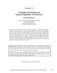
The Organism, Dictyostelium discoideum<br />
Dictyostelium discoideum is a eukaryotic microorganism popularly known as social ameba or<br />
slime mold. These individual cells will grow so long as a proper food source is available. Under<br />
starvation conditions, however,10 5 to 10 6 cells aggregate in an orderly manner to <strong>for</strong>m a<br />
multicellular body which eventually differentiates into spores or stalk cells (Figure 8.3). Because of<br />
this unique life-cycle feature <strong>and</strong> the simplicity with which this organism is grown <strong>and</strong> h<strong>and</strong>led, D.<br />
discoideum has become a model system <strong>for</strong> studying a variety of cellular processes. These include,<br />
but are not limited to, chemotaxis, signal transduction, pattern <strong>for</strong>mation <strong>and</strong> differentiation,<br />
cell-cell communication, <strong>and</strong> endocytosis.
134 <strong>Organelle</strong> <strong>Isolation</strong><br />
Figure 8.3. Life cycle of Dictyostelium discoideum. See text, Appendix B, <strong>and</strong> Loomis<br />
(1982) <strong>for</strong> details.<br />
Synopsis of the Experiment<br />
In this exercise, you will fractionate a homogenate of D. discoideum using differential<br />
centrifugation to separate mitochondria, lysosomes, <strong>and</strong> contractile vacuoles. The purified fractions<br />
will be assessed using marker enzymes specific to each of these cell organelles.<br />
In this exercise, cells grown on nutrient broth will be harvested, washed, <strong>and</strong> homogenized by<br />
passing them through two filters having pores 5 µm in diameter. Aliquots of the homogenate will<br />
then be subjected to three different centrifugal <strong>for</strong>ces <strong>and</strong> the pellets, as well as the supernatant<br />
fractions, will be assayed <strong>for</strong> marker enzymes specific to mitochondria, lysosomes, <strong>and</strong> contractile<br />
vacuoles. These enzymes <strong>and</strong> their associated organelles are given in Table 8.1. The ratio of these<br />
three markers in various fractions will give an indication about the relative enrichment of one<br />
organelle over another in a given fraction. The differential centrifugation steps used in the<br />
purification protocol make use of the fact that, in D. discoideum, mitochondria generally sediment<br />
faster than lysosomes, while contractile vacuoles sediment last.<br />
Table 8.1. <strong>Organelle</strong>s <strong>and</strong> associated marker enzymes used in this exercise.<br />
<strong>Organelle</strong> <strong>Marker</strong> enzyme<br />
Mitochondria Succinate dehydrogenase<br />
Lysosomes Acid phosphatase<br />
Contractile vacuoles Alkaline phosphodiesterase
Laboratory Protocol<br />
<strong>Organelle</strong> <strong>Isolation</strong> 135<br />
Note: Steps 1 through 3 will be per<strong>for</strong>med <strong>for</strong> you by a laboratory assistant. Students will<br />
there<strong>for</strong>e begin at step 4.<br />
Preparation of Homogenate<br />
1. Cells: Flask ____, cell density ____ × 10 6 cells/ml, centrifuge 60 ml cells at 500 RCF <strong>for</strong> 3<br />
minutes (model GLC-2B or similar centrifuge), resuspend cells in 20 ml of Buffer A (5 mM<br />
Na-glycinate, pH 8.5 + 100 mM sucrose at room temperature) <strong>and</strong> centrifuge again. Resuspend<br />
in 1.5 ml of the same buffer (cell density approximately 2 × 10 8 cells/ml) at room temperature.<br />
2. Homogenize cells by passing through 2 layers of 5 µm polycarbonate Nucleopore filters<br />
(diameter 25 mm) attached to a 3-ml plastic syringe (see Appendix B). Check homogenate<br />
under microscope <strong>for</strong> unlysed cells. Repeat homogenization if necessary.<br />
3. Spin as in step 1 to remove unlysed cells <strong>and</strong> undispersed cell fragments. Discard pellet <strong>and</strong><br />
save supernatant as homogenate. Dilute the homogenate 1:10 in ice-cold Buffer A (by adding 1<br />
ml of homogenate + 9 ml of ice-cold Buffer A).<br />
Centrifugation<br />
4. Obtain 5 ml of diluted homogenate in a tube. Obtain four (4) microfuge tubes (1.5 ml capacity)<br />
<strong>and</strong> label them A through D. Next, pipet 1 ml of the diluted homogenate into each of the four<br />
labelled microfuge tubes. Keep microfuge tube A on ice; it is a control <strong>for</strong> total homogenate<br />
enzyme activity.<br />
5. Using Table 8.2 as a guide, centrifuge tubes B through D in a Sorvall RC-5B containing an<br />
SS34 rotor precooled to 4°C. You will there<strong>for</strong>e need to per<strong>for</strong>m three separate centrifugations.<br />
Be<strong>for</strong>e starting any run, check that the rotor contains a tube from each group in the class. Also,<br />
make sure your tubes are in a balanced configuration in the rotor.<br />
Table 8.2. Centrifugation protocol.<br />
Tube Rpm Minutes RCF-min 1<br />
A – – 0<br />
B 4,200 10 1.0 × 10 4<br />
C 9,500 10 5.0 × 10 4<br />
D 18,000 25 5.0 × 10 5<br />
1 RCF-min is a measure of centrifugal <strong>for</strong>ce (RCF × time of centrifugation in<br />
minutes) generated during the centrifugation. Relative Centrifugal Force (RCF)<br />
can be calculated from rpm by the following equation: RCF = [(rpm × 2π)/60] 2<br />
× (r/g), where r = radius of rotor (5.00 cm <strong>for</strong> SS34), g = gravitational constant<br />
(= 980 cm/sec 2 ). For example, <strong>for</strong> tube B, centrifugation at 4200 rpm generates<br />
RCF of 1 × 10 3 , multiplied by 10 minutes, giving RCF-min of 1 × 10 4 .<br />
6. Label three microfuge tubes Bs, Cs, <strong>and</strong> Ds. Carefully separate (with eppendorf-type<br />
micropipets) the supernatants from tubes B through D as soon as they come out of centrifugation
136 <strong>Organelle</strong> <strong>Isolation</strong><br />
<strong>and</strong> place each into the three tubes labelled Bs, Cs, <strong>and</strong> Ds. Resuspend the pellets remaining in<br />
tubes B, C, <strong>and</strong> D in 0.9 ml of ice-cold Buffer A, <strong>and</strong> label them Bp, Cp, <strong>and</strong> Dp.<br />
7. <strong>Assay</strong> the contents of tubes A, Bs, Bp, Cs, Cp, Ds, <strong>and</strong> Dp according to the protocols <strong>for</strong>:<br />
(a) succinate dehydrogenase: use 0.5 ml/assay;<br />
(b) acid phosphatase: use 0.1 ml of 1:10 dilution (1:10 dilution prepared as follows: 0.1 ml of<br />
fractions + 0.9 ml of ice-cold Buffer A); <strong>and</strong><br />
(c) alkaline phosphodiesterase: use 0.10 ml/assay.<br />
<strong>Assay</strong> <strong>for</strong> Succinate Dehydrogenase<br />
The enzyme succinate dehydrogenase is an integral protein of the mitochondrial inner<br />
membrane. The major function of mitochondria is to generate energy (ATP) via oxidative<br />
phosphorylation. Succinate dehydrogenase, an FAD-containing enzyme, is involved in converting<br />
succinate to fumarate. In this assay, succinate is used as a substrate <strong>and</strong> nitroblue tetrazolium<br />
(NBT) as an artificial electron acceptor which changes to purple color when it accepts electrons.<br />
Thus, the <strong>for</strong>mation of purple color is directly proportional to enzyme activity.<br />
1. Label eight (8) glass tubes (13 × 100 mm) A, Bs, Bp, Cs, Cp, Ds, Dp, <strong>and</strong> BLANK. Pipet into<br />
each of these tubes:<br />
0.2 ml Buffer B [200 mM Na-Phosphate buffer, pH 7.4]<br />
0.1 ml 2.5 mg/ml NBT<br />
0.1 ml 1% Triton WR-1339<br />
0.1 ml Substrate B [100 mM Na succinate, pH > 7]<br />
2. When ready, add to each of the seven tubes (A, Bs, Bp, Cs, Cp, Ds, <strong>and</strong> Dp) 0.5 ml of the<br />
appropriate enzyme fraction. Add 0.5 ml Buffer A to the tube labelled BLANK. Note the<br />
starting time <strong>for</strong> each reaction.<br />
3. Incubate all tubes at 37°C <strong>for</strong> 30 minutes.<br />
4. Stop the reaction by adding 2.0 ml of 2% sodium dodecyl sulphate to each tube.<br />
5. Read Absorbance at 630 nm in a spectrophotometer adjusted to zero with the blank.<br />
<strong>Assay</strong> <strong>for</strong> Acid Phosphatase<br />
Acid phosphatase is present in lysosomes. The enzyme cleaves terminal phosphate groups<br />
<strong>and</strong> like other lysosomal enzymes operates maximally in acidic conditions. In this assay we will<br />
use a colorless compound, para-nitrophenol phosphate (pNPP) as the substrate <strong>for</strong> acid phosphatase.<br />
When the phosphate group of pNPP is cleaved, para-nitrophenol is generated. Para-nitrophenol is a<br />
yellow compound that is easily measured in a spectrophotometer.<br />
1. Label eight (8) glass tubes (13 × 100 mm) A, Bs, Bp, Cs, Cp, Ds, Dp, <strong>and</strong> BLANK. Pipet into<br />
each of these tubes:<br />
0.1 ml Buffer C [0.25 M glycine-HCl, pH 3.0, containing 0.5% Triton X-100]<br />
50 µl Substrate C [50 mM pNPP in water]
<strong>Organelle</strong> <strong>Isolation</strong> 137<br />
2. Make 1:10 dilution of A, Bs, Bp, Cs, Cp, Ds, <strong>and</strong> Dp [1:10 dilution prepared as follows: 0.1 ml<br />
of fractions + 0.9 ml of ice-cold Buffer A]. When ready, add to each of the seven tubes (A, Bs,<br />
Bp, Cs, Cp, Ds, <strong>and</strong> Dp) 0.1 ml of the 1:10 dilution of the appropriate enzyme fraction. Add 0.1<br />
ml Buffer A to the tube labelled BLANK. Note the starting time <strong>for</strong> each reaction.<br />
3. Incubate all tubes at 37°C <strong>for</strong> 30 minutes.<br />
4. Stop the reaction by adding 2.75 ml of ice-cold 0.2 M Na3PO4 (pH 12) to each tube.<br />
5. Read Absorbance at 410 nm in a spectrophotometer adjusted to zero with the blank.<br />
<strong>Assay</strong> <strong>for</strong> Alkaline Phosphodiesterase<br />
Alkaline phosphodiesterase (PDE) is present in contractile vacuoles. We will use a colorless<br />
substance, para-nitrophenol thymidine 5′ monophosphate, as the substrate <strong>for</strong> alkaline<br />
phosphodiesterase. Cleavage of thymidine 5′ monophosphate generates para-nitrophenol which is a<br />
yellow compound <strong>and</strong> easily measured in a spectrophotometer described above <strong>for</strong> the acid<br />
phosphatase assay.<br />
1. Pipet in eight (8) glass tubes (13 × 100 mm):<br />
0.1 ml Buffer D [0.25 M Tris-borate, pH 8.5, containing 0.5% Triton X-100, 20 mM MgCl2 <strong>and</strong><br />
20 µM ZnSO4]<br />
50 µl Substrate D [50 mM pNPT in water]<br />
2. When ready, add to each of the seven (7) tubes (A, Bs, Bp, Cs, Cp, Ds, <strong>and</strong> Dp) 0.1 ml of the<br />
appropriate enzyme fraction. Add 0.1 ml Buffer A to the tube labelled BLANK. Note the<br />
starting time <strong>for</strong> each reaction.<br />
3. Incubate all tubes at 37°C <strong>for</strong> 30 minutes.<br />
4. Stop the reaction by adding 2.75 ml of ice-cold 0.2 M Na3PO4 (pH 12) to each tube.<br />
5. Read Absorbance at 410 nm in a spectrophotometer adjusted to zero with the blank.<br />
Tabulation of Results<br />
Enter the data from your enzymes assays into Table 8.3. In addition to completing this<br />
table, you will need to plot a bar chart; <strong>for</strong> each RCF-min, plot f <strong>for</strong> each marker.
138 <strong>Organelle</strong> <strong>Isolation</strong><br />
Table 8.3. Tabulation of results.<br />
Succinate<br />
Acid<br />
Alkaline<br />
dehydrogenase phosphatase<br />
PDE<br />
Fraction A630 f* A410 f A410 f<br />
A<br />
Bp<br />
0 0 0<br />
Bs<br />
Cp<br />
– – –<br />
Cs<br />
Dp<br />
– – –<br />
Ds – – –<br />
* Calculation of f: Calculate the fraction (f) of total activity present in the respective pellets. For<br />
example, <strong>for</strong> tube Bp: fraction (f) = Bp/(Bp + Bs), where Bp is the enzyme activity in the pellet<br />
fraction <strong>and</strong> Bs is the enzyme activity in the supernatant fraction.<br />
Notes <strong>for</strong> the Instructor<br />
This exercise was developed <strong>for</strong> a biology major junior-level undergraduate class. Three<br />
hours time was sufficient when students worked in pairs. It is necessary that the students should<br />
have the outline at least 1 week in advance <strong>and</strong> that they study it be<strong>for</strong>e coming to the laboratory.<br />
The exercise described here could be shortened or elaborated depending on available time <strong>and</strong> need.<br />
For a shorter version, I suggest deleting the second spin (RCF-min of 5 × 10 4 <strong>for</strong> lysosomes) <strong>and</strong> the<br />
assay <strong>for</strong> acid phosphatase. To elaborate the exercise, protein <strong>and</strong> other marker enzymes could be<br />
assayed. In addition, density centrifugation or another suitable step can be added <strong>for</strong> even better<br />
purification of organelles.<br />
This exercise has been developed using an axenic Ax-3 strain of D. discoideum. The same<br />
principle applies when fractionating organelles from other cell-types. However, conditions should<br />
be worked out <strong>for</strong> different cell-types. The clear advantage here is that contractile vacuoles shows<br />
clean separation from mitochondria <strong>and</strong> lysosomes (as shown in Appendix C). Many cells which do<br />
not contain contractile vacuoles will not have such clear separation of organelles. The enzyme<br />
assays described here should work well with other cell-types.<br />
You will need about 10 ml of cell culture per student pair if cells are at 1 × 10 7 /ml. I<br />
homogenize cells in a large volume (up to 20 ml derived from 400 ml culture) <strong>for</strong> an entire class. I<br />
homogenize cells by passing them through polycarbonate filters (see step 2 in the Laboratory<br />
Protocol <strong>and</strong> Appendix B). Other methods of homogenization should also work well. However, it<br />
is important to use Buffer A because alkaline pH <strong>and</strong> a low ionic-strength buffer help dismantle<br />
cytoskeletons <strong>and</strong> there<strong>for</strong>e facilitates homogenization. See Das <strong>and</strong> Henderson (1983) <strong>for</strong><br />
homogenization procedure using polycarbonate filters.<br />
Expected results <strong>and</strong> examples of assignment questions <strong>and</strong> answers are given in Appendix<br />
A. Instructions <strong>for</strong> growing <strong>and</strong> maintaining D. discoideum are given in Appendix B. A table of<br />
the reagents that are required, instructions <strong>for</strong> the preparation of reagents, <strong>and</strong> the instruments,<br />
chemicals, <strong>and</strong> supplies that are required are given in Appendix C.<br />
Additional points to remember include the following:<br />
1. Keep all fractions on ice at all times.
<strong>Organelle</strong> <strong>Isolation</strong> 139<br />
2. Use of centrifuge: Tubes should be balanced, rotor lid should be tightly screwed on. Laboratory<br />
assistant should run centrifuges. A microfuge could be used with the appropriate speed (rpm)<br />
equivalent to the desired RCF-min.<br />
3. <strong>Assay</strong> buffers can be added to the assay tubes well in advance (while centrifugation is on). Add<br />
substrate to all tubes just be<strong>for</strong>e starting assay (less than 10 minutes be<strong>for</strong>e assay). Start<br />
reactions by adding enzyme solutions <strong>and</strong> stagger them as indicated below.<br />
4. It is imperative that each tube <strong>for</strong> the enzyme assay should be incubated <strong>for</strong> the indicated time.<br />
This is generally achieved by staggering tubes by definite intervals (1 minute). This means start<br />
the reaction in one tube. After 1 minute start the reaction in the next tube <strong>and</strong> so on. After 30<br />
minutes, terminate the reaction in the same sequence with 1-minute intervals between tubes.<br />
5. Colorimeter/spectrophotometer: Spectronic 20 is widely used <strong>for</strong> this purpose. However, other<br />
models should work well. The assay tubes (13 × 100 mm) described in the protocol will directly<br />
fit into a Spectronic 20 <strong>for</strong> reading color, eliminating the need to transfer to a cuvette <strong>for</strong> each<br />
assay tube.<br />
Acknowledgements<br />
I thank Dr. Theodore L. Steck <strong>for</strong> his constant encouragement, valuable advice, <strong>and</strong><br />
facilities; Ms. Malti Lavasa <strong>for</strong> her excellent technical expertise; <strong>and</strong> Dr. Robert P. George <strong>for</strong> his<br />
untiring help in running this workshop.<br />
Literature Cited<br />
Alberts, B., D. Bray, J. Lewis, M. Raff, K. Roberts, <strong>and</strong> J. D. Watson. 1989. How cells are studied.<br />
Pages 135–198 (Chapter 4), in Molecular biology of the cell (Second edition). Garl<strong>and</strong><br />
Publishing, New York, 1218 pages.<br />
Darnell, J., H. Lodish, <strong>and</strong> D. Baltimore. 1990. Principles of cellular organization <strong>and</strong> function.<br />
Pages 131–186 (Chapter 5), in Molecular cell biology (Second edition). W. H. Freeman, New<br />
York, 1105 pages.<br />
Das, O. P., <strong>and</strong> E. J. Henderson. 1983. A novel technique <strong>for</strong> gentle lysis of eukaryotic cells—<br />
isolation of plasma membrane from Dictyostelium discoideum. Biochimica Biophysica Acta,<br />
736:45–56.<br />
Loomis, W. F. 1982. The development of Dictyostelium discoideum. Academic Press, New York,<br />
551 pages.<br />
Novikoff, A. B., <strong>and</strong> E. Holtzman. 1976. Cell fractionation. Pages 28–31 (Chapter 1.2B), in Cells<br />
<strong>and</strong> organelles (Second edition). Holt, Rinehart <strong>and</strong> Winston, New York, 400 pages.
140 <strong>Organelle</strong> <strong>Isolation</strong><br />
Expected Results<br />
APPENDIX A<br />
Expected Results <strong>and</strong> Examples of Assignment Questions<br />
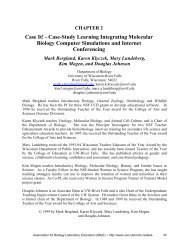
The results presented in Table 8.4 <strong>and</strong> Figure 8.4 indicate that the first spin (Sample B) brings<br />
down in the pellet most of the mitochondria without any significant amount of contractile vacuoles,<br />
giving excellent separation between these two organelles. The second spin (Sample C) pellets most<br />
of the lysosomes in addition to mitochondria while contractile vacuoles are still in the supernatant.<br />
The third spin pellets all markers indicating that they are pelletable organelles. This simple<br />
experiment implies that these three spins sequentially brings down mitochondria, lysosomes, <strong>and</strong><br />
contractile vacuoles, respectively.<br />
Table 8.4. Expected results (an example from a student's notebook).<br />
Succinate<br />
Acid<br />
Alkaline<br />
dehydrogenase phosphatase<br />
PDE<br />
Fraction A630 f A410 f A410 f<br />
A 0.7 0 0.58 0 0.83 0<br />
Bp 0.6 0.8 0.195 0.3 0.072 0.08<br />
Bs 0.15 – 0.455 – 0.83 –<br />
Cp 0.78 0.98 0.426 0.71 0.2 0.23<br />
Cs 0.02 – 0.174 – 0.67 –<br />
Dp 0.81 0.98 0.59 0.87 0.713 0.8<br />
Ds 0.02 – 0.09 – 0.167 –<br />
Examples of Assignment Questions<br />
1. Based on your results, how would you propose to separate organelles X <strong>and</strong> Y from<br />
mitochondria, lysosomes, <strong>and</strong> contractile vacuoles in one experiment employing not more than<br />
three steps of centrifugation? (Answer in 5–7 lines.)<br />
<strong>Organelle</strong> X sediments at 5.0 × 10 6 RCF-min<br />
<strong>Organelle</strong> Y sediments at 1.0 × 10 3 RCF-min<br />
Answer: Spin first at 1.0 × 10 3 RCF-min to pellet only organelle Y. The supernatant is then to be<br />
spun at 5 × 10 5 RCF-min to remove in the pellet mitochondria, lysosomes <strong>and</strong> contractile<br />
vacuoles (see Table 2). The supernatant which now contains only organelle X could be spun at<br />
5 × 10 6 RCF-min to pellet organelle X.<br />
2. As you know, materials ingested by cells are sent to lysosomes while mitochondria are involved<br />
in oxidative phosphorylation (ATP synthesis). How would you propose to separate them if they<br />
happen to sediment at the same RCF-min <strong>and</strong> have the same density on density gradients?<br />
(Answer in 3–4 lines.)
<strong>Organelle</strong> <strong>Isolation</strong> 141<br />
Figure 8.4. The data from Table 8.4 are plotted as f versus RCF-min <strong>for</strong> three markers.<br />
Answer: Cells could be fed lighter compounds like Triton WR-1339 which accumulate in<br />
lysosomes <strong>and</strong> in turn make them more buoyant than mitochondria. Separation should then be<br />
possible on a density gradient.<br />
3. A scientist using electron microscope has just observed a new organelle in Paramecium. This<br />
structure is 3 µm in size <strong>and</strong> has star-shaped morphology. The organelle resembles no other<br />
organelle <strong>and</strong> has no known marker enzyme activity. How would you proceed to characterize<br />
this organelle <strong>and</strong> what criteria would you use to monitor purification of this new organelle?<br />
(Answer in 5–7 lines.)<br />
Answer: Cell homogenate could be fractionated using a variety of conventional methods like<br />
differential, rate zonal, <strong>and</strong> density-equilibrium centrifugation. Since no marker enzyme is<br />
known <strong>for</strong> this organelle, unique morphology (star-shaped) under a microscope could be used to<br />
monitor the fractionation <strong>and</strong> purification of this organelle. An extra credit if student mentions<br />
light microscope since the organelle is large (3 µm) in size <strong>and</strong> should be visible in light<br />
microscope which is much faster <strong>and</strong> easier to operate than an electron microscope.
142 <strong>Organelle</strong> <strong>Isolation</strong><br />
APPENDIX B<br />
Growing Cells of Dictyostelium discoideum<br />
Loomis (1982) provides a comprehensive treatise on this organism. D. discoideum is a eukaryotic<br />
microorganism which can be easily grown in the laboratory. The axenic strain Ax-3 used in this exercise can<br />
be obtained either from the American Type Culture Collection or from any one of the numerous research<br />
laboratories throughout the world who work on this organism. Cells are grown in axenic medium (known as<br />
HL-5) in sterile flasks (250-ml flasks with 50–100 ml medium or 500-ml flask with 100–200 ml medium).<br />
Cells are grown on a rotary shaker (160 rpm) at room temperature. Cells will not survive<br />
temperatures higher than 25°C. Doubling time is 10–12 hours. Collect cells when density of cells is 5 × 10 6<br />
to 1.5 × 10 7 /ml. Cells can be harvested in clinical centrifuge at 500 g (about 1,500 rpm) <strong>for</strong> 5 minutes.<br />
Resuspend cells in Buffer A <strong>and</strong> centrifuge again.<br />
Cell viability could be tested by mixing 1 part of cell suspension with 1 part of 0.4% Trypan blue in<br />
buffer. Observe under a microscope. Healthy cells remain colorless while dead cells take up blue color.<br />
Homogenization of cells by passing them through polycarbonate nucleopore filters is a very<br />
convenient procedure (see Das <strong>and</strong> Henderson, 1983). A filter assembly using two filter membranes is<br />
prepared as shown in Figure 8.5. The assembly is attached to the end of a barrel of a syringe. Cell<br />
suspension is added into the barrel <strong>and</strong> the plunger is gently but firmly pressed to <strong>for</strong>ce cells through the filter<br />
membranes.<br />
For long-term preservation, vegetative cells can be developed into spores (see Figure 8.3): 2 × 10 7<br />
cells/ml in KK2Mg buffer are plated onto a 5.5-cm Whatman No. 50 filter resting on a buffer-soaked<br />
Whatman AP pad in a sterile petri dish. You can also use non-nutrient agar <strong>for</strong> this purpose. Fruiting bodies<br />
will develop in 2–3 days. Collect spores in 10% glycerol in KK2Mg buffer <strong>and</strong> store in the freezer. To<br />
germinate spores, thaw them quickly <strong>and</strong> add to axenic medium <strong>and</strong> grow as discussed above.<br />
HL-5 Axenic Medium<br />
D-glucose 10 g<br />
Proteose peptone No. 3 10 g<br />
Yeast extract 5 g<br />
Na2HPO4 600 mg<br />
KH2PO4 486 mg<br />
Deionized water to make 1 liter<br />
1. Dispense into flasks <strong>and</strong> autoclave <strong>for</strong> 45 minutes.<br />
2. Just be<strong>for</strong>e use add antibiotics to prevent bacterial growth: use a 100X mixture of penicillin (6 mg/ml)<br />
<strong>and</strong> streptomycin (10 mg/ml) available from GIBCO Laboratories (3175 Staley Rd., Gr<strong>and</strong> Isl<strong>and</strong>, NY<br />
14072 or 2260A Industrial St., Burlington, Ont. L7P 1A1). Filter sterilize. Add 1 ml per 100 ml of<br />
medium.<br />
KK2Mg Buffer<br />
1. 10X of K-phosphate: Add 23 g of KH2PO4 <strong>and</strong> 10 g of K2HPO4 in 1 liter of water <strong>and</strong> autoclave.<br />
2. Separately autoclave 5 g of MgSO4 in 100 ml of water.<br />
3. Add 100 ml of 10X K-phosphate solution + 10 ml of MgSO4 solution <strong>and</strong> 890 ml of water to make 1X<br />
KK2Mg buffer.
<strong>Organelle</strong> <strong>Isolation</strong> 143<br />
Figure 8.5. Schematic diagram of filtration assembly used to homogenize cells of D.<br />
discoideum.
144 <strong>Organelle</strong> <strong>Isolation</strong><br />
APPENDIX C<br />
Materials<br />
Reagents<br />
Buffer/solution Per student<br />
(ml)<br />
Per session<br />
[30 students]<br />
(ml)<br />
1 Buffer A 100 3000<br />
2 Buffer B 2 65<br />
3 1% Triton WR-<br />
1339<br />
1 30<br />
4 NBT solution 1 30<br />
5 Substrate B 1 30<br />
6 2% SDS 20 600<br />
7 Buffer C 1 30<br />
8 Substrate C 0.6 25<br />
9<br />
10<br />
0.2 M Na3PO4<br />
Buffer D<br />
60<br />
1<br />
2000<br />
30<br />
11 Substrate D 0.6 25<br />
Preparation of Reagents<br />
Note: Except 2% SDS, store all reagents on ice during the exercise. Dispense them into aliquots as<br />
indicated <strong>and</strong> give them to students. Keep 2% SDS at room temperature.<br />
1. Buffer A: 5 mM Na-glycinate, pH 8.5 + 100 mM sucrose<br />
As stock, make 100 ml of 1 M buffer (mol. wt. of glycine = 75); there<strong>for</strong>e take 7.5 g of glycine<br />
in about 70 ml water, adjust pH by dropwise addition of 10 N NaOH to pH 8.5. Make the<br />
volume to 100 ml. Freeze in 10 ml aliquots. Each day, thaw one tube. Dilute to 2 liter with<br />
water <strong>and</strong> add 68.4 g sucrose. This is Buffer A. Keep it ice-cold.<br />
2. Buffer B: 200 mM Na-phosphate buffer, pH 7.4<br />
Make 500 ml each of 0.2 M of NaH2PO4 <strong>and</strong> 0.2 M Na2HPO4. Add NaH2PO4 solution to<br />
Na2HPO4 solution to get pH 7.4. Store at 4°C.<br />
3. 1% Triton WR 1339: 2 g in 200 ml water. Store at 4°C.<br />
4. NBT Solution: Dissolve 50 mg of NBT in 20 ml of water. Prepare fresh everyday. Keep on ice.<br />
5. Substrate B: 100 mM Na-succinate, pH > 7.<br />
Na-succinate (<strong>for</strong>mula wt. = 270). Add 5.4 g in 200 ml water. Store frozen at -20°C.<br />
6. SDS 2%: 2% sodium dodecyl sulphate.<br />
Add 60 g of SDS to 3 liters of water. Store at room temperature.<br />
7. Buffer C: 0.25 M of glycine-HCl, pH 3.0 + 0.5% Triton X-100<br />
Prepare 300 ml of 0.25 M of glycine-HCl, pH 3.0 <strong>and</strong> add 1.5 g of Triton X-100. Store at 4°C.<br />
8. Substrate C: 50 mM pNPP (mol. wt. 263 + 108 (6 H2O) = 371)<br />
Dissolve 1.85 g in 100 ml of water. Store at -20°C in freezer in 10-ml aliquots.
<strong>Organelle</strong> <strong>Isolation</strong> 145<br />
9. 0.2 M Na3PO4, pH 12.0: Mol. wt. Na3PO4, 12H2O = 380.1<br />
Dissolve 608.1 g in 8 liters of water. pH should be about 12. Adjust if necessary. Store at 4°C.<br />
10. Buffer D: 0.25 M Tris-borate, pH 8.5 + additions<br />
Make 300 ml of 0.25 M Tris-borate, pH 8.5, <strong>and</strong> add 1.5 g of Triton X-100, 1.218 g of<br />
MgCl2,6H2O <strong>and</strong> 1.73 mg of ZnSO4,7H2O. Store at 4°C.<br />
11. Substrate D: 50 mM pNPT (<strong>for</strong>mula wt. = 443.3)<br />
Dissolve 2.217 g in 100 ml of water. Store at -20°C in freezer in 10-ml aliquots.<br />
Instruments, Chemicals, <strong>and</strong> Supplies<br />
Note: Common chemicals, such as Na3PO4, Na2HPO4, sodium dodecyl sulphate (SDS), <strong>and</strong><br />
instruments, such as pH meters, commonly employed in biochemistry <strong>and</strong> cell biology laboratories<br />
are not listed below.<br />
Items Supplier Catalog #<br />
13 × 100-mm glass tubes Baxter T1269-6<br />
1.5-ml microfuge tubes VWR 20170-331<br />
Polycarbonate membranes 25-mm diameter,<br />
5-µm pores, Nucleopore (#110613) VWR 28158-668<br />
Swin-Lok filter holder, 25-mm diameter,<br />
Nucleopore #420210) VWR 28163-045<br />
Difco dextrose (Difco #0155-17-4) VWR DF0155-17<br />
Proteose peptone No. 3 (Difco #0155-17-4) VWR DF0122-01<br />
Yeast extract (Difco #0127-01-7) VWR DF0127-01<br />
Sigma 104 phosphatase (pNPP) Sigma 104-O<br />
Thymidine 5-monophosphate<br />
PNP-ester ammonium Sigma T-5380<br />
Nitroblue tetrazolium Sigma N-6876<br />
Triton WR-1339 (tyloxapol) Sigma T-8761<br />
Plastic or glass syringes: 5- or 10-ml capacity<br />
Plastic or glass pipets: 5- <strong>and</strong> 10-ml capacity<br />
Pipet-Aids (or rubber bulbs to use with the pipets)<br />
200-µl <strong>and</strong> 1000-µl capacity eppendorf-type micropipets <strong>and</strong> pipet tips<br />
Ice buckets <strong>and</strong> crushed ice<br />
Baxter: Baxter Scientific Products, 1430 Waukegan Rd., McGraw Park, IL 60085-6787.<br />
Sigma: Sigma Chemical Co., P. O. Box 14508, St. Louis, MO 63178.<br />
VWR: VWR Scientific, 800 East Fabyan Park, Batavia, IL 60510.
146 <strong>Organelle</strong> <strong>Isolation</strong><br />
Instruments <strong>for</strong> the exercise:<br />
Clinical bench-top centrifuge<br />
Sorvall RC-5B/RC-2B-type or comparable refrigerated centrifuges which can use Sorvall SS34-type<br />
or similar rotors. (A microfuge could be used with appropriate speed [rpm] equivalent to the<br />
desired RCF-min.)<br />
SS34, or similar, rotors<br />
Water baths (37°C)<br />
Refrigerator (1)<br />
Spectronic 20 (or similar) spectrophotometers<br />
Instruments <strong>for</strong> growing cells:<br />
Gyratory shaker<br />
Incubator at 22°C (if room temperature is not controlled)