A Novel Meatoplasty Method in Canal Wall Down - KoreaMed ...
A Novel Meatoplasty Method in Canal Wall Down - KoreaMed ...
A Novel Meatoplasty Method in Canal Wall Down - KoreaMed ...

You also want an ePaper? Increase the reach of your titles
YUMPU automatically turns print PDFs into web optimized ePapers that Google loves.
Orig<strong>in</strong>al Article<br />
Cl<strong>in</strong>ical and Experimental Otorh<strong>in</strong>olaryngology Vol. 2, No. 4: 164-168, December 2009<br />
A <strong>Novel</strong> <strong>Meatoplasty</strong> <strong>Method</strong> <strong>in</strong> <strong>Canal</strong> <strong>Wall</strong> <strong>Down</strong><br />
Tympanomastoidectomy: A Perichondrial Posterior<br />
Fixation Technique<br />
Ik Joon Choi, MD 1 Jae-J<strong>in</strong> Song, MD 1 Jeong Hun Jang, MD 1 Sun O Chang, MD 1,2<br />
1 2 Department of Otorh<strong>in</strong>olaryngology, Seoul National University College of Medic<strong>in</strong>e, Sensory Organ Research Institute, Seoul National<br />
University Medical Research Center, Seoul, Korea<br />
Objectives. Although it is well recognized that a small meatus after canal wall down (CWD) tympanomastoidectomy can<br />
cause a lifetime problem, unsatisfactory results are frequently encountered. We here<strong>in</strong> <strong>in</strong>troduce a novel technique,<br />
perichondrial posterior fixation (PPF), to ma<strong>in</strong>ta<strong>in</strong> a wide external auditory canal (EAC), to m<strong>in</strong>imize postoperative<br />
wound <strong>in</strong>fection due to the smaller dead space and to improve the posterior auricular cosmetic outcome.<br />
<strong>Method</strong>s. A total of 73 patients who underwent CWD tympanomastoidectomy were <strong>in</strong>cluded. Interventions are CWD<br />
tympanomastoidectomy with the PPF technique. Review of the medical records and evaluation of the postoperative<br />
size of the meatus and the extent of the cavum conchal cartilage buried with<strong>in</strong> the mastoid cavity by tak<strong>in</strong>g<br />
digital photographs.<br />
Results. Thirty males and 43 females were <strong>in</strong>cluded and the mean age was 44.1 yr (range, 6 to 66 yr). The mean followup<br />
duration was 26 months (range, 12 to 56 months). All ears ma<strong>in</strong>ta<strong>in</strong>ed a clean and large external meatus. The<br />
posterior auricular cavum conchal cartilage was successfully prevented from be<strong>in</strong>g buried <strong>in</strong>to the mastoid cavity<br />
<strong>in</strong> all ears. The extent of cartilage buried <strong>in</strong>to the mastoid cavity was much reduced compared to the conventional<br />
technique.<br />
Conclusion. The PPF technique, which is a novel meatoplasty technique <strong>in</strong> CWD tympanomastoidectomy, seems to be<br />
effective <strong>in</strong> ma<strong>in</strong>ta<strong>in</strong><strong>in</strong>g a large external meatus and improv<strong>in</strong>g the cosmetic outcome with m<strong>in</strong>imal risk of complications.<br />
Key Words. Chronic otitis media, <strong>Meatoplasty</strong>, Tympanomastoideotomy<br />
INTRODUCTION<br />
<strong>Meatoplasty</strong> is rout<strong>in</strong>ely performed as an <strong>in</strong>tegral part of a canal<br />
wall down (CWD) mastoidectomy. It is typically performed at<br />
the end of the mastoidectomy to assist <strong>in</strong> ventilation and to pro-<br />
Received September 24, 2009<br />
Accepted after revision October 29, 2009<br />
Correspond<strong>in</strong>g author : Sun O Chang, MD, PhD<br />
Department of Otorh<strong>in</strong>olaryngology, Seoul National University<br />
College of Medic<strong>in</strong>e and Sensory Organ Research Institute, Seoul<br />
National University Medical Research Center, 101 Daehang-no,<br />
Jongno-gu, Seoul 110-744, Korea<br />
Tel : +82-2-2072-3649, Fax : +82-2-745-2387<br />
E-mail : suno@snu.ac.kr<br />
164<br />
DOI 10.3342/ceo.2009.2.4.164<br />
vide easy access for clean<strong>in</strong>g of the cavity postoperatively.<br />
Numerous methods for modified meatoplasty have been <strong>in</strong>troduced<br />
and used s<strong>in</strong>ce the first description by Stacke (1). However,<br />
conventional meatoplasty <strong>in</strong> CWD mastoidectomy results <strong>in</strong> slid<strong>in</strong>g<br />
of the cavum conchal portion from the posterior wall (Fig. 1).<br />
Therefore, the open<strong>in</strong>g of the external auditory canal (EAC) becomes<br />
slightly stenotic and auricular deformity may occur. Deformities<br />
of the EAC are especially more severe <strong>in</strong> children than<br />
<strong>in</strong> adults due to the th<strong>in</strong>ner, more elastic and less rigid nature of<br />
their cartilage.<br />
Hence, we <strong>in</strong>troduced a novel technique, perichondrial posterior<br />
fixation (PPF), to ma<strong>in</strong>ta<strong>in</strong> a wide EAC, to m<strong>in</strong>imize postoperative<br />
wound <strong>in</strong>fection due to the smaller dead space, and<br />
Copyright 2009 by Korean Society of Otorh<strong>in</strong>olaryngology-Head and Neck Surgery.<br />
This is an open-access article distributed under the terms of the Creative Commons Attribution Non-Commercial License (http://creativecommons.org/licenses/by-nc/3.0)<br />
which permits unrestricted non-commercial use, distribution, and reproduction <strong>in</strong> any medium, provided the orig<strong>in</strong>al work is properly cited.
Choi IJ et al.: A <strong>Novel</strong> <strong>Meatoplasty</strong> <strong>Method</strong> 165<br />
A B<br />
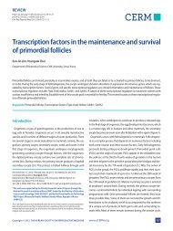
Fig. 1. A surgical outcome by conventional meatoplasty. (A) Lateral view. Narrow<strong>in</strong>g of the external auditory canal due to slid<strong>in</strong>g of the conchal<br />
cartilage (arrowhead) from the posterior wall. (B) Posterior view. The cavum conchal cartilage (arrowhead) is buried <strong>in</strong> the mastoid cavity (arrow).<br />
A B<br />
C D<br />
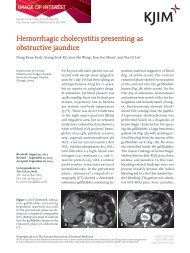
Fig. 2. The surgical technique of a novel meatoplasty <strong>in</strong> canal wall down tympanomastoidectomy us<strong>in</strong>g perichondrial posterior fixation. (A) Partial<br />
excision of the cavum cochal cartilage. (B) A wedge <strong>in</strong>cision is made <strong>in</strong> the Ko_rner flap. (C) Sutur<strong>in</strong>g the anterior edge (arrowhead) of the conchal<br />
cartilage to the periosteum of the mastoid bone (arrow) with 3-0 vicryl. (D) F<strong>in</strong>al result after f<strong>in</strong>ish<strong>in</strong>g our novel meatoplasty.
166 Cl<strong>in</strong>ical and Experimental Otorh<strong>in</strong>olaryngology Vol. 2, No. 4: 164-168, December 2009<br />
to improve the posterior auricular cosmetic outcome.<br />
MATERIALS AND METHODS<br />
Medical records of 88 patients diagnosed with chronic otitis media<br />
with/without cholesteatoma and who underwent CWD tympanomastoidectomy<br />
with the PPF technique from January 2003 to<br />
December 2006 were reviewed. Fifteen cases followed up for<br />
less than 12 months were excluded. The postoperative size of<br />
the meatus and the extent of the cavum conchal cartilage buried<br />
with<strong>in</strong> the mastoid cavity were evaluated on a regular basis. This<br />
technique was approved by <strong>in</strong>stitutional review board (IRB No:<br />
0905-030-281).<br />
Surgical technique<br />
<strong>Meatoplasty</strong> was performed as the f<strong>in</strong>al surgical procedure of<br />
CWD tympanomastoidectomy just before the closure of the subcutaneous<br />
tissue and sk<strong>in</strong>.<br />
To expose the conchal cartilage, soft tissue over the cartilage<br />
was elevated just above the perichondrial layer posterolaterally<br />
(Fig. 2). The excessive conchal cartilage was excised as <strong>in</strong> conventional<br />
meatoplasty. Then 2 stitches were used to pull the perichondrium<br />
of the cavum conchal cartilage posteriorly to the rem-<br />
a<br />
d<br />
c<br />
nant periostium of the mastoid bone with absorbable 3-0 vicryl<br />
thereafter (Fig. 3). After the subcutaneous suture was completed,<br />
the meatus was packed with nylon strips and antibiotics-soaked<br />
cotton wicks.<br />
RESULTS<br />
Sixty-five adults and 8 children underwent meatoplasty us<strong>in</strong>g the<br />
PPF technique. Thirty patients were male and 43 were female.<br />
The mean age of the patients at surgery was 44±14.1 yr old<br />
(range, 6 to 66 yr). The mean follow-up duration was 26±13.2<br />
months (range, 12 to 56 months). All ears ma<strong>in</strong>ta<strong>in</strong>ed a clean and<br />
large external meatus. Us<strong>in</strong>g our novel PPF technique, we could<br />
successfully prevent the posterior auricular cavum conchal cartilage<br />
from be<strong>in</strong>g buried <strong>in</strong> the mastoid cavity <strong>in</strong> all ears that were<br />
operated on. The extent of the cartilage buried with<strong>in</strong> the mastoid<br />
cavity was significantly reduced, result<strong>in</strong>g <strong>in</strong> an improved<br />
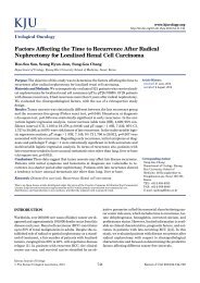
outcome compared to the conventional technique (Fig. 4). Five of<br />
73 patients (6.8%) had postoperative <strong>in</strong>fection of the retroauricular<br />
area, and of those five, <strong>in</strong>fections of four were easily controlled<br />
by topical antibiotics. Only one patient suffered from severe<br />
<strong>in</strong>fection, and was admitted and treated with <strong>in</strong>travenous<br />
antibiotics.<br />
A B<br />
C<br />
b<br />
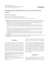
Fig. 3. Axial cross-section diagram at the level of the external auditory<br />
canal (EAC) of the right ear. (A) Narrowed EAC open<strong>in</strong>g (a) by conventional<br />
meatoplasty and reduced height of the external auricle by<br />
bor<strong>in</strong>g <strong>in</strong>to the mastoid cavity (B) Cutt<strong>in</strong>g the excessive anterior edge<br />
(b) of the conchal cartilage. (C) Sutur<strong>in</strong>g the anterior marg<strong>in</strong> of the conchal<br />
cartilage posteriorly to the periosteum of the mastoid bone (c),<br />
widened EAC open<strong>in</strong>g (d) by prevention of conchal cartilage slid<strong>in</strong>g.
DISCUSSION<br />
<strong>Meatoplasty</strong> is one of the key procedures <strong>in</strong> modified radical mastoidectomy.<br />
A large external auditory meatus allows ventilation<br />
of the mastoid cavity as well as easy access for exam<strong>in</strong>ation and<br />
debridement of the entire cavity. Removal of the soft tissue and<br />
cartilage of the EAC and concha could enlarge the meatus, but<br />
the meatoplasty procedures can be complicated by stenosis, <strong>in</strong>fection,<br />
and auricular deformity. Stenosis of the meatus has the potential<br />
to cause a number of problems <strong>in</strong>clud<strong>in</strong>g accumulation of<br />
cerumen, chronic otitis externa, hear<strong>in</strong>g impairment, and difficulty<br />
<strong>in</strong> exam<strong>in</strong><strong>in</strong>g the ear (2). Numerous surgical techniques to<br />
prevent and correct meatal stenosis of the ear canal have been<br />
described (3-6). Techniques of meatoplasty have <strong>in</strong>cluded a comb<strong>in</strong>ation<br />
of transposed sk<strong>in</strong> flaps, removal of the conchal cartilage,<br />
removal of the cartilage from the tragus or floor of the ear<br />
canal, and use of conchomastoid sutures or meatal pack<strong>in</strong>g to<br />
ma<strong>in</strong>ta<strong>in</strong> a large meatal diameter (7-9). The disadvantages of<br />
these techniques are that they are either excessively complicated<br />
or they fail to meet the need of remov<strong>in</strong>g both the excess cartilage<br />
and bone to achieve a proper meatus. Also, <strong>in</strong> patients who<br />
underwent conventional meatoplasty, stenoses of the external<br />
ear canal open<strong>in</strong>g were observed as the cavum conchal cartilage<br />
gradually collapsed <strong>in</strong>to the posterior wall. This also resulted <strong>in</strong><br />
adverse cosmetic outcomes. Eventually, stenosis of the meatus<br />
causes ventilation dysfunction, result<strong>in</strong>g <strong>in</strong> a wet cavity and thereby<br />
accumulation of cerumen. In particular, pediatric patients’ conchal<br />
cartilages are less elastic, and th<strong>in</strong>ner than those of adult patients.<br />
Hence pediatric patients’ conchal cartilages are easily pu-<br />
Choi IJ et al.: A <strong>Novel</strong> <strong>Meatoplasty</strong> <strong>Method</strong> 167<br />
A B<br />
Fig. 4. A surgical outcome by perichondrial posterior fixation. (A) Lateral view. Widen<strong>in</strong>g of the external auditory canal (arrowhead) by fixation<br />
of the conchal cartilage to the posterior wall. (B) Posterior view. Elevation of the cavum conchal cartilage (arrowhead) on the posterior mastoid<br />
wall (arrow).<br />
lled and displaced by the tension of the facial muscle. Consequently,<br />
the cartilag<strong>in</strong>ous portion of the EAC <strong>in</strong> pediatric patients<br />
tends to be more stenotic and tortuous than that of adult patients<br />
after conventional meatoplasty.<br />
An ideal meatoplasty for prevent<strong>in</strong>g stenosis of the EAC after<br />
CWD should be simple to perform, reliable, and cosmetically acceptable.<br />
S<strong>in</strong>ce 2003, we have designed and adopted a new method<br />
<strong>in</strong> which the perichondrium of the cavum conchal cartilage<br />
is anchored posteriorly to the periostium of the mastoid bone.<br />
There was no stenosis seen <strong>in</strong> our series and cosmetically successful<br />
outcomes were achieved <strong>in</strong> all cases. This technique allowed<br />
direct fixation of the cartilage to the posterior wall and resulted <strong>in</strong><br />
a decrease <strong>in</strong> postoperative <strong>in</strong>fection by reduc<strong>in</strong>g the dead space<br />
<strong>in</strong> the operation field. Perichondritis secondary to cartilage excision,<br />
although theoretically possible, has not been observed <strong>in</strong><br />
our cases. A previous study reported a 12 to 20% rate of revision<br />
meatoplasty <strong>in</strong> a series of patients who had undergone CWD<br />
mastoidectomy (10). In contrast, none of the patients required<br />
revision meatoplasty after perform<strong>in</strong>g our novel technique. In<br />
addition, the rate of postoperative <strong>in</strong>fection was not significantly<br />
different from the rate for conventional meatoplasty (1.9 to<br />
6.5%) found <strong>in</strong> a previous report (11). Furthermore, <strong>in</strong> cases <strong>in</strong><br />
which revision canaloplasty or CWD were required for stenosis<br />
of the EAC, or <strong>in</strong> cases of recurrent <strong>in</strong>fection after conventional<br />
meatoplasty, correction of the stenosis and a good cosmetic outcome<br />
were obta<strong>in</strong>ed by apply<strong>in</strong>g our PPF technique. However,<br />
cosmetic outcomes could not be improved when the cartilage or<br />
soft tissue had been removed excessively dur<strong>in</strong>g previous procedures.
168 Cl<strong>in</strong>ical and Experimental Otorh<strong>in</strong>olaryngology Vol. 2, No. 4: 164-168, December 2009<br />
The PPF technique, which is a novel method of meatoplasty<br />
<strong>in</strong> CWD tympanomastoidectomy, can be an effective treatment<br />
modality <strong>in</strong> ma<strong>in</strong>ta<strong>in</strong><strong>in</strong>g a large EAC and <strong>in</strong> improv<strong>in</strong>g the cosmetic<br />
outcome with m<strong>in</strong>imal risk of complications.<br />
REFERENCES<br />
1. Roulleau P. Development of the surgery of chronic otitis. Rev Laryngol<br />
Otol Rh<strong>in</strong>ol (Bord). 1993;114(2):87-91.<br />
2. Fisch U, Chang P, L<strong>in</strong>der T. <strong>Meatoplasty</strong> for lateral stenosis of the external<br />
auditory canal. Laryngoscope. 2002 Jul;112(7 Pt 1):1310-4.<br />
3. Paparella MM, Kurkjian JM. Surgical treatment for chronic stenos<strong>in</strong>g<br />
external otitis (Includ<strong>in</strong>g f<strong>in</strong>d<strong>in</strong>g of unusual canal tumor). Laryngoscope.<br />
1966 Feb;76(2):232-45.<br />
4. Soliman T, Fatt-Hi A, Abdel Kadir M. A simplified technique for the<br />
management of acquired stenosis of the external auditory canal. J Laryngol<br />
Otol. 1980 May;94(5):549-52.<br />
5. Hunsaker DH. Conchomeatoplasty for chronic otitis externa. Arch<br />
Otolaryngol Head Neck Surg. 1988 Apr;114(4):395-8.<br />
6. Mirck PG. The M-meatoplasty of the external auditory canal. Laryngoscope.<br />
1996 Mar;106(3 Pt 1):367-9.<br />
7. Eisenman DJ, Parisier SC. <strong>Meatoplasty</strong>: the cartilage of the floor of<br />
the ear canal. Laryngoscope. 1999 May;109(5):840-2.<br />
8. Susk<strong>in</strong>d DL, Bigelow CD, Knox GW. Y modification of the Fisch<br />
meatoplasty. Otolaryngol Head Neck Surg. 1999 Jul;121(1):126-7.<br />
9. Raut W, Rutka JA. The Toronto meatoplasty: enhanc<strong>in</strong>g one’s results<br />
<strong>in</strong> canal wall down procedures. Laryngoscope. 2002 Nov;112(11):<br />
2093-5.<br />
10. Garap JP, Dubey SP. <strong>Canal</strong>-down mastoidectomy: experience <strong>in</strong> 81<br />
cases. Otol Neurotol. 2001 Jul;22(4):451-6.<br />
11. Kos MI, Castrillon R, Montandon P, Guyot JP. Anatomic and functional<br />
long-term results of canal wall-down mastoidectomy. Ann Otol<br />
Rh<strong>in</strong>ol Laryngol. 2004 Nov;113(11):872-6.