a large pterodactyloidpterosaur from the late cretaceous - Western ...
a large pterodactyloidpterosaur from the late cretaceous - Western ...
a large pterodactyloidpterosaur from the late cretaceous - Western ...
You also want an ePaper? Increase the reach of your titles
YUMPU automatically turns print PDFs into web optimized ePapers that Google loves.
Rec Wen. Awl. Mu.\. 1991,15(2) 435443<br />
A <strong>large</strong> pterodactyloid pterosaur <strong>from</strong> <strong>the</strong> Late Cretaceous<br />
(Late Maastrichtian) of <strong>Western</strong> Australia<br />
S. Christopher Bennett*, John A. Long'f<br />
Abstract<br />
An incomplete right ulna of a <strong>large</strong> pterodactyloid pterosaur is described <strong>from</strong> <strong>the</strong> Late<br />
Cretaceous Miria Formation (Late Maastrichtian), Giralia Range, <strong>Western</strong> Australia. The<br />
bone represents <strong>the</strong> first pterosaur known <strong>from</strong> <strong>Western</strong> Australia, and <strong>the</strong> <strong>large</strong>st and<br />
geologically youngest pterosaur <strong>from</strong> Australia, and <strong>the</strong> youngest Mesozoic terrestrial<br />
tetrapod yet found in Australia. Comparisons with o<strong>the</strong>r pterosaurs give estimates of wingspan<br />
for <strong>the</strong> <strong>Western</strong> Australian pterosaur at between 3.6 (cL Santanadactylus) to 4.9 metres (cL<br />
Pteranodon). The presence of a ridge dividing <strong>the</strong> medial condyle on <strong>the</strong> proximal face of <strong>the</strong><br />
ulna suggests that <strong>the</strong> bone belongs to an azhdarchid; if so this is <strong>the</strong> first record of <strong>the</strong> family<br />
Azhdarchidae in Australia,<br />
Introduction<br />
The fossil reeord of pterosaurs in A ustralia is ra<strong>the</strong>r poor, a nd has been li mited to Early<br />
Cretaceous occurrences. A number of specimens, referred tentatively to ?Ornithocheirus<br />
sp. <strong>from</strong> <strong>the</strong> Albian Toolebuc Formation of Queensland, were described by Molnar and<br />
Thulborn (1980) and Molnar (1987). Rich and Rich ( 1989) described a small crushed<br />
pterosaur bone <strong>from</strong> <strong>the</strong> Aptian-Albian of south-<strong>Western</strong> Australia. The specimen was<br />
tentatively identified as a tibiotarsus of what would be a ra<strong>the</strong>r small pterosaur, but is<br />
similar to <strong>the</strong> crushed metatarsals of Pteranodon <strong>from</strong> North America and is a<br />
metatarsal of a <strong>large</strong> pterosaur. This paper describes <strong>the</strong> proximal end of <strong>the</strong> ulna of a<br />
pterosaur <strong>from</strong> <strong>the</strong> Miria Formation (Late Maastrichtian) of <strong>Western</strong> Australia. The<br />
specimen, which has been previously discussed and figured in Long (1990, p. 78), is <strong>the</strong><br />
first pterosaur <strong>from</strong> <strong>Western</strong> Australia, Australia's <strong>large</strong>st pterosaur, and <strong>the</strong> only Late<br />
Cretaceous pterosaur <strong>from</strong> Australia. It may also be <strong>the</strong> youngest known pteranodontid.<br />
Finally <strong>the</strong> specimen is also of interest in being Australia's only Late Maastrichtian<br />
terrestrial tetrapod as <strong>the</strong> o<strong>the</strong>rwise youngest Mesozoic vertebrates are sauropod<br />
dinosaur bones <strong>from</strong> <strong>the</strong> base of <strong>the</strong> <strong>late</strong> Cretaceous (Winton Formation, central north<br />
Queensland, Coombs and Molnar 1981).<br />
Discovery of <strong>the</strong> specimen<br />
The specimen (WAM 60.57) was found in 1960 by E. Car of <strong>the</strong> <strong>Western</strong> Australian<br />
Museum as three bone fragments washed out of <strong>the</strong> Miria Formation in its type section<br />
at Toothawarra Creek (Figure I). As this is <strong>the</strong> only Cretaceous unit exposed in <strong>the</strong> area<br />
and <strong>the</strong> rock still attached to <strong>the</strong> bone matches that of <strong>the</strong> Miria Formation. <strong>the</strong>re is no<br />
'Department of Systematics and Ecology. Museum of Natural History, University of Kansas. Lawrence.<br />
Kansas 66045. USA.<br />
+<strong>Western</strong> Australian Museum. Franeis Street. Perth. <strong>Western</strong> Australia 6000.<br />
435
Pterodactyloid pterosaur <strong>from</strong> western Australia<br />
doubt as to <strong>the</strong> exact age and stratigraphic source of <strong>the</strong> bone. In early 1990 one of us<br />
(J AL) recognised <strong>the</strong> bone fragments in <strong>the</strong> collection as belonging to one specimen and<br />
restored <strong>the</strong> bone. It was recognised as being unusual in its slender shaft proportions but<br />
an identification could not be assigned, so a cast of <strong>the</strong> bone was sent to Or. Ralph<br />
Molnar of <strong>the</strong> Queensland Museum. Molnar suggested it might be a pterosaur bone, so<br />
fur<strong>the</strong>r casts were sent to Or. Peter Wellnhofer of Munich and one of us (SCB), and<br />
shortly afterwards <strong>the</strong> bone was confirmed to be a pterosaur ulna.<br />
Geological setting<br />
The Miria Formation (Miria Marl, Condon et. al.. 1956; Hocking et. a11987) has been<br />
recently studied in detail by honours students in <strong>the</strong> Geology Department of <strong>the</strong><br />
University of <strong>Western</strong> Australia. The unpublished dissertations which describe<br />
foraminifers <strong>from</strong> <strong>the</strong> Miria Formation confirm its Late Maastrichtian age, earlier<br />
suggested by microfossil studies by several workers (Edgell 1957; Belford 1958,<br />
McGowan 1968, Apthorpe 1979) and <strong>from</strong> diverse ammonite faunas described by<br />
Henderson & McNamara (1985a). O<strong>the</strong>r fossils <strong>from</strong> <strong>the</strong> Miria Formation includes <strong>the</strong><br />
nautiloid Cimomia tenuicostata (Glenister et. al. 1956), as well as undescribed<br />
brachiopods, echinoids, sponges, corals, bryozoans and sharks teeth. O<strong>the</strong>r vertebrate<br />
remains which have come <strong>from</strong> <strong>the</strong> Miria Formation include one bone (WAM 90.10.2)<br />
N<br />
t<br />
Figure 1. Locality map showing outcrop of <strong>the</strong> Miria Formation and where <strong>the</strong> pterosaur bone was found<br />
(arrow). After Henderson and McNamara (1985a).<br />
436
se Bennen, J,A, Long<br />
tentatively assigned as a saurischian (possibly a <strong>the</strong>ropod humerus) as well as several<br />
unidentifiable lumps of reptilian bone. The Late Maastrichtian age of <strong>the</strong> Miria<br />
Formation makes it <strong>the</strong> youngest Mesozoic terrestrial vertebrate site in Australia.<br />
The deposition of <strong>the</strong> Miria Formation began in <strong>the</strong> wake of a Late Maastrichtian<br />
marine transgression (Apthorpe 1979), resulting in quiet shelf deposits in <strong>the</strong> nor<strong>the</strong>rn<br />
Carnarvon Basin. Its lithology is a cream coloured calcarenite, 0.6-2 metres thickness,<br />
with abundant phosphatic grains and nodules. These suggest <strong>the</strong> unit has been<br />
condensed, with fragmentary fossil preservation indicating periods of higher water<br />
energy which winnowed <strong>the</strong> sequence (Henderson & McNamara 1985b).<br />
Description of <strong>the</strong> pterosaur bone<br />
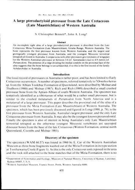
The specimen (Figure 2) is an incomplete right ulna consisting of <strong>the</strong> proximal cnd and<br />
some shaft. Parts of <strong>the</strong> proximal end arc broken and missing and as preserved it has a<br />
maximum width of 53.5 mm and a thickness of 31 mm. Thc shaft is broken at<br />
approximately <strong>the</strong> middle of <strong>the</strong> ulna where it is oval in cross-section and measures 22.5<br />
mm by 17.8 mm. The length of <strong>the</strong> specimen is 134 mm. The anterior surface (Figure 2c)<br />
is pitted by wea<strong>the</strong>ring. The cortical bone has flaked off in many places, and where it is<br />
missing <strong>the</strong> internal cast shows impressions of small ridges and struts. The cortical bone<br />
is up to 1.0 mm thick along <strong>the</strong> shaft and is thinner toward <strong>the</strong> expanded proximal cnd.<br />
[n proximal view <strong>the</strong> bone is roughly D-shaped (Figure 2a). The medial and <strong>late</strong>ral<br />
condyles extend across <strong>the</strong> entire anterior half of <strong>the</strong> proximal cnd. The posterior parts<br />
of both condyles are missing as is <strong>the</strong> tuberosity for <strong>the</strong> insertion of M. triceps brachii.<br />
The condyles face proximally and a little anteriorly but because <strong>the</strong>y are incomplete. it is<br />
difficult to accurately describe <strong>the</strong>ir orientations. The medial condyle has a slight ridge<br />
running diagonally across it. The anteromedial margin of <strong>the</strong> medial condyle and<br />
approximately 15 mm of bone distal to it arc broken away. Lateral to this is a depression<br />
for <strong>the</strong> proximal end of <strong>the</strong> radius. In <strong>the</strong> middle of <strong>the</strong> depression between <strong>the</strong> condyles<br />
arc <strong>the</strong> pneumatic foramina. It is not possible to determine if <strong>the</strong>re were foramina on <strong>the</strong><br />
proximal surface. The biceps tubercle is 18 mm <strong>from</strong> <strong>the</strong> medial condyle. [t is suboval 7<br />
mm by 3 mm, rises about 1.5 mm above <strong>the</strong> shaft, and is angled slightly. It is not possible<br />
to identify any o<strong>the</strong>r features on <strong>the</strong> anterior surface because of wea<strong>the</strong>ring and loss of<br />
cortical bone.rhe posterior surface of <strong>the</strong> ulna is not badly damaged, but does not show<br />
many features. There is a muscle scar on <strong>the</strong> postero<strong>late</strong>ral surface that is directed<br />
proximally and is part of <strong>the</strong> insertion of <strong>the</strong> M. triceps brachii. There is a similar scar on<br />
<strong>the</strong> posteromedial surface that is angled toward <strong>the</strong> medial epicondyle of <strong>the</strong> humerus<br />
and probably is <strong>from</strong> <strong>the</strong> medial col<strong>late</strong>ral ligament.<br />
Comparisons<br />
Hoolcy (1914) reviewed <strong>the</strong> pterosaur fauna of <strong>the</strong> Cambridge Greensand of England<br />
which includes at least four genera of pteranodontids and non-pteranodontids. He<br />
divided <strong>the</strong> proximal ulnae into three groups. Group A ulnae have a robust ridge on <strong>the</strong><br />
anterior surface of <strong>the</strong> shaft which provides a platform to support <strong>the</strong> radius and M.<br />
biceps brachii inserts on <strong>the</strong> side of <strong>the</strong> ridge ra<strong>the</strong>r than on <strong>the</strong> biceps tubercle. Group B<br />
437
Pterodactyloid pterosaur <strong>from</strong> western Australia<br />
A<br />
Figure 2. Right proximal ulna of ?azhdarchid pterosaur <strong>from</strong> <strong>Western</strong> Australia, WA M 60.57. A, in<br />
proximal view. B, in posterior view. C, in anterior view. Bar scale Icm.<br />
ulnae lack <strong>the</strong> ridge, have a biceps tubercle, and have a circular pit on <strong>the</strong> proximal<br />
surface posterior to <strong>the</strong> condyles. Group C ulnae, represented by a single specimen, are<br />
similar to Group B ulnae but lack <strong>the</strong> pit on <strong>the</strong> proximal end. All three types of ulnae<br />
have a pneumatic foramen on <strong>the</strong> anterior surface between <strong>the</strong> condyles. WAM. 60.57 is<br />
similar to Group B ulnae but <strong>the</strong> damage to <strong>the</strong> proximal end makes it impossible to<br />
determine if a pit was present. Unfortunately, it is not clear if Hooley's groupings<br />
correspond to c1adistic groups.<br />
438<br />
c
S.C. Bennett, l.A. Long<br />
Cretaceous pterodactyloids include four major clades, <strong>the</strong> Dsungaripteridae,<br />
Nyctosauridae, Pteranodontidae and Azhdarchidae (Bennett 1989). Comparisons of<br />
W A M 60.57 with representatives of those clades are more useful in determining <strong>the</strong><br />
relationships of W A M 60.57 than considering o<strong>the</strong>r pterosaur ulnae, but difficulties<br />
arise due to <strong>the</strong> scarcity ofcasts or good figures of o<strong>the</strong>r specimens. The proximal ulna of<br />
dsungaripterids is known only <strong>from</strong> Dsungaripterus weii. The ulna has a straight shaft,<br />
thick walls, and does not have noticeable pneumatic foramina. The condyles are<br />
extended anteriorly above <strong>the</strong> shaft and are angled anteriorly. The margins of <strong>the</strong><br />
condyles are massive and rounded. The condition of <strong>the</strong> biceps tubercle is not known<br />
because <strong>the</strong> anterior surface of <strong>the</strong> shaft is damaged. WA M 60.57 differs <strong>from</strong> <strong>the</strong> ulna of<br />
Dsungaripterus weii in having thin walls, <strong>the</strong> condyles do not extend as far above <strong>the</strong><br />
shaft and are angled more proximally, and <strong>the</strong> margins of <strong>the</strong> condyles are sharp, not<br />
massive and rounded. Ulnae of two indeterminate <strong>large</strong> pterodactyloids share certain<br />
similarities and differ <strong>from</strong> all o<strong>the</strong>r ulnae discussed here in certain details. They are<br />
proximal ulnae referred to Araripesaurus sp. (Wellnhofer 1985, figure 44, 45; 1988) and a<br />
proximal ulna referred to Ornithocheirus sp. <strong>from</strong> <strong>the</strong> Early Cretaceous of France<br />
(Buffetaut & Wellnhofer 1983). In both specimens <strong>the</strong> shaft is posterodorsally curved,<br />
<strong>the</strong> condyles appear to be angled anteriorly, and <strong>the</strong> biceps tubercle is ra<strong>the</strong>r indistinct,<br />
although this may be due to immaturity or abrasion. The Araripesaurus sp. ulnae have<br />
small foramina on <strong>the</strong> proximal surface and a <strong>large</strong> pneumatic foramen on <strong>the</strong> anterior<br />
surface between <strong>the</strong> condyles, while <strong>the</strong> Ornithocheirus sp. ulna has a number of small<br />
pneumatic foramina on <strong>the</strong> proximal surface posterior to <strong>the</strong> condyles. The<br />
Araripesaurus sp. ulnae are associated with humeri that display <strong>the</strong> primative<br />
morphology with a straight mid-section to <strong>the</strong> shaft. Therefore, <strong>the</strong> specimen cannot be<br />
pteranodontid or nyctosaurid (Bennett 1989), and it may well be dsungaripterid.<br />
Whatever taxa <strong>the</strong>se ulnae represent <strong>the</strong>y differ <strong>from</strong> W A M 60.57 in <strong>the</strong> posterodorsal<br />
curvature of <strong>the</strong> shaft, <strong>the</strong> anteriorly directed condyles, and <strong>the</strong> indistinct biceps tubercle.<br />
The proximal ulna of pteranodontids is known <strong>from</strong> Ornithodesmus latidens (Hooley<br />
1913), Santanadactylus pricei (Wellnhofer 1985) and Pteranodon (Eaton 1910). The<br />
shaft is relatively straight and <strong>the</strong> condyles are directed proximally. The ulnae of<br />
Santanadactylus pricei and Pteranodon have distinct biceps tubercles. That of<br />
Pteranodon has a longitudinal groove in <strong>the</strong> middle, while that of Santanadactylus pricei<br />
does not. The ulna of Ornithodesmus (Hooley 1913) has a robust ridge extending <strong>from</strong><br />
<strong>the</strong> foramen distally along <strong>the</strong> shaft. The ridge forms a platform that supports <strong>the</strong> radius.<br />
The ridge presumably takes <strong>the</strong> place of<strong>the</strong> biceps tubercle, and like <strong>the</strong> Greensand ulnae<br />
mentioned above, <strong>the</strong> M. biceps brachii inserted on <strong>the</strong> side of<strong>the</strong> ridge. Pteranodon and<br />
Ornithodesmus latidens have a <strong>large</strong> pneumatic foramen just distal to and between <strong>the</strong><br />
condyles, while Santanadactylus pricei lacks a pneumatic foramen on <strong>the</strong> anterior<br />
surface between <strong>the</strong> condyles, and instead has a <strong>large</strong> pneumatic foramen just proximal<br />
to <strong>the</strong> biceps tubercle. Pteranodon also has a nutrient foramen distal to <strong>the</strong> biceps<br />
tubercle.<br />
W A M 60.57 is like <strong>the</strong> ulna of pteranodontids in <strong>the</strong> relatively straight shaft and<br />
proximally directed condyles. It is like Pteranodon and Santanadactrlus pricei in<br />
439
Pterodacty)oid pterosaur <strong>from</strong> western Australia<br />
Figure 3. Right proximal ulna of ?azhdarchid pterosaur <strong>from</strong> <strong>Western</strong> Australia, WAM 60.57. A, in<br />
proximal view. B, in posterior view. C, in anterior view. Abbreviations: bi.t., biceps tubercle;<br />
coLs., col<strong>late</strong>ral ligament scar; I.con, <strong>late</strong>ral condyle; m.con., medial condyle; m.s., muscle scar;<br />
pn.f. pneumatic foramina; ri, ridge. Bar scale Icm.<br />
possessing a distinct biceps tubercle and lacking a ridge to support <strong>the</strong> radius. It is like<br />
Pteranodon and Ornithodesmus in <strong>the</strong> position of <strong>the</strong> pneumatic foramina, although it<br />
has a number of small foramina instead of a single <strong>large</strong> foramen.<br />
The ulna of nyctosaurids is not well known, and is represented only by badly crushed<br />
specimens of Nyctosaurus gracilis. The ulna has a straight shaft, proximally directed<br />
condyles, a distinct biceps tubercle, and a <strong>large</strong> single pneumatic foramen on <strong>the</strong> anterior<br />
surface between <strong>the</strong> condyles. The ulna is similar to that of Pteranodon except it lacks a<br />
440
se Rennet!, J.A. Long<br />
groove in <strong>the</strong> biceps tubercle. W A M 60,57 differs <strong>from</strong> <strong>the</strong> ulna of Nyctosaurus in that it<br />
has a number of pneumatic foramina on <strong>the</strong> anterior surface instead of single <strong>large</strong><br />
foramen, In addition, W AM 60.57 is considerably <strong>large</strong>r than <strong>the</strong> <strong>large</strong>st known<br />
nyctosaur, Nyctosaurus lamegoi, <strong>from</strong> <strong>the</strong> Maastrichtian of Brazil (Price 1953),<br />
The ulna of azhdarchids has not been described. However, specimens of<br />
Arambourgiania (Nessov and Jarkov 1989, = Titanopteryx Arambourg preoccupied)<br />
<strong>from</strong> Jordan and Quetzalcoatlus <strong>from</strong> Texas include proximal ulnae. The ulna of<br />
Arambourgiania (BMNH 9228) has a relatively straight shaft, proximally directed<br />
condyles, a distinct biceps tubercle, one or two ra<strong>the</strong>r small pneumatic foramina on <strong>the</strong><br />
anterior surface between <strong>the</strong> condyles, and a weak ridge running along <strong>the</strong> medial<br />
condyle. The ulna of Quetzalcoatlus is similar to that of Arambourgiania, but lacks<br />
pneumatic foramina on <strong>the</strong> anterior surface, and it is not known if it had a ridge on <strong>the</strong><br />
medial condyle. W A M 60.57 is very similar to <strong>the</strong> ulna of Arambourgiania and is<br />
virtually <strong>the</strong> same size. It differs <strong>from</strong> Arambourgiania in that it has a number of<br />
pneumatic foramina on <strong>the</strong> anterior surface just distal to <strong>the</strong> condyles, and its biceps<br />
tubercle is relatively smaller.<br />
The ulna of Pterodactylus has not been described in sufficient detail for comparisons,<br />
but <strong>the</strong> ulna of Rhamphorhynchus presumably displays <strong>the</strong> primitive condition of <strong>the</strong><br />
pterodactyloid ulna, The form is very similar to <strong>the</strong> general form seen in<br />
Santanadactylus, Pteranodon. NyclOsaurus, Quelzalcoatlus and W A M 60,57. The<br />
proximal end has condyles facing proximally, <strong>the</strong> shaft tapers to a relatively straight<br />
mid-section with a subcircular to suboval cross-section, and it does not have a ridge<br />
extending down <strong>the</strong> shaft supporting <strong>the</strong> radius. There are no visible pneumatic<br />
foramina and <strong>the</strong> shaft is relatively smaller in diameter and has relatively thicker walls<br />
than those of <strong>large</strong> pterodactyloids.<br />
Size<br />
It is difficult to estimate <strong>the</strong> size of a pterosaur <strong>from</strong> a single limb element, but it is<br />
routinely done. Comparisons of WAM 60.57 with complete ulnae of Pteranodon and<br />
Santanadactylus pricei suggest that <strong>the</strong> 134 mm long section is between one half to<br />
two-fifths of <strong>the</strong> total length. Therefore <strong>the</strong> complete ulna probably measured between<br />
27 and 34 cm. The wing proportions of <strong>large</strong> pterodactylids are variable. Extrapolating<br />
<strong>from</strong> an estimated length of 30 cm <strong>the</strong> wingspan in life (flexed as in flight) would be: 3.6<br />
m based on <strong>the</strong> proportions of Sanlanadactr/us araripensis (Wellnhoffer 1985), 3.8 m<br />
based on <strong>the</strong> proportions of Quetzalcoatlus (Langston 1981) or 4.9 metres based on <strong>the</strong><br />
proportions of Pteranodon (Bennett, unpublished data).<br />
Discussion<br />
On <strong>the</strong> basis of its <strong>large</strong> size and stratigraphic position it is clear that W A M 60.57 is a<br />
pterodactyloid. It is probably not a dsungaripterid because it is not thick-walled and <strong>the</strong><br />
condyles do not face anteriorly. It is probably not a nyctosaurid because it is much <strong>large</strong>r<br />
than <strong>the</strong> <strong>large</strong>st known nyctosaurid, and <strong>the</strong> group is not known outside <strong>the</strong> Americas.<br />
WA M 60.57 is very similar to Arambourgiania <strong>from</strong> Jordan and both have a ridge dividing<br />
441
Pterodactyloid pterosaur <strong>from</strong> western Australia<br />
<strong>the</strong> medial condyle that is not noted on <strong>the</strong> ulnae of dsungaripterids, pteranodontids or<br />
nyctosaurids. It is not certain that <strong>the</strong> ridge is a phylogentically important character. Ifit<br />
is not <strong>the</strong>re is nothing to suggest that <strong>the</strong> ulna is not a pteranodontid, however, on <strong>the</strong><br />
basis of<strong>the</strong> ridge dividing <strong>the</strong> medial condyle <strong>the</strong> Australian ulna is tentatively referred to<br />
<strong>the</strong> Azhdarchidae.<br />
This is <strong>the</strong> first record of a pterosaur <strong>from</strong> <strong>Western</strong> Australia, and <strong>the</strong> only <strong>late</strong><br />
Cretaceous occurrence <strong>from</strong> Australia. If <strong>the</strong> specimen is an azhdarchid, it is <strong>the</strong> first<br />
record of an azhdarchid <strong>from</strong> Australia. Azhdarchids are known <strong>from</strong> Early Cretaceous<br />
of England, and <strong>the</strong> Late Cretaceous of Central Asia, Jordan, Senegal, New Jersey,<br />
Texas and Wyoming in North America (Bennett 1989). If <strong>the</strong> specimen were a<br />
pteranodontid it would be <strong>the</strong> only member of that c1ade post mid-Campanian.<br />
The form of <strong>the</strong> pterodactyloid ulna changed little through <strong>the</strong> Cretaceous. As <strong>the</strong> size<br />
of <strong>the</strong> pterodactyloids increased, relatively <strong>large</strong> joints were needed and <strong>the</strong> shaft became<br />
pneumatic. Consequently <strong>the</strong> ulna became relatively stouter. The dsungaripterid ulna is<br />
derived in possessing a posterodorsal curvature of <strong>the</strong> shaft <strong>from</strong> <strong>the</strong> proximal end and<br />
<strong>the</strong> ra<strong>the</strong>r anteriorly directed condyles. The ulna of Ornithodesmus and Hooley's Group<br />
A ulnae are derived in <strong>the</strong> possession of a robust ridge supporting <strong>the</strong> radius and taking<br />
<strong>the</strong> place of a biceps tubercle. However, all o<strong>the</strong>r ulnae of <strong>large</strong>r pterodactyloids,<br />
including W AM 60.57, are similar and vary in what appear to be ra<strong>the</strong>r minor details. It<br />
is surprising that <strong>the</strong> ulnae of pteranodontids and azhdarchids would be similar because<br />
<strong>the</strong> humeri of <strong>the</strong> two clades are very different. Perhaps better knowledge of <strong>the</strong> two<br />
groups will reveal differences.<br />
Phylogenetically important morphological variation of pterosaur postcranials is not<br />
yet well understood, although recent attempts to characterise such variation have made<br />
progress (Padian 1984; Howse 1986; Wiffen & Molnar 1988; Bennett 1989). The above<br />
review of variation in <strong>the</strong> proximal ulna indicates that <strong>the</strong>re is considerable difference<br />
between taxa in <strong>the</strong> size and location of pneumatic foramina. It is not known whe<strong>the</strong>r<br />
this variation is of phylogenetic importance. In <strong>large</strong> samples of Pteranodon <strong>the</strong> size and<br />
position of <strong>the</strong> pneumatic foramina are relatively constant, however, fur<strong>the</strong>r study ofthis<br />
and o<strong>the</strong>r aspects of postcranial variation in pterosaurs is needed.<br />
Acknowledgements<br />
The authors thank Or. Ralph Molnar, Queensland Museum and Or. Tom Rich,<br />
Museum of Victoria, for helpful comments on <strong>the</strong> manuscript. Kris Brimmel is thanked<br />
for photography and casting of <strong>the</strong> specimen. Ors. Chang Mee-Mann, Angela Milner<br />
and Peter Wellnhofer are thanked for access to specimens in <strong>the</strong>ir care.<br />
References<br />
Apthorpe, M.C. (1979). Depositional history of <strong>the</strong> upper Cretaceous of <strong>the</strong> Northwest Shelf, based on<br />
Foraminifera. A PEA Journal 19: 74-89.<br />
Belford, D.J. (1958). Stratigraphyand micropalaeontology of <strong>the</strong> Upper Cretaceous of <strong>Western</strong> Australia.<br />
Geol. Rundschau 47: 629-647.<br />
Bennett, S. C. (1989). A pteranodontid pterosaur <strong>from</strong> <strong>the</strong> Early Cretaceous of Peru, with comments on <strong>the</strong><br />
relationships of Cretaceous pterosaurs. J. Paleont 63: 669-677.<br />
442


