Auxin: A major regulator of organogenesis - Department of Plant ...
Auxin: A major regulator of organogenesis - Department of Plant ...
Auxin: A major regulator of organogenesis - Department of Plant ...

Create successful ePaper yourself
Turn your PDF publications into a flip-book with our unique Google optimized e-Paper software.
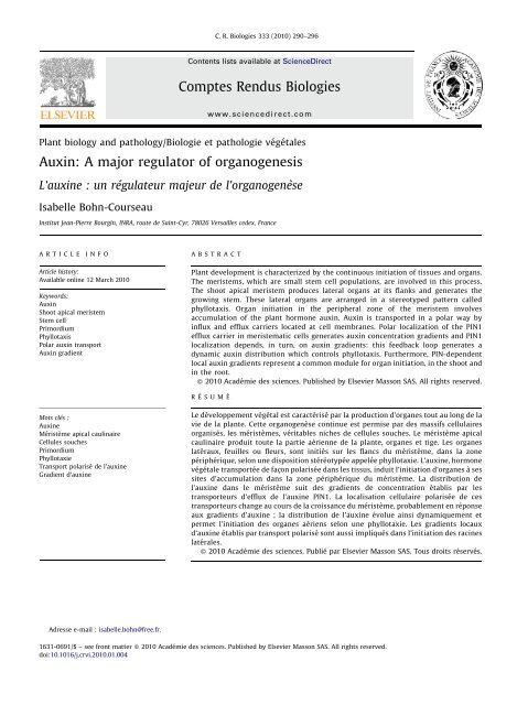
<strong>Plant</strong> biology and pathology/Biologie et pathologie végétales<br />
<strong>Auxin</strong>: A <strong>major</strong> <strong>regulator</strong> <strong>of</strong> <strong>organogenesis</strong><br />
L’auxine : un régulateur majeur de l’organogenèse<br />
Isabelle Bohn-Courseau<br />
C. R. Biologies 333 (2010) 290–296<br />
Institut Jean-Pierre Bourgin, INRA, route de Saint-Cyr, 78026 Versailles cedex, France<br />
ARTICLE INFO<br />
Article history:<br />
Available online 12 March 2010<br />
Keywords:<br />
<strong>Auxin</strong><br />
Shoot apical meristem<br />
Stem cell<br />
Primordium<br />
Phyllotaxis<br />
Polar auxin transport<br />
<strong>Auxin</strong> gradient<br />
Mots clés :<br />
<strong>Auxin</strong>e<br />
Méristème apical caulinaire<br />
Cellules souches<br />
Primordium<br />
Phyllotaxie<br />
Transport polarisé de l’auxine<br />
Gradient d’auxine<br />
Adresse e-mail : isabelle.bohn@free.fr.<br />
Contents lists available at ScienceDirect<br />
Comptes Rendus Biologies<br />
www.sciencedirect.com<br />
ABSTRACT<br />
<strong>Plant</strong> development is characterized by the continuous initiation <strong>of</strong> tissues and organs.<br />
The meristems, which are small stem cell populations, are involved in this process.<br />
The shoot apical meristem produces lateral organs at its flanks and generates the<br />
growing stem. These lateral organs are arranged in a stereotyped pattern called<br />
phyllotaxis. Organ initiation in the peripheral zone <strong>of</strong> the meristem involves<br />
accumulation <strong>of</strong> the plant hormone auxin. <strong>Auxin</strong> is transported in a polar way by<br />
influx and efflux carriers located at cell membranes. Polar localization <strong>of</strong> the PIN1<br />
efflux carrier in meristematic cells generates auxin concentration gradients and PIN1<br />
localization depends, in turn, on auxin gradients: this feedback loop generates a<br />
dynamic auxin distribution which controls phyllotaxis. Furthermore, PIN-dependent<br />
local auxin gradients represent a common module for organ initiation, in the shoot and<br />
in the root.<br />
ß 2010 Académie des sciences. Published by Elsevier Masson SAS. All rights reserved.<br />
RÉSUMÉ<br />
Le développement végétal est caractérisé par la production d’organes tout au long de la<br />
vie de la plante. Cette organogenèse continue est permise par des massifs cellulaires<br />
organisés, les méristèmes, véritables niches de cellules souches. Le méristème apical<br />
caulinaire produit toute la partie aérienne de la plante, organes et tige. Les organes<br />
latéraux, feuilles ou fleurs, sont initiés sur les flancs du méristème, dans la zone<br />
périphérique, selon une disposition stéréotypéeappelée phyllotaxie. L’auxine, hormone<br />
végétale transportéedefaçon polarisée dans les tissus, induit l’initiation d’organes à ses<br />
sites d’accumulation dans la zone périphérique du méristème. La distribution de<br />
l’auxine dans le méristème suit des gradients de concentration établis par les<br />
transporteurs d’efflux de l’auxine PIN1. La localisation cellulaire polarisée de ces<br />
transporteurs change au cours de la croissance du méristème, probablement en réponse<br />
aux gradients d’auxine ; la distribution de l’auxine évolue ainsi dynamiquement et<br />
permet l’initiation des organes aériens selon une phyllotaxie. Les gradients locaux<br />
d’auxine établis par transport polarisé sont aussi impliqués dans l’initiation des racines<br />
latérales.<br />
ß 2010 Académie des sciences. Publié par Elsevier Masson SAS. Tous droits réservés.<br />
1631-0691/$ – see front matter ß 2010 Académie des sciences. Published by Elsevier Masson SAS. All rights reserved.<br />
doi:10.1016/j.crvi.2010.01.004
Abbreviations<br />
SAM shoot apical meristem<br />
IAA indole-3-acetic-acid<br />
PIN pin-formed<br />
LFY LEAFY<br />
ANT AINTEGUMENTA<br />
CUC CUP-SHAPED COTYLEDONS<br />
NPA naphthylpthalamic acid<br />
LAX Like AUX1<br />
PID pinoid<br />
PLT plethora<br />
1. Introduction<br />
In higher plants, post-embryonic development is characterized<br />
by the continuous production <strong>of</strong> tissues and<br />
organs during the whole plant life. Histogenesis and<br />
<strong>organogenesis</strong> involve apical zones <strong>of</strong> cells established<br />
during embryogenesis at the opposite poles <strong>of</strong> the plant<br />
embryo which are called meristems. The mitotic activity <strong>of</strong><br />
pluripotent stem cells located in those meristems permits<br />
organ initiation. By comparison with animal stem cells,<br />
plant meristems are now considered as stem cell niches, i.e.<br />
cellular microenvironments that provide the signals and<br />
physical support to maintain stem cells [1]. Consequently,<br />
plant development depends on the organization, localization<br />
and maintenance <strong>of</strong> those stem cell niches, which<br />
involve several <strong>regulator</strong>y mechanisms including hormone<br />
signaling. The hormone auxin, from the Greek word ‘‘auxo’’<br />
to increase (grow), induces various growth responses in<br />
plants. It has been identified at the end <strong>of</strong> the XIX century by<br />
the Darwins studying the phototropism <strong>of</strong> canary grass<br />
coleoptiles. <strong>Auxin</strong> is implied in many developmental<br />
processes by inducing cellular and molecular modifications<br />
such as cell expansion, cell proliferation or transcription. In<br />
this short review, we will focus on the role <strong>of</strong> auxin in<br />
<strong>organogenesis</strong> at the SAM. Hereby, we will focus on the<br />
angiosperms, which have been best characterised.<br />
2. Organogenesis at the SAM<br />
The SAM produces lateral organs, such as leaves, and a<br />
growing stem. The SAM is a multilayered structure<br />
composed <strong>of</strong> up to three (sometimes even more) clonally<br />
distinct layers <strong>of</strong> cells, L1 to L3. The descendants <strong>of</strong> the L1<br />
cells preferentially generate the epidermis, L2 cells<br />
produce sub-epidermal tissues and gametes whereas the<br />
L3 cell lineage will form inner stem tissues and lateral<br />
organs (for a review, see [2]). Organogenesis relies on<br />
spatially and temporally coordinated cell proliferation in<br />
the SAM. Differences in mitotic activities underline the<br />
existence <strong>of</strong> three cytological zones in the SAM which<br />
overlap the layered partitioning [3,4]: the peripheral zone<br />
on the sides, the central zone at the apex and the rib zone<br />
formed by the internal cells beneath the apex. These zones<br />
have different functions. Organ initiation takes place in the<br />
peripheral zone and the internal tissues <strong>of</strong> the stem (pith<br />
I. Bohn-Courseau / C. R. Biologies 333 (2010) 290–296 291<br />
and vascular system) are formed in the rib zone. Stem cells<br />
located in the central zone divide and part <strong>of</strong> their<br />
daughter-cells are displaced into the peripheral and rib<br />
zones so as to replenish those regions; the other daughtercells<br />
stay in the central zone and maintain a stem cell pool<br />
for future growth. The size <strong>of</strong> the stem cell pool is<br />
controlled by an underlying organizing center, marked by<br />
the expression <strong>of</strong> the transcription factor WUSCHEL, and a<br />
<strong>regulator</strong>y loop involving WUSCHEL and the CLAVATA<br />
receptor kinase pathway signaling cascade. ([1,5–7], for<br />
detailed reviews on the SAM).<br />
Organ initiation begins with the selection <strong>of</strong> a group <strong>of</strong><br />
founder cells in the peripheral zone, followed by the<br />
outgrowth <strong>of</strong> the incipient primordium and then the<br />
morphogenesis into a differentiated organ. Studies on<br />
living meristems revealed that initial primordium outgrowth<br />
is associated with oriented cell divisions; cell<br />
proliferation and cell expansion increase once the primordium<br />
outgrowth has started [4]. Organ boundaries with the<br />
SAM are marked by reduced cell expansion and division<br />
[4,8]. These patterns <strong>of</strong> cellular behavior are correlated<br />
with patterns <strong>of</strong> gene expression. In the organ founder<br />
cells, the KNOX meristematic genes, in particular SHOOT-<br />
MERISTEMLESS (STM), are down-regulated [9,10]; these<br />
founder cells then start to express the ANT and the LFY<br />
transcription factors respectively associated with organ<br />
outgrowth and identity [11,12]. In the same time, the three<br />
CUC transcription factors are expressed in the boundary<br />
[13–15]. (for reviews, see [16] and [17,18] in this issue).<br />
Lateral organs are arranged in regular patterns around<br />
the stem, a phenomenon called phyllotaxis from the Greek<br />
words ‘‘phyllo’’, leaf and ‘‘taxis’’, order. Various phyllotactic<br />
patterns are encountered in nature, namely alternate,<br />
opposite and spiral. Spiralled phyllotaxis, the most<br />
widespread one, follows a mathematical rule: consecutive<br />
lateral organs are formed with an angle close to 137.58, the<br />
golden angle defined by the Fibonacci numbers [19]. At the<br />
Arabidopsis SAM, the first four leaves are formed as<br />
opposite pairs and then leaves and flowers follow such a<br />
spiraled phyllotaxis (Fig. 1).<br />
What is the mechanism behind this regular spacing<br />
between primordia? Historical surgical experiments, consisting<br />
in removing an existing primordium, demonstrated<br />
that existing primordia determine future sites <strong>of</strong> organ<br />
initiation in adjacent regions <strong>of</strong> the SAM (for a review, see<br />
[20]). These results and others support a lateral inhibition<br />
model where developing organs and the centre <strong>of</strong> the<br />
meristem produce an inhibitory signal that acts locally to<br />
prevent organ formation. Due to the growth, existing organs<br />
move away from the meristem; new primordia can develop,<br />
far from the repelling effect (Fig. 1). This inhibitory field<br />
theory <strong>of</strong> phyllotaxis was postulated 80 years ago but the<br />
nature <strong>of</strong> the signal or signals involved remained unknown<br />
until recently. In this short review, we focus on the function<br />
<strong>of</strong> a very important signal, the plant hormone auxin (for<br />
detailed reviews on phyllotaxis [21–24]).<br />
3. <strong>Auxin</strong> synthesis and transport<br />
The <strong>major</strong> form <strong>of</strong> auxin identified in plants is<br />
IAA. Meristems, young primordia, vascular tissues and
292<br />
Fig. 1. Phyllotaxis at the shoot apical meristem. A. Scanning electron microscopy <strong>of</strong> a young Arabidopsis inflorescence meristem. The meristem is the central<br />
dome. P indicates a floral meristem primordium, P1 being the youngest and P7 the oldest. Two consecutive primordia are separated by an angle close to<br />
137.58. Bar = 100 mm. From [3]. B. The inhibitory field theory <strong>of</strong> phyllotaxis. Shoot apical meristem is depicted in pink and organ primordia (P) in blue. Each<br />
preexisting primordium and the meristem central zone are surrounded by a hypothetical inhibitory field (light zone around primordia, dashed white line<br />
around the central zone). The future organ, called initium (I1), can only develop in the region without inhibitor.<br />
reproductive organs produce IAA [25]. IAA is then<br />
transported in the plant by two pathways: a passive<br />
one, in the phloem with the sap flow, and an active and<br />
directional one. This second process is called polar auxin<br />
transport; in stems, it occurs from the shoot apex towards<br />
the base <strong>of</strong> the plant.<br />
The chemiosmotic hypothesis, proposed by Rubery and<br />
Sheldrake in 1974, provides an explanation for the<br />
molecular basis <strong>of</strong> polar auxin transport. Following the<br />
pH, IAA is present in its protonated form IAAH or in its<br />
anionic form IAA . The pKa <strong>of</strong> this weak acid is 4.7. The pH<br />
in the cell wall is around 5.5; about 15% <strong>of</strong> IAA is in its IAAH<br />
form, an electrically neutral molecule which can easily<br />
diffuse into the cell through the membrane. In the<br />
cytoplasm the pH is around 7: IAAH totally dissociates<br />
into IAA , a charged molecule which cannot pass through<br />
the membrane. As a result IAA needs active efflux carriers<br />
to flow from cell to cell. It was proposed that the<br />
asymmetric membranous localization <strong>of</strong> these carriers<br />
would generate the polarity <strong>of</strong> auxin transport. The<br />
identification <strong>of</strong> polar auxin transport inhibitors such as<br />
the naphthylpthalamic acid (NPA) helped to confirm this<br />
hypothesis [26]. The observation <strong>of</strong> a saturable component<br />
for auxin uptake [27,28] and the identification <strong>of</strong> auxin<br />
influx inhibitors such as 1-naphthoxyacetic acid (1-NOA)<br />
[29] suggested that auxin influx carriers are also involved<br />
in polar auxin transport (Fig. 2); for reviews: [30–32].<br />
Molecular and genetic approaches subsequently permitted<br />
to identify and characterize the auxin influx and<br />
efflux carriers. Influx is under the control <strong>of</strong> the AUX1<br />
protein, composed <strong>of</strong> 485 amino acids and 11 transmembrane<br />
domains [33]. The gene is expressed in the root and<br />
SAMs [34]; its expression in a heterologous system<br />
(Xenopus oocytes) leads to a saturable influx <strong>of</strong> auxin<br />
[35]. The lack <strong>of</strong> shoot phenotype in the aux1 mutant<br />
suggests that redundant factors could also be involved,<br />
probably LAX1, LAX2, LAX3 (Like AUX1) ([34]; for a review,<br />
see [27]).<br />
The Arabidopsis pin-formed1 mutant (pin1), named in<br />
reference to its knitting-needle like inflorescence stem,<br />
I. Bohn-Courseau / C. R. Biologies 333 (2010) 290–296<br />
shows a strongly decreased polar auxin transport. The<br />
culture <strong>of</strong> wild-type plants on the auxin efflux inhibitor<br />
NPA provokes the same phenotype [36]. The PIN1 protein,<br />
composed <strong>of</strong> 622 amino acids with eight to 12 putative<br />
transmembrane domains, shows similarities with certain<br />
bacterian and eucaryotes carrier proteins [37]. PIN1 is<br />
localized in a polarized way at the basal end <strong>of</strong> cells in stem<br />
tissues, as predicted for the efflux carriers by the<br />
chemiosmotic hypothesis [38]. PIN expression in yeast<br />
and mammalian cells induces an auxin efflux [39]. Genetic<br />
studies then demonstrated that a <strong>major</strong>ity <strong>of</strong> the eight<br />
Arabidopsis genes (PIN1-PIN8) are auxin efflux carriers but<br />
differ in their expressions patterns [30,40,41]. Last, it has<br />
been directly demonstrated that PIN polarity directly<br />
determines the auxin flow [42].<br />
Fig. 2. Polar auxin transport at the cellular level. IAAH enters the cell by<br />
diffusion or via the influx carrier AUX1. In the cytoplasm, IAAH tends to<br />
dissociate into IAA and H + . IAA can only exit the cell via the efflux<br />
carrier PIN, which has a polar localization. Polar auxin transport direction<br />
is controlled by PIN localization. NPA is an inhibitor <strong>of</strong> auxin efflux.
Polar auxin transport is a highly regulated mechanism,<br />
although the molecular basis <strong>of</strong> PIN localization within the<br />
cell remains poorly understood. One <strong>of</strong> the most characterized<br />
<strong>regulator</strong>s is the PINOID (PID) serine–threonine<br />
protein kinase, identified on the basis <strong>of</strong> the pin-like<br />
phenotype <strong>of</strong> the Arabidopsis pid mutants [43–45]. The<br />
analysis <strong>of</strong> PIN localization in the presence or absence <strong>of</strong><br />
PID provided insight in its role. At the shoot apex, PID loss<br />
<strong>of</strong> function leads to an apical-to-basal shift in PIN1 polarity<br />
[46]. These results indicate that PID regulates polar auxin<br />
transport via a direct control <strong>of</strong> PIN localization. More<br />
recently it was shown that this involves the direct<br />
phosphorylation <strong>of</strong> PIN itself by PID [47]; for detailed<br />
reviews on polar auxin transport [31,32,48].<br />
As briefly described above, the identification <strong>of</strong> the<br />
auxin efflux carrier PIN1 points to a role for auxin in<br />
<strong>organogenesis</strong> at the SAM. PIN1 gene expression is<br />
upregulated in the peripheral zone <strong>of</strong> the SAM at very<br />
early stages <strong>of</strong> <strong>organogenesis</strong>, even before the incipient<br />
primordium outgrowth has started: so far this is one <strong>of</strong> the<br />
earliest markers for organ formation [12,49].<br />
4. <strong>Auxin</strong> and phyllotaxis<br />
Inhibition <strong>of</strong> polar auxin transport, either by NPA or by<br />
mutations in PIN1 or PID, stops organ initiation at the<br />
flanks <strong>of</strong> the SAM but does not affect its other functions: it<br />
results in the formation <strong>of</strong> a naked vegetative or<br />
inflorescence stem [36,43,49,50] (Fig. 3). Exogenous<br />
microapplication <strong>of</strong> IAA to the apex <strong>of</strong> these naked stems<br />
restores organ initiation, showing that auxin is necessary<br />
and sufficient for inducing <strong>organogenesis</strong>. Another striking<br />
result is that organ initiation always takes place at the<br />
meristem flank, whether the IAA is applied to the<br />
peripheral zone or on the meristem summit [50] (Fig. 3).<br />
I. Bohn-Courseau / C. R. Biologies 333 (2010) 290–296 293<br />
This suggests that the cells at the flank <strong>of</strong> the peripheral<br />
zone are competent to react to auxin, whereas cells at the<br />
central zone on top <strong>of</strong> the meristematic dome are not.<br />
Moreover, in the pin1 mutant, the two early markers <strong>of</strong><br />
organ initiation LFY and ANT and the boundary marker<br />
CUC2 are co-expressed in the whole meristem periphery,<br />
suggesting that these competent cells have a hybrid organ/<br />
boundary identity. It is subsequently the amount <strong>of</strong> auxin<br />
applied that will decide whether or not a cell adopts organ<br />
or boundary identity [49]. If we interpret these results in<br />
the context <strong>of</strong> a wild type, a picture emerges, where local<br />
auxin distribution set up by polar auxin transport at the<br />
meristem periphery controls organ positioning and<br />
separation. In the next paragraphs we will further discuss<br />
what controls auxin distribution at meristem flanks.<br />
Application <strong>of</strong> synthetic auxins as 2,4-D (2,4-dichlorophenoxyacetic<br />
acid) and NAA (naphthalene-1-acetic acid)<br />
at naked meristem flanks also provokes organ initiation<br />
but the primordia are ring-shaped [51]. These synthetic<br />
auxins have different properties regarding the polar auxin<br />
transport machinery: NAA does not need the influx carrier<br />
to enter into cells and 2,4-D does not use the efflux carrier<br />
[52]. The severely compromised localization and delimitation<br />
<strong>of</strong> primordia induced by those synthetic auxins<br />
confirm the importance <strong>of</strong> auxin local distribution for<br />
phyllotaxis and reveal the active implication <strong>of</strong> influx and<br />
efflux carriers in setting up local auxin gradients at the<br />
meristem periphery.<br />
Powerful microscopic tools permit to visualize precise<br />
PIN1 localization in the meristem, either by immunolocalization<br />
with a PIN1 antibody or with the pPIN1::PIN1-GFP<br />
reporter gene. PIN1 is located at anticlinal sides <strong>of</strong> L1 cells<br />
and is also expressed in L2 and vascular tissues [53]. At<br />
presumptive sites <strong>of</strong> organ initiation, PIN1 is directed<br />
towards the initium center where it is strongly expressed<br />
Fig. 3. Importance <strong>of</strong> polar auxin transport in the shoot apical meristem for <strong>organogenesis</strong>. A. Arabidopsis wilt-type plant. B. Arabidopsis pin1 mutant; the<br />
stem has no cauline leaf, lateral branche and flower. C. Scanning electron micrograph <strong>of</strong> a pin1 inflorescence apex. The meristem is completely naked<br />
compared with a wild type SAM (see Fig. 1 A). Bar = 200 mm. D. Microapplication <strong>of</strong> IAA (1 mM, paste on right) at the flank <strong>of</strong> the SAM induces the initiation<br />
<strong>of</strong> a flower (result 4 days after treatment). Bar = 100 mm. A to C: from [49]. D: from [50].
294<br />
Fig. 4. PIN1 localization in Arabidopsis shoot apical meristem. A. PIN1 immunolabeling on a meristem cross section showing L1. Asterisks indicate young<br />
primordia. PIN1 is localized on cell membranes and is mostly polarized. Bar = 20 mm. B. Detail <strong>of</strong> PIN1 immunolobaling showing PIN1 concentric orientation<br />
towards the centre <strong>of</strong> a future organ. White arrows indicate presumed local auxin fluxes. Bar = 10 mm. From [55].<br />
[12] (Fig. 4). At the same time, DR5::GFP, an auxinresponsive<br />
reporter gene, is also strongly expressed,<br />
suggesting that auxin accumulates at these sites. This<br />
DR5::GFP expression pattern is abolished when auxin<br />
transport is inhibited by NPA treatment [12,54]. The<br />
complex patterns <strong>of</strong> PIN localization at the meristem were<br />
interpreted using computer simulations which confirmed<br />
earlier observations: PIN1 directs auxin to the sites <strong>of</strong><br />
organ initiation [55]. Together, the observations suggest<br />
that auxin accumulation generated by PIN1 concentric<br />
distribution around incipient primordia is at the basis <strong>of</strong><br />
organ initiation. Monitoring <strong>of</strong> PIN1 localization and<br />
DR5::GFP expression in living meristems reveals stereotypical<br />
PIN1 polarity changes correlated with auxin<br />
distribution changes and with the different stages <strong>of</strong><br />
primordium development [12]. These results further<br />
indicate that PIN1 polarity, directing auxin accumulation<br />
and depletion in the meristem, controls phyllotaxis. Thus,<br />
an auxin maximum in the meristem periphery would<br />
provide the instructive signal for primordium initiation.<br />
<strong>Auxin</strong> would then be drained away from the surrounding<br />
cells by PIN1 via the young vascular tissues inside the<br />
primordium. As a result <strong>of</strong> this auxin depletion the<br />
neighboring L1 cells would not be able to initiate a new<br />
primordium at proximity. Instead <strong>of</strong> both an inductive and<br />
inhibitory signal as proposed by the inhibitory field theory,<br />
phyllotaxis would thus be controlled by a single positive<br />
signal, auxin. In this scenario, the postulated inhibitory<br />
fields around the primordia arise from draining away a<br />
positive signal (Fig. 5).<br />
A <strong>major</strong> question concerns the mechanisms controlling<br />
the auxin fluxes within the meristem. Recent computer<br />
models, developed to simulate phyllotactic patterns,<br />
provided interesting insights. Two models proposed that<br />
cells check the auxin concentrations in their direct<br />
neighbourhood, and subsequently PIN1 gets polarized so<br />
as to pump auxin towards the highest auxin concentration.<br />
This simple positive feedback loop is sufficient to generate<br />
I. Bohn-Courseau / C. R. Biologies 333 (2010) 290–296<br />
different phyllotactic patterns [56,57] (for a detailed<br />
review, see [58]). Other models have since then been<br />
proposed. Stoma et al. showed that a model where cells<br />
sense auxin fluxes rather than auxin concentrations can<br />
equally reproduce the observed phyllotactic patterns [59].<br />
More recently, Bayer and Kuhlemeier showed that a hybrid<br />
model, where both flux based and auxin concentration<br />
based PIN localization could also produce realistic<br />
patterning [60]. With regard to the role <strong>of</strong> the AUX1<br />
importer, it has been proposed these could play a more<br />
discrete but nonetheless crucial role in phyllotaxis, by<br />
stabilizing auxin maxima generated by PIN1 distribution.<br />
Indeed, interference with auxin influx prevents proper<br />
localization and delimitation <strong>of</strong> primordia [51,53,61]. The<br />
quadruple mutant aux1-lax1-lax2-lax3 presents an altered<br />
phyllotactic pattern and a diffuse auxin distribution in the<br />
meristem peripheral zone; PIN1 distribution is also<br />
disturbed [62]. These results reveal a role for influx<br />
carriers in L1 auxin distribution, in interaction with PIN1.<br />
Fig. 5. Model <strong>of</strong> the regulation <strong>of</strong> phyllotaxis by polar auxin transport in<br />
the shoot apical meristem. A. PIN1 directs auxin fluxes (grey arrows).<br />
<strong>Auxin</strong> accumulates (red) in the peripheral zone at the presumptive<br />
initiation site (I1, initium). B. PIN1 expression in vascular tissues inside<br />
the primordium evacuates auxin into inner layers, leading to an auxin<br />
minimum around P1. Another auxin maximum appears in the peripheral<br />
zone, determining the future organ initiation site following phyllotaxis.<br />
From [57].
Even if some mechanisms are still not completely<br />
understood, it seems now clear that phyllotaxis is mainly<br />
controlled by auxin distribution in the peripheral zone <strong>of</strong><br />
the meristem. Polar auxin transport, in particular the<br />
dynamic PIN1 localization, generates auxin gradients<br />
which are then interpreted by meristematic cells in terms<br />
<strong>of</strong> differential growth and cell differentiation, leading to<br />
organ outgrowth and set up <strong>of</strong> boundaries.<br />
Other factors, such as the hormones cytokinin and<br />
gibberellins, and mechanical constraints also influence<br />
phyllotaxis by modulating SAM size, tissue growth and cell<br />
differentiation [63].<br />
5. Outlook: auxin and <strong>organogenesis</strong> at the root apical<br />
meristem<br />
<strong>Auxin</strong> is not only crucial for shoot development but also<br />
in root development and patterning [64]. The organization<br />
<strong>of</strong> the root stem cell niche organization is different from<br />
the one in the shoot. The stem cells, called initials,<br />
surround an organizing center, called the quiescent center,<br />
which positions and maintains the stem cell niche. Stem<br />
cells asymmetrically divide and give rise to the various<br />
root tissues. In the embryo, specification <strong>of</strong> the basal root<br />
pole depends on the auxin gradient established by PIN1<br />
and PIN7 [40]. During and after embryogenesis, PIN protein<br />
activity in the root leads to auxin accumulation at the root<br />
tip, which promotes the expression <strong>of</strong> the PLETHORA (PLT)<br />
transcription factors, <strong>major</strong> determinants for quiescent<br />
centre specification [65,66]. <strong>Auxin</strong> thus contributes to root<br />
stem cell niche specification and positioning (for reviews,<br />
see [67–69]).<br />
The positioning <strong>of</strong> a new root does not follow a regular<br />
pattern as phyllotaxis. Lateral roots emerge in the<br />
differentiation zone from the pericycle, an internal cell<br />
layer. Pericycle cells locally re-initiate cell division and<br />
form root primordia with new stem cell niches, expressing<br />
PLT genes. Lateral root initiation depends on local auxin<br />
accumulation and detection in the pericycle [54]. Periodic<br />
fluctuations in auxin distribution and signaling along the<br />
root seem to regulate lateral root positioning [70].([1], for<br />
a review).<br />
To conclude, it is worth noticing that local auxin<br />
gradients established by the dynamic polar auxin transport,<br />
especially the PIN efflux carriers, are involved in<br />
formation <strong>of</strong> all plant organs, lateral roots, leaves, and<br />
flowers, during post-embryonic development [54]. Interesting<br />
breakthroughs will now come from understanding<br />
auxin perception and cell competence.<br />
Acknowledgements<br />
I am grateful to Dr Jan Traas for a critical reading <strong>of</strong> the<br />
manuscript.<br />
References<br />
[1] B. Scheres, Stem-cell niches: nursery rhymes across kingdoms, Nat Rev<br />
Mol Cell Biol 8 (2007) 345–354.<br />
[2] E.J. Szymkowiak, I.M. Sussex, What Chimeras Can Tell Us About <strong>Plant</strong><br />
Development, Annu Rev <strong>Plant</strong> Physiol <strong>Plant</strong> Mol Biol 47 (1996) 351–<br />
376.<br />
I. Bohn-Courseau / C. R. Biologies 333 (2010) 290–296 295<br />
[3] P. Laufs, O. Grandjean, C. Jonak, K. Kieu, J. Traas, Cellular parameters <strong>of</strong><br />
the shoot apical meristem in Arabidopsis, <strong>Plant</strong> Cell 10 (1998) 1375–<br />
1390.<br />
[4] G.V. Reddy, M.G. Heisler, D.W. Ehrhardt, E.M. Meyerowitz, Real-time<br />
lineage analysis reveals oriented cell divisions associated with morphogenesis<br />
at the shoot apex <strong>of</strong> Arabidopsis thaliana, Development 131<br />
(2004) 4225–4237.<br />
[5] J.L. Bowman, Y. Eshed, Formation and maintenance <strong>of</strong> the shoot apical<br />
meristem, Trends <strong>Plant</strong> Sci 5 (2000) 110–115.<br />
[6] J. Traas, T. Vernoux, The shoot apical meristem: the dynamics <strong>of</strong> a stable<br />
structure, Philos Trans R Soc Lond B Biol Sci 357 (2002) 737–747.<br />
[7] A. Bleckmann, R. Simon, Interdomain signaling in stem cell maintenance<br />
<strong>of</strong> plant shoot meristems, Mol Cells 27 (2009) 615–620.<br />
[8] S. Breuil-Broyer, P. Morel, J. de Almeida-Engler, V. Coustham, I. Negrutiu,<br />
C. Trehin, High-resolution boundary analysis during Arabidopsis<br />
thaliana flower development, <strong>Plant</strong> J 38 (2004) 182–192.<br />
[9] J.A. Long, E.I. Moan, J.I. Medford, M.K. Barton, A member <strong>of</strong> the KNOT-<br />
TED class <strong>of</strong> homeodomain proteins encoded by the STM gene <strong>of</strong><br />
Arabidopsis, Nature 379 (1996) 66–69.<br />
[10] M. Lenhard, G. Jurgens, T. Laux, The WUSCHEL and SHOOTMERISTEM-<br />
LESS genes fulfil complementary roles in Arabidopsis shoot meristem<br />
regulation, Development 129 (2002) 3195–3206.<br />
[11] R.C. Elliott, A.S. Betzner, E. Huttner, M.P. Oakes, W.Q. Tucker, D. Gerentes,<br />
P. Perez, D.R. Smyth, AINTEGUMENTA, an APETALA2-like gene <strong>of</strong><br />
Arabidopsis with pleiotropic roles in ovule development and floral<br />
organ growth, <strong>Plant</strong> Cell 8 (1996) 155–168.<br />
[12] M.G. Heisler, C. Ohno, P. Das, P. Sieber, G.V. Reddy, J.A. Long, E.M.<br />
Meyerowitz, Patterns <strong>of</strong> auxin transport and gene expression during<br />
primordium development revealed by live imaging <strong>of</strong> the Arabidopsis<br />
inflorescence meristem, Curr Biol 15 (2005) 1899–1911.<br />
[13] M. Aida, T. Ishida, H. Fukaki, H. Fujisawa, M. Tasaka, Genes involved in<br />
organ separation in Arabidopsis: an analysis <strong>of</strong> the cup-shaped cotyledon<br />
mutant, <strong>Plant</strong> Cell 9 (1997) 841–857.<br />
[14] S. Takada, K. Hibara, T. Ishida, M. Tasaka, The CUP-SHAPED COTYLE-<br />
DON1 gene <strong>of</strong> Arabidopsis regulates shoot apical meristem formation,<br />
Development 128 (2001) 1127–1135.<br />
[15] C.W. Vroemen, A.P. Mordhorst, C. Albrecht, M.A. Kwaaitaal, S.C. De<br />
Vries, The CUP-SHAPED COTYLEDON3 Gene Is Required for Boundary<br />
and Shoot Meristem Formation in Arabidopsis, <strong>Plant</strong> Cell 15 (2003)<br />
1563–1577.<br />
[16] N. Carraro, A. Peaucelle, P. Laufs, J. Traas, Cell differentiation and organ<br />
initiation at the shoot apical meristem, <strong>Plant</strong> Mol Biol 60 (2006) 811–826.<br />
[17] O. Hamant, V. Pautot, Dissecting TALE Homeoproteins functions during<br />
plant ontogeny. <strong>Plant</strong> Development: a TALE story, C. R. Biologies 333<br />
(2010) 371–381.<br />
[18] A. Hasson, T. Blein, P. Laufs, Leaving the meristem behind: the genetic<br />
and molecular control <strong>of</strong> leaf patterning and morphogenesis, C. R.<br />
Biologies 333 (2010) 350–360.<br />
[19] J.D. Callos, J.I. Medford, Organ positions and pattern formation in the<br />
shoot apex, <strong>Plant</strong> J. 6 (1994) 1–7.<br />
[20] J.F. Golz, Signalling between the shoot apical meristem and developing<br />
lateral organs, <strong>Plant</strong> Mol Biol 60 (2006) 889–903.<br />
[21] I. Adler, D. Barabe, R.V. Jean, A History <strong>of</strong> the Study <strong>of</strong> Phyllotaxis,<br />
Annals <strong>of</strong> Botany 80 (1997) 231–244.<br />
[22] D. Reinhardt, Regulation <strong>of</strong> phyllotaxis, Int J Dev Biol 49 (2005) 539–<br />
546.<br />
[23] D. Reinhardt, Phyllotaxis–a new chapter in an old tale about beauty and<br />
magic numbers, Curr Opin <strong>Plant</strong> Biol 8 (2005) 487–493.<br />
[24] C. Kuhlemeier, Phyllotaxis, Trends <strong>Plant</strong> Sci 12 (2007) 143–150.<br />
[25] Y. Cheng, X. Dai, Y. Zhao, <strong>Auxin</strong> biosynthesis by the YUCCA flavin<br />
monooxygenases controls the formation <strong>of</strong> floral organs and vascular<br />
tissues in Arabidopsis, Genes Dev 20 (2006) 1790–1799.<br />
[26] P.H. Rubery, Phytotropins: receptors and endogenous ligands, Symp<br />
Soc Exp Biol 44 (1990) 119–146.<br />
[27] G. Parry, A. Marchant, S. May, R. Swarup, K. Swarup, N. James, N.<br />
Graham, T. Allen, T. Martucci, A. Yemm, R. Napier, K. Manning, G. King,<br />
M. Bennett, Quick on the Uptake: Characterization <strong>of</strong> a Family <strong>of</strong> <strong>Plant</strong><br />
<strong>Auxin</strong> Influx Carriers, Journal <strong>of</strong> <strong>Plant</strong> Growth Regulation 20 (2001)<br />
217–225.<br />
[28] G. Parry, A. Delbarre, A. Marchant, R. Swarup, R. Napier, C. Perrot-<br />
Rechenmann, M.J. Bennett, Novel auxin transport inhibitors phenocopy<br />
the auxin influx carrier mutation aux1, <strong>Plant</strong> J 25 (2001) 399–406.<br />
[29] V. Imh<strong>of</strong>f, P. Muller, J. Guern, A. Delbarre, Inhibitors <strong>of</strong> the carriermediated<br />
influx <strong>of</strong> auxin in suspension-cultured tobacco cells, <strong>Plant</strong>a<br />
210 (2000) 580–588.<br />
[30] J. Friml, K. Palme, Polar auxin transport–old questions and new concepts?<br />
<strong>Plant</strong> Mol Biol 49 (2002) 273–284.<br />
[31] H. Tanaka, P. Dhonukshe, P.B. Brewer, J. Friml, Spatiotemporal asymmetric<br />
auxin distribution: a means to coordinate plant development,<br />
Cell Mol Life Sci 63 (2006) 2738–2754.
296<br />
[32] W.D. Teale, I.A. Paponov, K. Palme, <strong>Auxin</strong> in action: signalling, transport<br />
and the control <strong>of</strong> plant growth and development, Nat Rev Mol Cell Biol<br />
7 (2006) 847–859.<br />
[33] M.J. Bennett, A. Marchant, H.G. Green, S.T. May, S.P. Ward, P.A. Millner,<br />
A.R. Walker, B. Schulz, K.A. Feldmann, Arabidopsis AUX1 gene: a<br />
permease-like <strong>regulator</strong> <strong>of</strong> root gravitropism, Science 273 (1996)<br />
948–950.<br />
[34] A. Marchant, J. Kargul, S.T. May, P. Muller, A. Delbarre, C. Perrot-<br />
Rechenmann, M.J. Bennett, AUX1 regulates root gravitropism in Arabidopsis<br />
by facilitating auxin uptake within root apical tissues, Embo J<br />
18 (1999) 2066–2073.<br />
[35] Y. Yang, U. Hammes, C. Taylor, D. Schachtman, E. Nielsen, High-Affinity<br />
<strong>Auxin</strong> Transport by the AUX1 Influx Carrier Protein, Curr Biol 16 (2006)<br />
1123–1127.<br />
[36] K. Okada, J. Ueda, M.K. Komaki, C.J. Bell, Y. Shimura, Requirement <strong>of</strong> the<br />
<strong>Auxin</strong> Polar Transport System in Early Stages <strong>of</strong> Arabidopsis Floral Bud<br />
Formation, <strong>Plant</strong> Cell 3 (1991) 677–684.<br />
[37] K. Palme, L. Galweiler, PIN-pointing the molecular basis <strong>of</strong> auxin<br />
transport, Curr Opin <strong>Plant</strong> Biol 2 (1999) 375–381.<br />
[38] L. Galweiler, C. Guan, A. Muller, E. Wisman, K. Mendgen, A. Yephremov,<br />
K. Palme, Regulation <strong>of</strong> polar auxin transport by AtPIN1 in Arabidopsis<br />
vascular tissue, Science 282 (1998) 2226–2230.<br />
[39] J. Petrasek, J. Mravec, R. Bouchard, J.J. Blakeslee, M. Abas, D. Seifertova, J.<br />
Wisniewska, Z. Tadele, M. Kubes, M. Covanova, P. Dhonukshe, P. Skupa,<br />
E. Benkova, L. Perry, P. Krecek, O.R. Lee, G.R. Fink, M. Geisler, A.S.<br />
Murphy, C. Luschnig, E. Zazimalova, J. Friml, PIN proteins perform a<br />
rate-limiting function in cellular auxin efflux, Science 312 (2006) 914–<br />
918.<br />
[40] J. Friml, A. Vieten, M. Sauer, D. Weijers, H. Schwarz, T. Hamann, R.<br />
Offringa, G. Jurgens, Efflux-dependent auxin gradients establish the<br />
apical-basal axis <strong>of</strong> Arabidopsis, Nature 426 (2003) 147–153.<br />
[41] J. Friml, E. Benkova, I. Blilou, J. Wisniewska, T. Hamann, K. Ljung, S.<br />
Woody, G. Sandberg, B. Scheres, G. Jurgens, K. Palme, AtPIN4 mediates<br />
sink-driven auxin gradients and root patterning in Arabidopsis, Cell<br />
108 (2002) 661–673.<br />
[42] J. Wisniewska, J. Xu, D. Seifertova, P.B. Brewer, K. Ruzicka, I. Blilou, D.<br />
Rouquie, E. Benkova, B. Scheres, J. Friml, Polar PIN localization directs<br />
auxin flow in plants, Science 312 (2006) 883.<br />
[43] S.R.M. Bennett, J. Alvarez, G. Bossinger, D.R. Smyth, Morphogenesis in<br />
pinoid mutants <strong>of</strong> Arabidopsis thaliana, The <strong>Plant</strong> Journal 8 (1995)<br />
505–520.<br />
[44] S.K. Christensen, N. Dagenais, J. Chory, D. Weigel, Regulation <strong>of</strong> auxin<br />
response by the protein kinase PINOID, Cell 100 (2000) 469–478.<br />
[45] R. Benjamins, A. Quint, D. Weijers, P. Hooykaas, R. Offringa, The PINOID<br />
protein kinase regulates organ development in Arabidopsis by enhancing<br />
polar auxin transport, Development 128 (2001) 4057–4067.<br />
[46] J. Friml, X. Yang, M. Michniewicz, D. Weijers, A. Quint, O. Tietz, R.<br />
Benjamins, P.B. Ouwerkerk, K. Ljung, G. Sandberg, P.J. Hooykaas, K.<br />
Palme, R. Offringa, A PINOID-dependent binary switch in apical-basal<br />
PIN polar targeting directs auxin efflux, Science 306 (2004) 862–865.<br />
[47] M. Michniewicz, M.K. Zago, L. Abas, D. Weijers, A. Schweigh<strong>of</strong>er, I.<br />
Meskiene, M.G. Heisler, C. Ohno, J. Zhang, F. Huang, R. Schwab, D.<br />
Weigel, E.M. Meyerowitz, C. Luschnig, R. Offringa, J. Friml, Antagonistic<br />
regulation <strong>of</strong> PIN phosphorylation by PP2A and PINOID directs auxin<br />
flux, Cell 130 (2007) 1044–1056.<br />
[48] A.W. Woodward, B. Bartel, <strong>Auxin</strong>: regulation, action, and interaction,<br />
Ann Bot (Lond) 95 (2005) 707–735.<br />
[49] T. Vernoux, J. Kronenberger, O. Grandjean, P. Laufs, J. Traas, PIN-<br />
FORMED 1 regulates cell fate at the periphery <strong>of</strong> the shoot apical<br />
meristem, Development 127 (2000) 5157–5165.<br />
[50] D. Reinhardt, T. Mandel, C. Kuhlemeier, <strong>Auxin</strong> regulates the initiation and<br />
radial position <strong>of</strong> plant lateral organs, <strong>Plant</strong> Cell 12 (2000) 507–518.<br />
I. Bohn-Courseau / C. R. Biologies 333 (2010) 290–296<br />
[51] P.A. Stieger, D. Reinhardt, C. Kuhlemeier, The auxin influx carrier is<br />
essential for correct leaf positioning, <strong>Plant</strong> J 32 (2002) 509–517.<br />
[52] A. Delbarre, P. Muller, V. Imh<strong>of</strong>f, J. Guern, Comparison <strong>of</strong> mechanisms<br />
controlling uptake and accumulation <strong>of</strong> 2,4-dichlorophenoxy acetic<br />
acid, naphthalene-1-acetic acid, and indole-3-acetic acid in suspension-cultured<br />
tobacco cells, <strong>Plant</strong>a 198 (1996) 532–541.<br />
[53] D. Reinhardt, E.R. Pesce, P. Stieger, T. Mandel, K. Baltensperger, M.<br />
Bennett, J. Traas, J. Friml, C. Kuhlemeier, Regulation <strong>of</strong> phyllotaxis by<br />
polar auxin transport, Nature 426 (2003) 255–260.<br />
[54] E. Benkova, M. Michniewicz, M. Sauer, T. Teichmann, D. Seifertova, G.<br />
Jurgens, J. Friml, Local, efflux-dependent auxin gradients as a common<br />
module for plant organ formation, Cell 115 (2003) 591–602.<br />
[55] P.B. de Reuille, I. Bohn-Courseau, K. Ljung, H. Morin, N. Carraro, C.<br />
Godin, J. Traas, Computer simulations reveal properties <strong>of</strong> the cell-cell<br />
signaling network at the shoot apex in Arabidopsis, Proc Natl Acad Sci U<br />
S A 103 (2006) 1627–1632.<br />
[56] H. Jonsson, M.G. Heisler, B.E. Shapiro, E.M. Meyerowitz, E. Mjolsness, An<br />
auxin-driven polarized transport model for phyllotaxis, Proc Natl Acad<br />
Sci U S A 103 (2006) 1633–1638.<br />
[57] R.S. Smith, S. Guyomarc’h, T. Mandel, D. Reinhardt, C. Kuhlemeier, P.<br />
Prusinkiewicz, A plausible model <strong>of</strong> phyllotaxis, Proc Natl Acad Sci U S A<br />
103 (2006) 1301–1306.<br />
[58] E.M. Kramer, Computer models <strong>of</strong> auxin transport: a review and<br />
commentary, J Exp Bot (2007).<br />
[59] S. Stoma, M. Lucas, J. Chopard, M. Schaedel, J. Traas, C. Godin, Fluxbased<br />
transport enhancement as a plausible unifying mechanism for<br />
auxin transport in meristem development, PLoS Comput Biol 4 (2008)<br />
e1000207.<br />
[60] E.M. Bayer, R.S. Smith, T. Mandel, N. Nakayama, M. Sauer, P. Prusinkiewicz,<br />
C. Kuhlemeier, Integration <strong>of</strong> transport-based models for<br />
phyllotaxis and midvein formation, Genes Dev 23 (2009) 373–384.<br />
[61] M.G. Heisler, H. Jonsson, Modeling <strong>Auxin</strong> Transport and <strong>Plant</strong> Development,<br />
J <strong>Plant</strong> Growth Regul 25 (2006) 302–312.<br />
[62] K. Bainbridge, S. Guyomarc’h, E. Bayer, R. Swarup, M. Bennett, T.<br />
Mandel, C. Kuhlemeier, <strong>Auxin</strong> influx carriers stabilize phyllotactic<br />
patterning, Genes Dev 22 (2008) 810–823.<br />
[63] O. Hamant, M.G. Heisler, H. Jonsson, P. Krupinski, M. Uyttewaal, P.<br />
Bokov, F. Corson, P. Sahlin, A. Boudaoud, E.M. Meyerowitz, Y. Couder, J.<br />
Traas, Developmental patterning by mechanical signals in Arabidopsis,<br />
Science 322 (2008) 1650–1655.<br />
[64] S. Sabatini, D. Beis, H. Wolkenfelt, J. Murfett, T. Guilfoyle, J. Malamy, P.<br />
Benfey, O. Leyser, N. Bechtold, P. Weisbeek, B. Scheres, An auxindependent<br />
distal organizer <strong>of</strong> pattern and polarity in the Arabidopsis<br />
root, Cell 99 (1999) 463–472.<br />
[65] M. Aida, D. Beis, R. Heidstra, V. Willemsen, I. Blilou, C. Galinha, L.<br />
Nussaume, Y.S. Noh, R. Amasino, B. Scheres, The PLETHORA genes<br />
mediate patterning <strong>of</strong> the Arabidopsis root stem cell niche, Cell 119<br />
(2004) 109–120.<br />
[66] I. Blilou, J. Xu, M. Wildwater, V. Willemsen, I. Paponov, J. Friml, R.<br />
Heidstra, M. Aida, K. Palme, B. Scheres, The PIN auxin efflux facilitator<br />
network controls growth and patterning in Arabidopsis roots, Nature<br />
433 (2005) 39–44.<br />
[67] R. Sablowski, The dynamic plant stem cell niches, Curr Opin <strong>Plant</strong> Biol<br />
(2007).<br />
[68] J.R. Dinneny, P.N. Benfey, <strong>Plant</strong> stem cell niches: standing the test <strong>of</strong><br />
time, Cell 132 (2008) 553–557.<br />
[69] T. Vernoux, P.N. Benfey, Signals that regulate stem cell activity during<br />
plant development, Curr Opin Genet Dev 15 (2005) 388–394.<br />
[70] I. De Smet, T. Tetsumura, B. De Rybel, N.F. Frey, L. Laplaze, I. Casimiro, R.<br />
Swarup, M. Naudts, S. Vanneste, D. Audenaert, D. Inze, M.J. Bennett, T.<br />
Beeckman, <strong>Auxin</strong>-dependent regulation <strong>of</strong> lateral root positioning in<br />
the basal meristem <strong>of</strong> Arabidopsis, Development 134 (2007) 681–690.