Programmed Cell Death by hok/sok of Plasmid - Department of Plant ...
Programmed Cell Death by hok/sok of Plasmid - Department of Plant ...
Programmed Cell Death by hok/sok of Plasmid - Department of Plant ...

You also want an ePaper? Increase the reach of your titles
YUMPU automatically turns print PDFs into web optimized ePapers that Google loves.
<strong>Programmed</strong> <strong>Cell</strong> <strong>Death</strong> <strong>by</strong> <strong>hok</strong>/<strong>sok</strong> <strong>of</strong> <strong>Plasmid</strong> R1:<br />
Processing at the <strong>hok</strong> mRNA 3 0 -end Triggers<br />
Structural Rearrangements that Allow Translation and<br />
Antisense RNA Binding<br />
Thomas Franch 1 , Alexander P. Gultyaev 2 and Kenn Gerdes 1 *<br />
1 <strong>Department</strong> <strong>of</strong> Molecular<br />
Biology, Odense University<br />
Campusvej 55, DK-5230<br />
Odense M, Denmark<br />
2 Leiden Institute <strong>of</strong> Chemistry<br />
Leiden University, P.O. Box<br />
9502, 2300 RA, Leiden<br />
The Netherlands<br />
*Corresponding author<br />
Introduction<br />
The <strong>hok</strong>/<strong>sok</strong> system <strong>of</strong> plasmid R1 codes for two<br />
RNAs, <strong>hok</strong> mRNA and Sok antisense RNA (Gerdes<br />
et al., 1988, 1990). The mRNA encodes two reading<br />
frames, denoted <strong>hok</strong> (host killing) and mok (modulation<br />
<strong>of</strong> killing), that overlap extensively (Figure 1).<br />
Translation <strong>of</strong> <strong>hok</strong> is coupled to translation <strong>of</strong> mok<br />
(Thisted & Gerdes, 1992). Sok-RNA represses<br />
translation <strong>of</strong> mok via binding to the translational<br />
initiation region (TIR) <strong>of</strong> mok (Thisted et al., 1994a).<br />
Therefore, Sok-RNA regulates <strong>hok</strong> translation<br />
Abbreviations used: SD, Shine-Dalgarno; ucb,<br />
upstream complementary box; dcb, downstream<br />
complementary box; tac, translational activator; TIR,<br />
translational initiation region; DMS, dimethyl sulphate;<br />
wt, wild-type; IPTG, isopropyl-b,Dthiogalactopyranoside.<br />
J. Mol. Biol. (1997) 273, 38±51<br />
The <strong>hok</strong>/<strong>sok</strong> locus <strong>of</strong> plasmid R1 mediates plasmid stabilization <strong>by</strong> killing<br />
<strong>of</strong> plasmid-free cells. The locus speci®es two RNAs, <strong>hok</strong> mRNA and Sok<br />
antisense RNA. The post-segregational killing mediated <strong>by</strong> <strong>hok</strong>/<strong>sok</strong> is governed<br />
<strong>by</strong> a complicated control mechanism that involves both post-transcriptional<br />
inhibition <strong>of</strong> translation <strong>by</strong> Sok-RNA and activation <strong>of</strong> <strong>hok</strong><br />
translation <strong>by</strong> mRNA 3 0 processing. Sok-RNA inhibits translation <strong>of</strong> a<br />
reading frame (mok) that overlaps with <strong>hok</strong>, and translation <strong>of</strong> <strong>hok</strong> is<br />
coupled to translation <strong>of</strong> mok. In the inactive full-length <strong>hok</strong> mRNA, the<br />
translational activator element at the mRNA 5 0 -end (tac) is sequestered <strong>by</strong><br />
the fold-back-inhibitory element located at the mRNA 3 0 -end (fbi). The 5 0<br />
to 3 0 pairing locks the RNA in an inert con®guration in which the SD mok<br />
and Sok-RNA target regions are sequestered. Here we show that the 3 0<br />
processing leads to major structural rearrangements in the mRNA 5 0 -end.<br />
The structure <strong>of</strong> the refolded RNA explains activation <strong>of</strong> translation and<br />
antisense RNA binding. The refolded RNA contains an antisense RNA<br />
target stem-loop that presents the target nucleotides in a single-stranded<br />
conformation. The stem <strong>of</strong> the target hairpin contains SD mok and AUG mok<br />
in a paired con®guration. Using toeprinting analysis, we show that this<br />
pairing keeps SD mok in an accessible con®guration. Furthermore, a mutational<br />
analysis shows that an internal loop in the target stem is prerequisite<br />
for ef®cient translation and antisense RNA binding.<br />
# 1997 Academic Press Limited<br />
Keywords: Sok; <strong>hok</strong>; antisense RNA; translational initiation; RNA<br />
rearrangements<br />
indirectly through mok. The 64 nt Sok-RNA consists<br />
<strong>of</strong> a stem±loop with an 11 nucleotide 5 0<br />
single-stranded extension (Figure 2A). The 5 0 extension<br />
is complementary to the part <strong>of</strong> the mok TIR<br />
that we denote <strong>sok</strong>T 0 (Figure 2) and is responsible<br />
for the initial recognition reaction between the<br />
RNAs (Thisted et al., 1994a). After initial recognition,<br />
more extensive duplex formation progresses<br />
from the Sok-RNA 5 0 -end <strong>by</strong> a zippering-like<br />
mechanism. Subsequently, such RNA duplexes are<br />
cleaved <strong>by</strong> RNase III (Gerdes et al., 1992). Thus,<br />
binding <strong>of</strong> Sok-RNA to <strong>hok</strong> mRNA occurs <strong>by</strong> a<br />
one-step binding mechanism similar to the RNA-<br />
IN/RNA-OUT pairing in Tn10 (Kittle et al., 1989;<br />
Case et al., 1989).<br />
The complex RNA metabolism prerequisite for<br />
activation <strong>of</strong> <strong>hok</strong> translation in plasmid-free cells is<br />
described in detail in the accompanying paper<br />
(Gultyaev et al., 1997) and is brie¯y outlined<br />
0022±2836/97/410038±14 $25.00/0/mb971294 # 1997 Academic Press Limited
Structure and Function <strong>of</strong> <strong>hok</strong> mRNA <strong>of</strong> <strong>Plasmid</strong> R1 39<br />
Figure 1. Overview <strong>of</strong> the structural elements <strong>of</strong> the <strong>hok</strong>/<br />
<strong>sok</strong> system from plasmid R1. FL-1 and FL-2 denote the<br />
full-length <strong>hok</strong> mRNA-1 and mRNA-2, respectively. TR<br />
denotes the 3 0 truncated <strong>hok</strong> mRNA. Other abbreviations<br />
used: <strong>hok</strong>, host killing; mok, modulation <strong>of</strong> killing; Sok,<br />
suppression <strong>of</strong> killing;, <strong>sok</strong>T, Sok-RNA target; fbi, foldback-inhibition;<br />
and tac, translational activation.<br />
below. We showed previously that <strong>hok</strong> mRNA<br />
exists in two variants in vivo (Gerdes et al., 1990;<br />
Thisted et al., 1994a). A truncated, translationally<br />
active mRNA is generated <strong>by</strong> slow exonucleolytic<br />
removal <strong>of</strong> 40 nt from the 3 0 -end <strong>of</strong> the full-length<br />
mRNA. Thus, the full-length mRNA acts as a reservoir<br />
from which the active mRNA is continuously<br />
generated. In plasmid-carrying cells, the presence<br />
<strong>of</strong> Sok-RNA prevents translation <strong>of</strong> the truncated<br />
mRNA. However, in plasmid-free cells, in which<br />
Sok-RNA has decayed, the continuous slow processing<br />
<strong>of</strong> the full-length mRNA leads to accumulation<br />
<strong>of</strong> the translatable truncated mRNA.<br />
Eventually, this leads to synthesis <strong>of</strong> the toxic Hok<br />
protein, and killing <strong>of</strong> the plasmid-free cells<br />
ensues.<br />
Recently, we showed that the stable, inactive <strong>hok</strong><br />
mRNA is compactedly folded with only a few<br />
single-stranded regions, apart from loops (Franch<br />
& Gerdes, 1996). A genetic analysis established<br />
that the full-length mRNA contains a peculiar 5 0 to<br />
3 0 long-range pairing in which the very ®rst<br />
nucleotides pair with the very last ones (Figure 2B).<br />
This interaction is prerequisite for the post-segregational<br />
killing mechanism, since its disruption (<strong>by</strong><br />
mutation) leads to mRNA instability and depletion<br />
<strong>of</strong> the activatable pool <strong>of</strong> full-length mRNA<br />
(Franch & Gerdes, 1996). The 3 0 -element responsible<br />
for inhibition <strong>of</strong> translation and antisense<br />
RNA binding was called fbi (fold-back-inhibition).<br />
The 5 0 element that pairs with fbi was denoted tac<br />
(translational activation). The tac element is<br />
required for activation <strong>of</strong> translation (Franch &<br />
Gerdes, 1996).<br />
In full-length <strong>hok</strong> mRNA, the SD mok element is<br />
sequestered <strong>by</strong> an upstream sequence that we<br />
denote ucb (upstream complementary box). The<br />
ucb element is shown in red in Figure 2. Previously<br />
we suggested that pairing between SD mok and ucb<br />
prevents ribosome entry at SD mok and there<strong>by</strong> inhibits<br />
translation <strong>of</strong> mok (and <strong>hok</strong>; Nielsen & Gerdes,<br />
1995). A nearly perfect repeat <strong>of</strong> the 9 nt ucb<br />
element is located 58 nt downstream <strong>of</strong> ucb<br />
(Figure 2). This element, which pairs with the<br />
translation initiation region <strong>of</strong> <strong>hok</strong> (TIR <strong>hok</strong>) in fulllength<br />
<strong>hok</strong> mRNA, was denoted dcb (downstream<br />
complementary box). This nomenclature is introduced<br />
to illustrate the complex structural<br />
rearrangements that occur in <strong>hok</strong> mRNA (see<br />
below). As a consequence <strong>of</strong> their repetitive nature,<br />
the ucb and dcb elements are both complementary<br />
to SD mok (compare Figure 2B and D).<br />
The structure <strong>of</strong> the antisense RNA target in <strong>hok</strong><br />
mRNA is yet unknown. However, the genetic<br />
algorithm calculations and phylogenetic comparisons<br />
presented in the accompanying paper suggest<br />
that the exonucleolytic removal <strong>of</strong> fbi triggers<br />
major structural rearrangements at the mRNA 5 0 -<br />
end. More precisely, the computer-simulated RNA<br />
foldings predict that in truncated mRNA tac pairs<br />
with ucb, and SD mok with dcb. The latter interaction<br />
is involved in the formation <strong>of</strong> an antisense RNA<br />
target hairpin (Figure 2D). The target hairpin is<br />
topped <strong>by</strong> a 7 nt loop complementary to the very<br />
5 0 -end <strong>of</strong> Sok-RNA.<br />
Here, we investigate the biological properties<br />
and secondary structures <strong>of</strong> the full-length and<br />
truncated <strong>hok</strong> mRNAs. We show that the binding<br />
<strong>of</strong> Sok-RNA to full-length <strong>hok</strong> mRNA is negligible<br />
both in vivo and in vitro. Using in vitro toeprint<br />
analyses, we show that the mok TIR in full-length<br />
<strong>hok</strong> mRNA is unable to bind ribosomal 30 S subunits,<br />
in accordance with the observation that<br />
full-length <strong>hok</strong> mRNA is not translated in vivo.<br />
Most importantly, we obtain ®rm evidence for<br />
the postulated structural rearrangements triggered<br />
<strong>by</strong> 3 0 processing <strong>of</strong> the full-length mRNA. In the<br />
truncated mRNA, tac was found to pair with ucb,<br />
and SD mok with dcb, thus con®rming the presence<br />
<strong>of</strong> the tac and target hairpins. A mutational analysis<br />
shows that the structure <strong>of</strong> the target hairpin<br />
is crucial for rapid antisense RNA binding and<br />
for translation <strong>of</strong> mok, and that even modest<br />
changes in its structure have pr<strong>of</strong>ound effects on<br />
these parameters. We further show that the pairing<br />
between SD mok and dcb is required to maintain<br />
truncated <strong>hok</strong> mRNA in a translatable<br />
con®guration.<br />
Results<br />
Sok antisense RNA binds to truncated but not<br />
to full-length <strong>hok</strong> mRNA in vivo<br />
We have previously examined the metabolism <strong>of</strong><br />
the <strong>hok</strong> mRNAs in vivo (Gerdes et al., 1990, 1992;<br />
Thisted & Gerdes, 1992; Thisted et al., 1994a). Indirect<br />
evidence from Northern analyses suggested<br />
that Sok-RNA binds to truncated <strong>hok</strong> mRNA much<br />
more ef®ciently than to the full-length RNA. To<br />
test this inference more directly, we constructed a<br />
plasmid vector carrying a <strong>hok</strong>/<strong>sok</strong> system in which<br />
P <strong>hok</strong> was precisely replaced <strong>by</strong> the strong, LacI<br />
repressed P A1/04 promotor (Lanzer & Bujard, 1988).<br />
In this system, synthesis <strong>of</strong> <strong>hok</strong> mRNA is halted <strong>by</strong><br />
the removal <strong>of</strong> isopropyl-b,D-thiogalactopyranoside<br />
(IPTG) from the growth-medium. Thus, synthesis
40 Structure and Function <strong>of</strong> <strong>hok</strong> mRNA <strong>of</strong> <strong>Plasmid</strong> R1<br />
<strong>of</strong> <strong>hok</strong> mRNA could be blocked without the<br />
addition <strong>of</strong> rifampicin.<br />
The in vivo processing pattern <strong>of</strong> the <strong>hok</strong> mRNAs<br />
was analysed <strong>by</strong> Northern blotting in the presence<br />
(<strong>sok</strong> ‡ ) or absence (<strong>sok</strong> ) <strong>of</strong> antisense RNA. In its<br />
presence, two <strong>hok</strong> mRNAs, denoted full-length<br />
mRNA-1 and mRNA-2, were observed (Figure 3A,<br />
®rst panel). Both mRNAs were quite stable with a<br />
half-life <strong>of</strong> approximately 20 minutes. Previous<br />
analyses using rifampicin to inhibit transcription<br />
yielded half-lives <strong>of</strong> the order <strong>of</strong> 20 to 40 minutes<br />
(Gerdes et al., 1990, 1992; Thisted et al., 1994a). <strong>hok</strong><br />
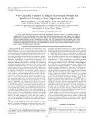
Figure 2. Secondary structures <strong>of</strong> Sok-RNA and <strong>hok</strong> mRNAs. A, Secondary structure <strong>of</strong> Sok-RNA. B, Secondary structure<br />
<strong>of</strong> full-length <strong>hok</strong> mRNA. C, Con®guration <strong>of</strong> non-refolded truncated <strong>hok</strong> mRNA as predicted <strong>by</strong> computerassisted<br />
structure calculations. D, Putative truncated, refolded mRNA supported <strong>by</strong> the structure-probing analyses<br />
presented in Figure 4 and the RNase H cleavage pattern shown in Figure 5. Shine-Dalgarno and start codons <strong>of</strong> the<br />
mok and <strong>hok</strong> reading frames are shown in blue, and the ucb and dcb repetitive sequences are shown in red. C74 in<br />
ucb, marked in green, emphasizes a heterogeneity between the ucb and dcb elements. Thin numbered lines mark the<br />
nucleotides complementary to the three DNA oligonucleotides used in the RNase H cleavage assay. The antisense<br />
RNA target that is complementary to the 5 0 -tail <strong>of</strong> Sok-RNA is denoted <strong>sok</strong>T 0 (B). The sequence and structure <strong>of</strong><br />
super-tac truncated RNA is shown below the wt tac/ucb pairing in D. The non-refolded (C) and refolded (D) con®gurations<br />
are proposed to exist in a dynamic equilibrium.
Structure and Function <strong>of</strong> <strong>hok</strong> mRNA <strong>of</strong> <strong>Plasmid</strong> R1 41<br />
Figure 3. A, Effect <strong>of</strong> Sok antisense RNA on the stability<br />
and processing pattern <strong>of</strong> <strong>hok</strong> mRNAs as shown <strong>by</strong><br />
Northern blotting. The ®rst panel shows <strong>hok</strong> mRNAs<br />
after arrest <strong>of</strong> <strong>hok</strong> transcription in the presence <strong>of</strong> Sok-<br />
RNA, the second panel shows <strong>hok</strong> mRNAs after arrest<br />
<strong>of</strong> transcription in the absence <strong>of</strong> Sok-RNA. <strong>Cell</strong>s<br />
(CSH50 containing either pKG1540 (<strong>sok</strong> ‡ ) or pKG1541<br />
(<strong>sok</strong> )) were grown in LB medium containing 1 mM<br />
IPTG to an A 450 <strong>of</strong> ca 0.2, harvested, and resuspended in<br />
prewarmed medium without IPTG. <strong>Cell</strong> samples for<br />
total RNA preparation were taken at the intervals indicated.<br />
The mRNAs contained stop codon mutations in<br />
the <strong>hok</strong> gene to prevent detrimental <strong>hok</strong> expression (from<br />
Thisted & Gerdes, 1992). B, In vitro binding assay<br />
between 32 P-labelled Sok-RNA and unlabelled ( 3 H) truncated<br />
<strong>hok</strong> mRNA (®rst panel) and full-length <strong>hok</strong><br />
mRNA-1 (second panel). Labelled antisense RNA was<br />
added to a tenfold excess <strong>of</strong> target RNA. For details, see<br />
Materials and Methods.<br />
mRNA-2 is generated from <strong>hok</strong> mRNA-1 <strong>by</strong> an as<br />
yet unknown mechanism (Nikolaj Dam Mikkelsen,<br />
unpublished results). The absence <strong>of</strong> Sok-RNA<br />
caused the appearance <strong>of</strong> a third band corresponding<br />
to truncated <strong>hok</strong> mRNA (Figure 3A, second<br />
panel). This RNA is generated <strong>by</strong> the exonucleolytic<br />
removal <strong>of</strong> 40 nt at the 3 0 -end <strong>of</strong> <strong>hok</strong> mRNA-2<br />
(Gerdes et al., 1990). Comparison <strong>of</strong> the two panels<br />
<strong>of</strong> Figure 3A yields valuable new information on<br />
the in vivo metabolism <strong>of</strong> <strong>hok</strong> mRNA: ®rst, the<br />
absence <strong>of</strong> Sok-RNA has no in¯uence on the<br />
amounts or the stabilities <strong>of</strong> the two full-length<br />
species. Since antisense RNA binding leads to<br />
duplex formation and rapid RNase III cleavage,<br />
this observation indicates that Sok-RNA does not<br />
bind to <strong>hok</strong> mRNA-1 or mRNA-2 at a signi®cant<br />
rate. Secondly, the absence <strong>of</strong> Sok-RNA resulted in<br />
the accumulation <strong>of</strong> the truncated mRNA. Since<br />
this RNA is absent from the ®rst panel, this observation<br />
indicates that Sok-RNA binds avidly to<br />
truncated <strong>hok</strong> mRNA and there<strong>by</strong> confers its rapid<br />
decay via RNase III-mediated hydrolysis. Taken<br />
together, these results show that Sok-RNA binds<br />
full-length <strong>hok</strong> mRNA at a negligible rate in vivo,<br />
whereas the truncated version <strong>of</strong> <strong>hok</strong> mRNA is scavenged<br />
rapidly.<br />
Sok-RNA binds 100-fold more rapidly to<br />
truncated than to full-length <strong>hok</strong> mRNA<br />
in vitro<br />
The above results indicate that Sok-RNA binds<br />
much more rapidly to truncated than to full-length<br />
<strong>hok</strong> mRNA in vivo. Using a standard in vitro binding<br />
assay, we showed previously that full-length<br />
<strong>hok</strong> mRNA bound Sok-RNA with an approximately<br />
30% reduced rate (Thisted et al., 1994b). To<br />
resolve this apparent discrepancy, we repeated the<br />
in vitro binding assay using a more physiologically<br />
correct binding buffer (i.e. 200 mM potassium glutamate<br />
was included). The resulting gel-shift experiment<br />
is shown in Figure 3B. As seen, Sok-RNA<br />
bound readily to the truncated RNA (Figure 3A).<br />
However, binding to the full-length RNA was<br />
inhibited (Figure 3B). The apparent second-order<br />
binding-rate constants (K app) were estimated to<br />
be 3 10 5 M 1 s 1 for truncated and less than<br />
3 10 3 M 1 s 1 for full-length <strong>hok</strong> mRNA, corresponding<br />
to a more than 100-fold difference in<br />
binding-rates (for calculations, see Materials and<br />
Methods). This result suggests that the <strong>sok</strong>T 0 region<br />
exists in different con®gurations in the two RNAs<br />
(see Figure 2B and D). Now we can address this<br />
important question experimentally.<br />
Secondary structure analyses <strong>of</strong> fulllength,<br />
truncated and super-tac truncated<br />
<strong>hok</strong> mRNAs<br />
Mutational analyses, computer calculations and<br />
phylogenetic comparisons suggested that translational<br />
activation <strong>of</strong> <strong>hok</strong> mRNA involves a substantial<br />
structural rearrangement at the mRNA 5 0 -end,<br />
and that this rearrangement is triggered <strong>by</strong> the 3 0<br />
processing <strong>of</strong> full-length <strong>hok</strong> mRNA (Franch &<br />
Gerdes, 1996; Gultyaev et al., 1997, accompanying<br />
paper). More speci®cally, our analyses predicted<br />
that in truncated RNA, tac preferentially pairs with<br />
ucb there<strong>by</strong> leading to formation <strong>of</strong> the extended<br />
tac-stem (see Figure 2D). Although a large internal<br />
loop destabilizes the tac/ucb interaction, it contains<br />
the possibility <strong>of</strong> non-canonical GA/AG base-pairing<br />
(Turner & Bevilacqua, 1993), which may add to<br />
the energy <strong>of</strong> the tac stem. Most importantly,<br />
the 5 0 refolding was accompanied <strong>by</strong> structural<br />
rearrangements further downstream in the molecule.<br />
Thus, a stem±loop that we coin the antisense
42 Structure and Function <strong>of</strong> <strong>hok</strong> mRNA <strong>of</strong> <strong>Plasmid</strong> R1<br />
Figure 4. Secondary structure probing <strong>by</strong> DMS (®rst panel), RNase T 2 (second panel) and RNase V 1 (third panel) <strong>of</strong><br />
full-length, truncated and super-tac truncated mRNA. The sequence and secondary structure <strong>of</strong> the super-tac mutation<br />
are shown in Figure 2D. Modi®cation/cleavage bands mentioned in the text are marked <strong>by</strong> arrowheads.<br />
RNA target hairpin was formed (Figure 2D and<br />
the accompanying paper, Gultyaev et al., 1997).<br />
Construction <strong>of</strong> a ``perfect'' tac stem <strong>by</strong> the introduction<br />
<strong>of</strong> the super-tac mutation (see Figure 2D)<br />
increased translation <strong>of</strong> the truncated <strong>hok</strong> mRNA<br />
tw<strong>of</strong>old (Franch & Gerdes, 1996). The super-tac<br />
mutation greatly increases the stability <strong>of</strong> the tac<br />
stem. Consequently, the presence <strong>of</strong> super-tac predictably<br />
would lead to refolding <strong>of</strong> the entire<br />
population <strong>of</strong> <strong>hok</strong> mRNA molecules present in a<br />
pool <strong>of</strong> in vitro prefolded RNA. This inference is in<br />
accordance with the increased translation rate <strong>of</strong><br />
super-tac truncated RNA.<br />
In order to verify the predicted secondary structure<br />
transitions, structure probing analyses were<br />
conducted on full-length, truncated and super-tac<br />
truncated RNAs (Figure 4). Chemical modi®cation<br />
<strong>by</strong> dimethyl sulphate (modi®es N1 <strong>of</strong> unpaired<br />
A > N3 <strong>of</strong> unpaired C), cleavage <strong>by</strong> RNase T 2<br />
(cleaves 3 0 <strong>of</strong> unpaired nucleotides) and RNase V 1<br />
(cleaves paired and stacked nucleotides) were<br />
detected <strong>by</strong> primer extension using the <strong>hok</strong>1 primer.<br />
For convenience, only the regulatory region<br />
covering nucleotides 70 to 160 is shown in<br />
Figure 4. However, the entire mRNA 5 0 -ends<br />
were also extensively analysed during this work<br />
(data not shown).<br />
Full-length <strong>hok</strong> mRNA<br />
In the full-length mRNA, the proposed position<br />
<strong>of</strong> the loop in the ucb/SD mok stem±loop structure
Structure and Function <strong>of</strong> <strong>hok</strong> mRNA <strong>of</strong> <strong>Plasmid</strong> R1 43<br />
was supported <strong>by</strong> T 2 cuts in the region U89-U91<br />
and DMS modi®cation at C92. The DMS modi®cation<br />
at A81 supports the internal loop at the top<br />
<strong>of</strong> the stem. Furthermore, V 1 cuts at 110 to 112<br />
could support the double-stranded bottom stem.<br />
Three A-C mismatches (C74-A107, A78-C103 and<br />
A82-C100) were predicted in the ucb/SD mok stem.<br />
No signi®cant modi®cation <strong>of</strong> these nucleotides<br />
was observed, probably due to the formation <strong>of</strong><br />
A-C reverse wobble pairings (Saenger, 1984). The<br />
modi®cations at C115 and A117, and to a lesser<br />
extent at C113 and C114, suggest that this region is<br />
at least partially single-stranded. The secondary<br />
structure probing <strong>of</strong> the full-length <strong>hok</strong> mRNA is<br />
consistent with the structure presented in Figure 2B<br />
in which <strong>sok</strong>T 0 is in a double-stranded conformation.<br />
Wild-type and super-tac truncated <strong>hok</strong> mRNAs<br />
The wild-type and super-tac truncated mRNAs<br />
exhibited nearly identical modi®cation/cleavage<br />
patterns and comparison with that <strong>of</strong> the fulllength<br />
RNA revealed important differences. Cleavage<br />
<strong>by</strong> T 2 at A78 and A81-A82 in the truncated<br />
mRNA suggests the presence <strong>of</strong> a single-stranded<br />
region not present in the full-length transcript.<br />
These cuts appeared enhanced in super-tac mRNA.<br />
In the SD mok region signi®cant DMS modi®cations<br />
were observed in super-tac mRNA compared to the<br />
weak probing <strong>of</strong> the wt truncated mRNA, consistent<br />
with incomplete 5 0 -end refolding <strong>of</strong> the latter<br />
species. However, DMS and T 2 probing at C120-<br />
U124 con®rmed the single-stranded loop <strong>of</strong> the target<br />
stem. In addition, V 1 cuts at C125, C126 and<br />
A127 are consistent with stacking <strong>of</strong> the CC loop<br />
dinucleotide on a stem adenine nucleotide. The<br />
super-tac mutation increased the T 2 cleavages in the<br />
loop region (C120 to U124), whereas it reduced<br />
cleavage at U117, G118 and A127-A in the stem.<br />
Thus, the super-tac mutation seems to stabilize the<br />
target hairpin, consistent with incomplete refolding<br />
<strong>of</strong> the wt truncated RNA. This conclusion was corroborated<br />
<strong>by</strong> the V 1 cuts at C139 in the dcb<br />
element. These cuts increase gradually in the order:<br />
full-length < truncated < super-tac truncated mRNA<br />
consistent with the proposed transition <strong>of</strong> the dcb/<br />
TIR <strong>hok</strong> interaction in full-length RNA to the SD mok/<br />
dcb interaction in truncated RNA.<br />
The secondary structure model <strong>of</strong> truncated <strong>hok</strong><br />
mRNA shown in Figure 2D predicts that <strong>sok</strong>T 0 and<br />
the mok TIR are located within the same hairpin.<br />
The validity <strong>of</strong> our secondary structure model <strong>of</strong><br />
the truncated mRNA was further investigated<br />
using an RNase H cleavage assay that tests the<br />
propensity <strong>of</strong> an RNA to hybridize with different<br />
DNA oligonucleotides (Zarrinkar & Williamson,<br />
1994). The DNA oligonucleotides used here are<br />
indicated in Figure 2.<br />
The cleavage pattern <strong>of</strong> full-length <strong>hok</strong> mRNA is<br />
shown in the ®rst panel <strong>of</strong> Figure 5. Oligo 1<br />
resulted in 6% cleavage only, and oligos 2 and 3<br />
both yielded negligible cleavage. This pattern is<br />
Figure 5. RNase H cleavage patterns <strong>of</strong> 32 P-labelled fulllength<br />
(®rst panel), truncated (second panel) and supertac<br />
truncated mRNA (third panel). The regions <strong>of</strong> complementarity<br />
to the oligonucleotides used are shown in<br />
Figure 2. Cleavage products are marked <strong>by</strong> arrowheads.<br />
consistent with a compact secondary structure,<br />
especially in the region <strong>of</strong> <strong>sok</strong>T 0 . In contrast, all<br />
three oligonucleotides yielded signi®cant RNase H<br />
cleavage <strong>of</strong> truncated <strong>hok</strong> mRNA (second panel). In<br />
this case, 38%, 14% and 50% cleavage was<br />
observed with oligonucleotides 1, 2 and 3, respectively.<br />
The third panel shows the cleavage pattern<br />
<strong>of</strong> super-tac truncated mRNA. Here, oligos 1, 2 and<br />
3 yielded 77%, 19% and 65% cleavage, respectively.<br />
These two latter sets <strong>of</strong> mapping data are consistent<br />
with the formation <strong>of</strong> the antisense target<br />
stem±loop presented in Figure 2D. In addition,<br />
oligo 3, which is complementary to <strong>sok</strong>T 0 , yielded<br />
more than a 100-fold difference in cleavage activity<br />
between full-length and truncated mRNA. This<br />
pattern is consistent with the difference in antisense<br />
RNA binding described above. Oligo 1,<br />
which is complementary to SD mok, showed an<br />
increased cleavage in super-tac truncated mRNA as<br />
compared to the wild-type, consistent with the<br />
observed tw<strong>of</strong>old increase in translation (Franch &<br />
Gerdes, 1996). In addition, the increased cleavage<br />
<strong>of</strong> the super-tac mRNA with all three oligonucleotides<br />
corroborates the suggestion that the<br />
wild-type truncated mRNA in vitro exists in two<br />
con®gurations, a refolded and a non-refolded. This<br />
further suggests that the antisense RNA target<br />
stem±loop favoured <strong>by</strong> the tac/ucb and SD mok/dcb<br />
pairings may be in a dynamic equilibrium with the<br />
non-refolded structure shown in Figure 2C.
44 Structure and Function <strong>of</strong> <strong>hok</strong> mRNA <strong>of</strong> <strong>Plasmid</strong> R1<br />
Figure 6. Primary and proposed local secondary structures <strong>of</strong> wild-type and mutated antisense RNA target hairpins.<br />
The SD mok and AUG mok sequences are marked <strong>by</strong> a black line and the dcb box <strong>by</strong> a hairline. Mutations are shown in<br />
bold. The energy <strong>of</strong> the local structures is given below each structure (calculated <strong>by</strong> MFOLD). The in vitro binding<br />
assays and translation data from Figures 7 and 8 are presented here in a quantitative form.<br />
The internal loop <strong>of</strong> the target hairpin is<br />
required for antisense RNA binding<br />
The loop <strong>of</strong> the target hairpin in truncated <strong>hok</strong><br />
mRNA is complementary to the 5 0 single-stranded<br />
end <strong>of</strong> Sok-RNA (Figure 2A and D). This suggests<br />
that the stem±loop structure is crucial for optimal<br />
antisense RNA binding. Therefore, we tested the<br />
function <strong>of</strong> the internal loop in antisense RNA<br />
Figure 7. In vitro binding between truncated <strong>hok</strong> mRNA<br />
containing the tst mutations and 32 P-labelled Sok-RNA<br />
shown <strong>by</strong> gel-shift. The Sok-RNAs used to bind tst2 and<br />
tst1,2 target RNAs carried cognate mutations to maintain<br />
complete antisense/target complementarity.<br />
binding. Structure-closing mutations yielding a<br />
non-interrupted upper stem were introduced at<br />
both sides <strong>of</strong> the target stem (tst1 and tst2). The<br />
double tst1,2 mutation was constructed in order to<br />
regain the internal loop. The secondary structures<br />
<strong>of</strong> the mutated target RNAs are shown in Figure 6.<br />
Antisense binding assays were accomplished as<br />
described above using antisense RNAs with 5 0 -<br />
ends completely complementary to the mutated<br />
<strong>hok</strong> target RNAs. Typical binding assays are shown<br />
in Figure 7, and the relative binding-rates calculated<br />
from three independent experiments are<br />
given in Figure 6. The observed binding rates for<br />
the truncated mRNA carrying tst1 and tst2 were<br />
reduced approximately eightfold and 16-fold,<br />
respectively. The double mutation (tst1,2) completely<br />
restored the binding rate to that <strong>of</strong> the wildtype<br />
RNAs. These results show (i) that interrupted<br />
helicity in the target stem is required for optimal<br />
antisense RNA binding and (ii) that the postulated<br />
secondary structure <strong>of</strong> the target hairpin is valid.<br />
The internal loop <strong>of</strong> the target hairpin is<br />
required for translation <strong>of</strong> <strong>hok</strong> (and mok)<br />
The effect <strong>of</strong> the tst mutations on <strong>hok</strong> expression<br />
was tested <strong>by</strong> in vitro translation <strong>of</strong> truncated<br />
mRNAs. Note, that this assay detects translation <strong>of</strong><br />
<strong>hok</strong>, not mok. As shown in Figure 8A, the tst1 and
Structure and Function <strong>of</strong> <strong>hok</strong> mRNA <strong>of</strong> <strong>Plasmid</strong> R1 45<br />
Figure 8. A and B, SDS-PAGE <strong>of</strong> in vitro translation<br />
reactions <strong>of</strong> truncated <strong>hok</strong> mRNA. Relative translation<br />
ef®ciencies are normalized to LacZ(a) and b-lactamase<br />
internal standards, and their quanti®cations are given in<br />
Figure 6.<br />
tst2 mutations abolished <strong>hok</strong> translation. The tst1,2<br />
mutation partially restored translation (to 9% <strong>of</strong><br />
that <strong>of</strong> the wild-type). The tst1,2 mutation increases<br />
the G value <strong>of</strong> the hairpin <strong>by</strong> 3.4 kcal/mol<br />
because <strong>of</strong> the G-U base-pairing partially closing<br />
the internal loop (Figure 6). Therefore, an<br />
additional mutation containing an inverted internal<br />
loop sequence was constructed and tested (tst-inv).<br />
This mutation reduced the <strong>hok</strong> translation rate to<br />
17% (Figure 8A). These results are consistent with<br />
the notion that the internal loop is responsible for a<br />
suf®ciently low hairpin stability as to allow loading<br />
<strong>of</strong> ribosomes at the mok TIR.<br />
Mutations in the downstream complementary<br />
box (dcb) reduce <strong>hok</strong> expression<br />
In the target hairpin, the SD mok element is proposed<br />
to base-pair with the downstream complementary<br />
box, dcb (Figures 2D and 6). This<br />
sequestration was puzzling, since it is known that<br />
secondary structures tend to reduce or even prevent<br />
translation (de Smit & van Duin, 1990a,b),<br />
and <strong>hok</strong> mRNA is translated at a signi®cant rate<br />
(Figure 8). To investigate this apparent discre-<br />
pancy, mutations that reduce the pairing to SD mok<br />
were introduced into the dcb element (Figure 6).<br />
Surprisingly, both dcb mutations resulted in a<br />
severe reduction <strong>of</strong> <strong>hok</strong> translation (Figure 8A).<br />
Thus, the SD mok/dcb interaction appears to be<br />
required to maintain the wild-type translation rate<br />
<strong>of</strong> <strong>hok</strong> mRNA.<br />
A straightforward explanation for this unexpected<br />
result could be that the SD mok/dcb interaction<br />
is required for 5 0 refolding and therefore for<br />
translational activation. Hence, weakening <strong>of</strong> the<br />
SD mok/dcb interaction would favour the nonrefolded<br />
ucb/SD mok con®guration, thus explaining<br />
the effect <strong>of</strong> the dcb mutations on translation. To<br />
test this inference, the super-tac mutation was<br />
combined with the dcb1 and dcb2 mutations and<br />
the respective RNAs subjected to translation<br />
(Figure 8B). The tst1,2 and tst-inv mutations were<br />
also combined with super-tac and translation <strong>of</strong> the<br />
respective RNAs included for comparison. The<br />
relative translation-rates <strong>of</strong> tst1,2 and tst-inv were<br />
not signi®cantly increased <strong>by</strong> the super-tac refolding<br />
mutation. This was expected, since the tst1,2<br />
and tst-inv mutations stabilize the target stem (see<br />
Figure 6). In contrast, the super-tac mutation<br />
resulted in a three- to sixfold increase in <strong>hok</strong> translation<br />
<strong>of</strong> RNAs carrying the dcb1 and dcb2<br />
mutations (Figure 6). Thus, the super-tac mutation<br />
could, at least partially, reverse the translational<br />
defect conferred <strong>by</strong> the dcb mutations. However,<br />
the super-tac dcb mRNAs were still translated at a<br />
rate signi®cantly lower than that <strong>of</strong> the wild-type<br />
truncated RNA (Figure 8B).<br />
Toeprinting <strong>of</strong> the mok TIR in full-length and<br />
truncated <strong>hok</strong> mRNAs<br />
To investigate the accessibility <strong>of</strong> SD mok more<br />
directly, we employed the toeprinting technique.<br />
The toeprint assay semi-quantitatively monitors<br />
the binding af®nity between a TIR and the ribosomal<br />
30 S subunit (Hartz et al., 1988). Our toeprint<br />
analysis <strong>of</strong> the mok TIR is shown in Figure 9. In<br />
truncated <strong>hok</strong> mRNA, a weak, but clearly discernible<br />
band corresponding to a reverse transcriptase<br />
stop at ‡15 relative to the mok AUG start-codon<br />
was detected (lane 4). The position <strong>of</strong> this toeprint<br />
is consistent with a ternary complex formed at the<br />
mok AUG codon. We were not able to detect a<br />
similar toeprint in full-length <strong>hok</strong> mRNA (lane 2).<br />
This correlates well with the lack <strong>of</strong> translation <strong>of</strong><br />
full-length <strong>hok</strong> mRNA in vivo and in vitro (Gerdes<br />
et al., 1990; Thisted et al., 1994a). Furthermore, the<br />
weak mok toeprint in truncated <strong>hok</strong> mRNA<br />
suggests that it is translated relatively infrequently,<br />
consistent with the pairing <strong>of</strong> the mok<br />
TIR to the dcb element in the target RNA hairpin.<br />
Similar results were obtained <strong>by</strong> the use <strong>of</strong><br />
another oligonucleotide as primer in the toeprinting<br />
assay, indicating that the quantitative aspects<br />
described here are independent <strong>of</strong> the actual<br />
experimental setup.
46 Structure and Function <strong>of</strong> <strong>hok</strong> mRNA <strong>of</strong> <strong>Plasmid</strong> R1<br />
Figure 9. In vitro toeprinting analysis <strong>of</strong> full-length and truncated <strong>hok</strong> mRNAs. The <strong>hok</strong>1 primer was used in all reactions<br />
(see Materials and Methods). Reverse transcription stops (toeprints) on wild-type and mutated <strong>hok</strong> mRNAs at<br />
AUG mok ‡ 15 are marked <strong>by</strong> squares. Lanes ‡ contain mRNA incubated with tRNA fMet and 30 S subunits. Lanes<br />
are control reactions without tRNA fMet and 30 S subunits. The type <strong>of</strong> RNA used in each toeprint is indicated for each<br />
pair <strong>of</strong> lanes. All mRNAs used are truncated <strong>hok</strong> mRNA derivatives, except for lanes 1 and 2. The tst and dcb<br />
mutations are shown in Figure 6, and super-tac in Figure 2.<br />
The accessibility <strong>of</strong> the mok TIR determines<br />
the rate <strong>of</strong> <strong>hok</strong> translation<br />
To investigate whether the translatability <strong>of</strong> <strong>hok</strong><br />
mRNA and toeprinting at the mok TIR appear to<br />
be correlated, we performed toeprinting analyses<br />
<strong>of</strong> mutated <strong>hok</strong> mRNAs. As seen from lanes 6<br />
and 8 <strong>of</strong> Figure 9, the presence <strong>of</strong> the tst1 and<br />
tst2 structure-closing mutations completely abolished<br />
toeprinting at the mok TIR, consistent with<br />
the lack <strong>of</strong> translation <strong>of</strong> these RNAs (Figure 6).<br />
The tst1,2 and tst-inv mRNAs yielded reduced<br />
toeprinting signals (lanes 10 and 12), again consistent<br />
with the degree <strong>of</strong> translation <strong>of</strong> the<br />
mRNAs. The mRNAs containing the dcb1 and<br />
dcb2 mutations yielded slightly reduced toeprint<br />
signals (lanes 14 and 16) and their translation<br />
rates were also clearly reduced (Figure 6). Furthermore,<br />
the super-tac mutation that previously<br />
was shown to enhance translation <strong>of</strong> <strong>hok</strong> <strong>by</strong> tw<strong>of</strong>old,<br />
yielded a threefold increase in the mok toeprinting<br />
signal (lane 18). These results indicate<br />
that the strength <strong>of</strong> the toeprinting signal from<br />
the mok TIR and the <strong>hok</strong> translation rate are correlated.<br />
We next asked if the combination <strong>of</strong> the<br />
super-tac mutation and the tst1,2 and tst-inv<br />
mutations would enhance the toeprinting signal<br />
(lanes 20 and 22, respectively). As seen, the<br />
super-tac mutation in these cases did not lead to<br />
an increased toeprinting signal. This is consistent<br />
with the lack <strong>of</strong> enhanced <strong>hok</strong> translation <strong>by</strong><br />
super-tac in these two cases (Figure 6). Thus, all<br />
our observations indicate that within limits, the<br />
rate <strong>of</strong> ribosome loading at the mok TIR determines<br />
the rate <strong>of</strong> <strong>hok</strong> translation.<br />
The SD mok/dcb interaction is required for<br />
loading <strong>of</strong> ribosomes at the mok TIR<br />
Even though super-tac to a signi®cant degree<br />
reverted the negative effect <strong>of</strong> the dcb mutations,<br />
the double super-tac dcb mutants were not translated<br />
as ef®ciently as the wild-type <strong>hok</strong> mRNA<br />
(Figure 6). One explanation could be that in the<br />
double super-tac dcb mutant mRNA mok translation<br />
is so intense that it reduces translation <strong>of</strong> <strong>hok</strong>.<br />
Examples <strong>of</strong> occluding overlapping translation<br />
have been described (Berkhout et al., 1985). In the<br />
latter case, toeprinting at the mok TIR yields much<br />
more direct information than the <strong>hok</strong> translationrate.<br />
As seen from Figure 9, the super-tac mutation<br />
enhanced the toeprinting signals <strong>of</strong> both dcb<br />
mutant mRNAs more than tenfold (compare lanes<br />
14 and 16 with lanes 24 and 26), and these RNAs<br />
clearly yielded the strongest signals <strong>of</strong> all the<br />
mutants. These results are most readily explained<br />
<strong>by</strong> the assumption that super-tac forced refolding <strong>of</strong><br />
the mRNA into the SD mok/dcb con®guration, combined<br />
with the reduced base-pairing between<br />
SD mok and dcb together cause the severe increase in<br />
the toeprinting signals <strong>of</strong> the doubly mutated<br />
mRNAs. Taken together, our results show that the<br />
antisense RNA hairpin is ®nely tuned to its two<br />
functions, translation and antisense RNA binding,<br />
since virtually any change in its structure has detrimental<br />
effects.<br />
Discussion<br />
Here, we obtain direct evidence for the refolding<br />
model proposed in the accompanying paper
Structure and Function <strong>of</strong> <strong>hok</strong> mRNA <strong>of</strong> <strong>Plasmid</strong> R1 47<br />
(Gultyaev et al., 1997). We predict therein that the<br />
3 0 processing <strong>of</strong> the inactive full-length <strong>hok</strong> mRNA<br />
triggers major structural rearrangements at its 5 0 -<br />
end that activate translation and antisense RNA<br />
binding. Accordingly, we show here that the two<br />
versions <strong>of</strong> <strong>hok</strong> mRNA have highly different properties<br />
with respect to translation and antisense<br />
RNA binding, and subsequently correlate the biological<br />
properties <strong>of</strong> the molecules with their foldings.<br />
Finally, we dissect the antisense RNA target<br />
hairpin and show that this structure is required for<br />
optimal antisense RNA binding and for proper<br />
translation.<br />
Full-length <strong>hok</strong> mRNA bound Sok-RNA at a negligible<br />
rate in vivo (Figure 3A) and in vitro<br />
(Figure 3B). These results are consistent with the<br />
secondary structure model <strong>of</strong> the RNA as shown<br />
in Figure 2B: the Sok-RNA target region (<strong>sok</strong>T 0 ) is<br />
base-paired with the 3 0 -end <strong>of</strong> the RNA and is<br />
therefore unable to react with the 5 0 -end <strong>of</strong> the<br />
antisense RNA. Furthermore, the fbi element<br />
located at the very 3 0 -end <strong>of</strong> the mRNA pairs with<br />
the 5 0 tac element. This pairing prevents tac stem<br />
formation and forces the ucb element to pair with<br />
SD mok. By toeprinting analyses (Figure 9), we show<br />
that this pairing prevented access <strong>of</strong> the ribosomes<br />
at SD mok, which is consistent with the lack <strong>of</strong> translation<br />
<strong>of</strong> full-length <strong>hok</strong> mRNA (Thisted et al.,<br />
1994a).<br />
In contrast, truncated <strong>hok</strong> mRNA bound Sok-<br />
RNA avidly (Figure 3) and was translated<br />
ef®ciently (Figure 6). Truncated <strong>hok</strong> mRNA is generated<br />
<strong>by</strong> slow continuous 3 0 processing <strong>of</strong> the<br />
full-length mRNA (Gerdes et al., 1990; Thisted et al.,<br />
1994a) in both plasmid-carrying and plasmid-free<br />
cells. In the former case, the presence <strong>of</strong> Sok-RNA<br />
prevents translation <strong>of</strong> truncated <strong>hok</strong> mRNA and<br />
mediates its rapid decay via RNase III-mediated<br />
hydrolysis (Figure 3A, ®rst panel). <strong>Plasmid</strong>-free<br />
cells, however, do not contain antisense RNA, and<br />
truncated <strong>hok</strong> mRNA therefore accumulates<br />
(Figure 3A, second panel). We have shown previously<br />
that the 3 0 processing leads to Hok protein<br />
synthesis and killing <strong>of</strong> the plasmid-free cells<br />
(Gerdes et al., 1990; Thisted et al., 1994a). Thus, the<br />
post-segregational killing mechanism is dependent<br />
on the formation <strong>of</strong> truncated <strong>hok</strong> mRNA.<br />
In the accompanying paper we suggest that the<br />
exonucleolytic removal <strong>of</strong> the <strong>hok</strong> mRNA 3 0 -end<br />
triggers refolding <strong>of</strong> the 5 0 -end with formation <strong>of</strong><br />
the tac stem and the antisense RNA target hairpin.<br />
Figure 2 shows the structural rearrangements proposed<br />
to activate both translation and antisense<br />
RNA binding. In truncated RNA, tac now prefers<br />
to pair with the ucb element (Figure 2D). This interaction<br />
favours the formation <strong>of</strong> the antisense RNA<br />
target hairpin. In this hairpin, SD mok pairs with the<br />
dcb element (Figure 2D). The AUG mok start codon is<br />
also paired, but an internal loop is located between<br />
the start codon and SD mok. The 5 0 -end <strong>of</strong> Sok-RNA<br />
is complementary to six <strong>of</strong> the seven nucleotides in<br />
the loop <strong>of</strong> the target hairpin. We have shown<br />
recently that these nucleotides are involved in the<br />
initial recognition reaction between the antisense<br />
RNA and the mRNA, consistent with their singlestranded<br />
conformation (unpublished results). The<br />
analysis <strong>of</strong> the antisense RNA recognition reaction<br />
will be presented elsewhere.<br />
To obtain direct evidence for the proposed structural<br />
rearrangements, we accomplished secondary<br />
structure analyses (Figure 4) and RNase H cleavage<br />
mapping (Figure 5) <strong>of</strong> the two RNAs. Data<br />
obtained from both types <strong>of</strong> experiments are largely<br />
consistent with the structure models shown in<br />
Figure 2B and D. Clearly, the introduction <strong>of</strong> the<br />
super-tac mutation (Figure 2D) into truncated <strong>hok</strong><br />
mRNA changed the cleavage pattern further<br />
downstream in the molecule (Figure 4). The altered<br />
cleavage pattern is best explained <strong>by</strong> the assumption<br />
that wild-type truncated RNA exists in two<br />
con®gurations, a refolded and a non-refolded<br />
form, and that the super-tac mutation forces refolding<br />
<strong>of</strong> the entire population <strong>of</strong> molecules. This<br />
interpretation is consistent with our translation<br />
and toeprinting data (see below).<br />
Phylogeny and genetic algorithm calculations<br />
support the suggestion that all the <strong>hok</strong>-homologous<br />
gene systems form antisense RNA target hairpins<br />
<strong>of</strong> similar sizes and energies (see the accompanying<br />
paper, Gultyaev et al., 1997). Therefore, we investigated<br />
the function <strong>of</strong> the target hairpin with<br />
respect to translation and antisense RNA binding.<br />
To this end, we introduced a number <strong>of</strong> speci®c<br />
mutations in the stem and assayed their effects<br />
(summarized in Figure 6). First, the stem-closing<br />
mutations (tst1 and tst2) completely abolished<br />
translation and severely impeded antisense RNA<br />
binding. The double tst1,2 re-opening mutation<br />
fully restored antisense RNA binding, and translation<br />
was partially regained. These results show<br />
that the internal loop is required for the proper<br />
function <strong>of</strong> the target hairpin. The tst-inv mutation,<br />
in which the nucleotides <strong>of</strong> the internal loop were<br />
inverted, also exhibited a reduced translation rate<br />
(Figure 6). The latter two mutations show that the<br />
wild-type stem is accurately tuned to its particular<br />
translation rate, since even modest changes in its<br />
stability pr<strong>of</strong>oundly in¯uenced the translation rate.<br />
In the RNAs described until now, the ( G values<br />
<strong>of</strong> the target hairpin and the translation-rates are<br />
inversely correlated with the stability <strong>of</strong> the hairpin<br />
(Figure 6).<br />
The pairing <strong>of</strong> SD mok with dcb was puzzling,<br />
since sequestration <strong>of</strong> SD elements is known to<br />
reduce or prevent translation (de Smit & van Duin,<br />
1990a,b, 1994). However, phylogeny and structureprobing<br />
data strongly support the postulated interaction.<br />
Point mutations in dcb that reduced the<br />
SD mok/dcb interaction therefore should lead to an<br />
increased translation rate. However, the dcb1 and<br />
dcb2 mutations both exhibited severely decreased<br />
rates <strong>of</strong> <strong>hok</strong> translation (eightfold and 16-fold,<br />
respectively; see Figure 6). To some extent this<br />
result was surprising and indicated that the point<br />
mutations perhaps shift the mok TIR into an<br />
alternative, non-translatable con®guration. One
48 Structure and Function <strong>of</strong> <strong>hok</strong> mRNA <strong>of</strong> <strong>Plasmid</strong> R1<br />
straight-forward possibility would be that the dcb<br />
mutations favour the ucb/SD mok interaction present<br />
in the full-length RNA. As shown <strong>by</strong> toeprinting,<br />
this RNA con®guration prevents access <strong>of</strong> ribosomes<br />
at SD mok (Figure 9). To test this assumption,<br />
we combined the super-tac mutation with the dcb<br />
mutations, since super-tac predictably would shift<br />
the equilibrium towards the SD mok/dcb interaction<br />
and there<strong>by</strong> stimulate ribosome-binding at the mok<br />
TIR and thus, translation <strong>of</strong> <strong>hok</strong>. The super-tac<br />
mutation was also combined with the tst1,2, and<br />
tst-inv mutations. The super-tac mutation stimulated<br />
<strong>hok</strong> translation <strong>of</strong> the wild-type mRNA tw<strong>of</strong>old,<br />
consistent with the assumption that super-tac<br />
forces refolding <strong>of</strong> the entire population <strong>of</strong> molecules<br />
(Figure 6). In contrast, super-tac did not<br />
stimulate translation <strong>of</strong> RNAs containing tst1,2 and<br />
tst-inv. The latter result shows that translation <strong>of</strong><br />
these RNAs is refractory to stimulation <strong>by</strong> supertac.<br />
The target hairpins containing tst1,2 and tst-inv<br />
are more energy-rich than the wt hairpin (Figure 6).<br />
Thus, the lack <strong>of</strong> stimulation <strong>by</strong> super-tac suggests<br />
that these RNAs are already fully shifted towards<br />
the refolded, active con®guration.<br />
In the cases <strong>of</strong> dcb1 and dcb2, super-tac stimulated<br />
translation threefold and sixfold, respectively (i.e.<br />
the stimulation was signi®cant). This result is consistent<br />
with the proposed structural change conferred<br />
<strong>by</strong> the dcb mutations. However, even<br />
though super-tac stimulated <strong>hok</strong> translation <strong>of</strong> the<br />
dcb mutant RNAs, the translation rates <strong>of</strong> the doubly<br />
mutated RNAs never exceeded that <strong>of</strong> the<br />
wild-type RNA. This was surprising, since supertac<br />
forces formation <strong>of</strong> the translatable con-<br />
®guration and the dcb mutations reduce the<br />
sequestration <strong>of</strong> SD mok. However, these observations<br />
are indirect, since we change the environment<br />
<strong>of</strong> the mok TIR and measure <strong>hok</strong> translation.<br />
This is, <strong>of</strong> course, a technical caveat.<br />
To gain more direct insight into the status <strong>of</strong> the<br />
mok TIR, we turned to the toeprinting technique<br />
(Figure 9). Most importantly, we found that in all<br />
but two cases there was a correlation between the<br />
in vitro translation rates <strong>of</strong> <strong>hok</strong> and the strength <strong>of</strong><br />
the corresponding SD mok toeprinting signal. In the<br />
cases exempt, dcb1 and dcb2, respectively, super-tac<br />
severely enhanced the toeprinting signals relative<br />
to the translation rates. These results show that the<br />
dcb mutations lead, as expected, to a highly accessible<br />
SD mok element when combined with super-tac.<br />
The relatively slow translation rates from the<br />
double super-tac dcb mutant RNAs may be caused<br />
<strong>by</strong> the highly increased mok translation rate, which<br />
inhibits <strong>hok</strong> translation (Berkhout et al., 1985). This<br />
inference is consistent with the presence <strong>of</strong> unexpected<br />
Mok protein bands appearing in the translation<br />
reactions <strong>of</strong> these RNAs (data not shown).<br />
The results discussed above suggest that the<br />
SD mok/dcb interaction prevents the RNA from folding<br />
into the non-translatable con®guration. Only<br />
when the non-translatable con®guration is disfavoured<br />
<strong>by</strong> other structural mutations (i.e. supertac),<br />
the dcb element appears, as expected, to have<br />
a negative effect on translation. These assumptions<br />
are supported <strong>by</strong> foldings <strong>of</strong> the mutated RNAs<br />
using the genetic algorithm (not shown). Thus, the<br />
toeprinting data explain the somewhat surprising<br />
observation that an anti-SD element (dcb) is<br />
required to maintain an SD element in a translatable<br />
con®guration. Our results are still compatible<br />
with and actually support the paradigm that secondary<br />
structure interactions at SD elements certainly<br />
reduce translation (de Smit & van Duin,<br />
1990a, 1994).<br />
In bacteria, exonucleolytic 3 0 processing is a<br />
major pathway <strong>of</strong> mRNA inactivation and decay<br />
(Higgins et al., 1988; Belasco & Higgins, 1988). We<br />
believe that the <strong>hok</strong> and <strong>hok</strong>-homologous mRNAs<br />
represent a unique class <strong>of</strong> mRNAs that are activated<br />
<strong>by</strong> 3 0 processing and we know <strong>of</strong> no other<br />
example in which an RNA is activated <strong>by</strong> 5 0 refolding<br />
triggered <strong>by</strong> the removal <strong>of</strong> its 3 0 -end. However,<br />
several other mRNAs contain alternative<br />
competing structures involved in translational control.<br />
The rpsO mRNA encoding the ribosomal protein<br />
S15 and the exoribonuclease PNPase is<br />
autoregulated <strong>by</strong> S15 that binds to the rpsO TIR.<br />
The rpsO mRNA leader region exists as a translational-competent<br />
pseudoknot structure in equilibrium<br />
with a double hairpin structure occluding<br />
SD rpsO (Phillipe et al., 1990). Interestingly, the S15<br />
protein binds and sequesters the pseudoknot structure<br />
and there<strong>by</strong> inhibits initiation <strong>of</strong> translation<br />
(Phillipe et al., 1993). Also, the leader region <strong>of</strong> cIII<br />
mRNA from phage l adopts two alternative secondary<br />
structures with almost equal energies but<br />
different translational capacities (Altuvia et al.,<br />
1989). Here, the RNase III protein is believed to<br />
trap, without cleavage, the active mRNA con®guration<br />
(Altuvia et al., 1991).<br />
Many antisense RNAs contain upper-stem mismatches<br />
in order to accommodate stable antisense<br />
RNA/target interaction (Hjalt & Wagner, 1995;<br />
Kittle et al., 1989; Wilson et al., 1997). The structure<br />
<strong>of</strong> the regulatory hairpin in <strong>hok</strong> mRNA contains a<br />
four nucleotide internal loop separated from the<br />
seven nucleotide loop <strong>by</strong> four base-pairs. The<br />
stem-closing mutations tst1 and tst2 severely<br />
impaired Sok-RNA binding. A similar effect was<br />
observed for stem-closing mutations in both CopA<br />
<strong>of</strong> the R1 plasmid replication system (Hjalt &<br />
Wagner, 1995) and the RNA-OUT <strong>of</strong> Tn10 (Kittle<br />
et al., 1989). In the <strong>hok</strong>/<strong>sok</strong> system, restoring the<br />
internal loop <strong>by</strong> the tst1,2 mutation regained binding<br />
kinetics, thus indicating the requirement <strong>of</strong><br />
helix imperfection for ef®cient Sok-RNA/target<br />
pairing in vitro. Most likely, the initial binding <strong>of</strong><br />
the 5 0 -leader <strong>of</strong> Sok-RNA to the complementary six<br />
nucleotides in the target loop creates the ®rst<br />
unstable binding intermediate. Our results suggest<br />
that subsequent progression to more complete<br />
duplex formation requires interrupted helicity to<br />
favour intrastrand opening.<br />
In conclusion, we show here that the processing<br />
at the <strong>hok</strong> mRNA 3 0 -end confers dynamic<br />
rearrangements at its 5 0 -end that mediate activation
Structure and Function <strong>of</strong> <strong>hok</strong> mRNA <strong>of</strong> <strong>Plasmid</strong> R1 49<br />
<strong>of</strong> translation and antisense binding. Phylogeny<br />
and genetic algorithm calculations indicate that the<br />
other <strong>hok</strong>-homologous mRNAs exhibit similar complex<br />
rearrangements (Gultyaev et al., 1997). We<br />
believe that this is a unique example <strong>of</strong> a group <strong>of</strong><br />
phylogenetically related mRNAs that are activated<br />
<strong>by</strong> structural rearrangements triggered <strong>by</strong> 3 0 processing.<br />
Materials and Methods<br />
Enzymes and chemicals<br />
Antibiotics and chemicals was added at the following<br />
concentrations: ampicillin, 100 mg/ml; IPTG, 1 mM. All<br />
enzymes were purchased from Boehringer Mannheim<br />
unless stated otherwise.<br />
Bacterial strains<br />
The Escherichia coli K-12 strain CSH50 [ (lac pro) rpsL]<br />
(Miller, 1972) was used in the Northern transfer analysis.<br />
<strong>Plasmid</strong>s<br />
The plasmids used and constructed are as follows:<br />
plasmids pKG1540 and pKG1541 are pGEM3 (Promega)<br />
derivatives encoding the bla and lacI q genes. The plasmids<br />
carry a mutant 450 bp <strong>hok</strong>/<strong>sok</strong> system cloned into<br />
the EcoRI-BamHI sites <strong>of</strong> the polylinker. <strong>Plasmid</strong><br />
pKG1540 is constructed <strong>by</strong> PCR on a template carrying a<br />
stop codon in <strong>hok</strong> (Thisted et al., 1992) using the oligos (i)<br />
and (ii). Oligo (i) is 5 0 -CGGATCCAAAAAGAGTG-<br />
TTGACTTGTGAGCGGATAACAATGATACTTAGAT-T-<br />
CGGCGCTTGAGGCTTTCTGCCTCATG, italics mark<br />
the BamHI restriction site and the underlined region is<br />
complementary to ‡131 to ‡ 156 in the <strong>hok</strong>/<strong>sok</strong> locus.<br />
This oligo contains the sequence for the strongly LacI<br />
repressed P A1/04 promoter. Oligo (ii) is 5 0 -CCCCGAATTC<br />
ACAACATCAGCAAGGAGAAA, italics mark the EcoRI<br />
restriction site and the underlined region is complementary<br />
to ‡580 to ‡561 in the 3 0 -end <strong>of</strong> the <strong>hok</strong>/<strong>sok</strong> system.<br />
<strong>Plasmid</strong> pKG1541 was constructed accordingly <strong>by</strong> PCR<br />
with the same oligos on a template containing a double<br />
mutation in the 10 sequence <strong>of</strong> the Sok promoter in<br />
addition to the <strong>hok</strong> mutation described above (Thisted &<br />
Gerdes, 1992).<br />
Total RNA preparation for Northern transfer analysis<br />
Preparation <strong>of</strong> total RNA from E. coli and Northern<br />
analysis were performed as described (Gerdes et al.,<br />
1990). A culture carrying a high copy number plasmid<br />
(pUC derivative; Yanisch-Perron et al., 1985) containing<br />
the P A1/04::<strong>hok</strong> fusion and either the <strong>sok</strong> ‡ or <strong>sok</strong> genotype<br />
was grown exponentially at 37 C. At A 450 0.2,<br />
1 mM IPTG was added to induce <strong>hok</strong> transcription. After<br />
one hour the cells were pelleted and resuspended in prewarmed<br />
medium without IPTG. Samples were collected<br />
at the time-points indicated. In order to determine the<br />
precise half-life for the <strong>hok</strong> mRNAs, the dilution <strong>of</strong><br />
mRNA after IPTG withdrawal (<strong>by</strong> cell growth) was compensated<br />
for <strong>by</strong> increasing the amount <strong>of</strong> total RNA<br />
loaded onto the gel as determined from the A 450<br />
measurements. The 32 P-labelled RNA probe used was<br />
generated <strong>by</strong> phage T7 RNA polymerase and the pGEMbased<br />
plasmid pGEM342 (<strong>hok</strong> probe; Thisted & Gerdes,<br />
1992). Calculation <strong>of</strong> mRNA half-lives was accomplished<br />
<strong>by</strong> evaluation <strong>of</strong> band intensity using an LKB Ultroscan<br />
laser densitometer and the GelScan XL s<strong>of</strong>tware package<br />
(version 2.1) provided <strong>by</strong> Pharmacia.<br />
Site-directed mutagenesis<br />
The mutation shown in Figure 6 was introduced in<br />
the <strong>hok</strong>/<strong>sok</strong> system <strong>by</strong> double PCR as described <strong>by</strong><br />
Nielsen & Gerdes (1995). PCR was performed using the<br />
pBR322 vector carrying the 580 bp wild-type <strong>hok</strong>/<strong>sok</strong> system,<br />
inserted in the EcoRI - BamHI sites, as template and<br />
the pBR322 EcoRI CW and pBR322 BamHI CCW external<br />
primers. Mutant primers are: tst1, 5 0 -GGACTAGACA-<br />
TAGGGGATGC (TF17) and 5 0 -GAGGCATCCCTATG-<br />
TCTAGTCCACATAGGGATAGCCTCTTACC (TF17b);<br />
tst1,2, 5 0 -ATCCTGATGTGGACTAGACATCAGGATG-<br />
CCTC (TF17-5) and 5 0 -GAGGCATCCTGATGTCTAGTC-<br />
CACATAGGGATAGCCTCTTACC, tst-inv, 5 0 - GGCA-<br />
TCCACATGTCTAGTCCACATTCGGATAGCCTCTTA-<br />
CC (TF20a) and 5 0 -GCTATCCGAATGTGGACTAGA-<br />
CATGTGGATGCCTCGTGGTGG (TF20b); dcb1, 5 0 -<br />
CACATCAGGATAGGCTCTTACCGCGC-3 0 (TF25a) and<br />
5 0 -GCGCGGTAAGAGCCTATCCTGATGTG-3 0 (TF24b);<br />
dcb2, 5 0 - CACATCAGGATAGCGTCTTACCGCGC<br />
(TF25a) and 5 0 -GCGCGGTAAGACGCTATCCTGATGTG<br />
(TF25b). The tst2 mutation was introduced <strong>by</strong> double<br />
template PCR as described (Franch & Gerdes, 1996)<br />
using the primer 5 0 -ATCCCTATGTGGACTAGACATA<br />
(TF17-4). Mutant <strong>hok</strong>/<strong>sok</strong> fragments were all cloned in the<br />
EcoRI-BamHI restriction-sites in pUC19 or mini-R1<br />
derivatives. The plasmids carrying a mutant <strong>hok</strong>/<strong>sok</strong> system<br />
were used for PCR to generate templates for in vitro<br />
synthesis <strong>of</strong> 32 P and 3 H-labelled truncated mRNA. These<br />
transcripts were used for in vitro experiments.<br />
Synthesis <strong>of</strong> <strong>hok</strong> mRNAs in vitro<br />
Large-scale wild-type and mutant in vitro <strong>hok</strong> transcripts<br />
with different lengths were synthesized using T7<br />
RNA polymerase and DNA templates generated <strong>by</strong> PCR<br />
according to Franch & Gerdes (1996).<br />
Secondary structure probing using DMS, T 2 and V 1<br />
DMS, T 2 and V 1 structure probings were conducted<br />
according to Thisted et al. (1995), with the following<br />
exceptions. The buffer used for DMS modi®cation was<br />
as follows; 20 mM Hepes (pH 7.8), 100 mM NH 4Cl,<br />
10 mM magnesium acetate, 1 mM DTT and 10% (v/v)<br />
glycerol. RNase T 2 and V 1 cleavage reactions were carried<br />
out in TMK-glutamate buffer (20 mM Tris-acetate<br />
(pH 7.5), 10 mM magnesium acetate, 200 mM potassium<br />
glutamate).<br />
Secondary structure probing <strong>by</strong> RNase H cleavage<br />
Uniformly 32 P-labelled full-length or truncated <strong>hok</strong><br />
mRNA (20 fmol) was incubated in TMK-glutamate buffer<br />
(see above) supplemented with 1 M DTT, for three<br />
minutes at 37 C to equilibrate RNA folding. DNA oligo<br />
(10 pmol) and one unit <strong>of</strong> RNase H (Gibco) were added<br />
to give a total reaction volume <strong>of</strong> 10 ml. The reaction was<br />
incubated at 30 C for an additional 30 minutes and<br />
stopped <strong>by</strong> addition <strong>of</strong> Formamid Dye (FD) and withdrawal<br />
to 0 C. In the control reaction the oligo was<br />
omitted. Samples were heated at 80 C for three minutes<br />
prior to separation on a 5.5% polyacrylamide gel con-
50 Structure and Function <strong>of</strong> <strong>hok</strong> mRNA <strong>of</strong> <strong>Plasmid</strong> R1<br />
taining 8 M urea. The oligonucleotides used were: oligo<br />
1 (SD), 5 0 -CCTCGTGGTG; oligo 2 (stem), 5 0 -CATAGG-<br />
GATG; oligo 3 (loop), 5 0 -GTGGACTAGAC.<br />
in vitro binding reactions between <strong>hok</strong> mRNA<br />
and Sok-RNA<br />
All binding reactions were performed according<br />
to Thisted et al. (1994b), except that the <strong>hok</strong> mRNA<br />
and Sok-RNA concentrations were 3 10 8 M and<br />
3 10 9 M, respectively. Also, the buffer used was TMK<br />
(see above). Band intensity quanti®cation was done <strong>by</strong><br />
Phosphor Imager and the Imagequant s<strong>of</strong>tware package<br />
(Molecular Dynamics).<br />
In vitro translation<br />
The E. coli coupled transcription/translation system<br />
(Zubay, 1973) was purchased from Promega. The translation<br />
reactions contained the following in a total volume<br />
<strong>of</strong> 20 ml: 3 pmol <strong>of</strong> 3 H-labelled truncated <strong>hok</strong> mRNA in<br />
5 ml <strong>of</strong> TE, 1 mg <strong>of</strong> pUC19 plasmid (internal standard),<br />
0.5 mM each <strong>of</strong> the 20 amino acids minus methionine,<br />
0.2 mM L-[ 35 S]methionine (1000 Ci/mmol; NEN), 2 mM<br />
ATP, 0.5 mM each <strong>of</strong> GTP, UTP and CTP, 210 mM potassium<br />
glutamate, 20 mM phosphoenolpyruvate, 35 mM<br />
Tris-acetate (pH 8.0), 27 mM ammonium acetate, 1 mM<br />
cAMP, 20 g/ml folinic acid, 100 mg/ml E. coli tRNAs,<br />
9 mM magnesium acetate, 0.8 mM IPTG, 2 mM DTT,<br />
35 mg/ml PEG 8000 and 6 ml <strong>of</strong> E. coli S30 extract. The<br />
reactions were allowed to proceed for 30 minutes. The<br />
samples were precipitated with four volumes <strong>of</strong> acetone<br />
at 0 C, dried and redissolved in 2 SDS sample buffer<br />
(per 10 ml: 2 ml <strong>of</strong> glycerol, 2 ml <strong>of</strong> 10% (w/v) SDS,<br />
0.25 mg <strong>of</strong> bromophenol blue, 2.5 ml <strong>of</strong> 0.5 mM Tris-HCl<br />
(pH 6.8), 0.4% SDS, 0.5 ml <strong>of</strong> b-mercaptoethanol) and<br />
denatured at 100 C for ®ve minutes prior to loading<br />
onto SDS/polyacrylamide gels according to Thisted et al.<br />
(1994a).<br />
Toeprinting analyses<br />
The 30 S ribosomal subunits were puri®ed as follows:<br />
100 ml <strong>of</strong> mid-log culture <strong>of</strong> the E. coli strain MRE600<br />
(RNase I-de®cient) was snap-cooled and cells were harvested.<br />
The cell pellet was resuspended in 1 ml <strong>of</strong> ribosome<br />
buffer (50 mM Tris-HCl (pH 7.5), 1 mM MgCl 2,<br />
100 mM NH 4Cl). <strong>Cell</strong> membrane fracturing was accomplished<br />
<strong>by</strong> three cycles <strong>of</strong> snap-freezing and thawing (on<br />
ice). The suspension was pelleted and the supernatant<br />
containing uncoupled ribosome subunits was ultracentrifuged<br />
(18,000 rpm, SW28 buckets) in a 5% to 20% (w/v)<br />
sucrose gradient in ribosome buffer at 4 C for 16 hours.<br />
The 30 S ribosome fractions were collected and pelleted<br />
<strong>by</strong> ultracentrifugation. The 30 S ribosome subunits were<br />
dissolved in ribosome buffer containing 10 mM MgCl 2.<br />
The toeprinting analysis was conducted as follows:<br />
0.4 pmol <strong>of</strong> 5 0 -end labelled <strong>hok</strong>1 primer was annealed to<br />
0.4 pmol <strong>of</strong> <strong>hok</strong> mRNA in a buffer containing 60 mM<br />
NH 4Cl, 10 mM Tris-acetate (pH 7.5), 8.5 mM b-mercaptoethanol,<br />
10 mM MgCl 2 supplemented with ten units <strong>of</strong><br />
RNAguard (Pharmacia) and 100 mM <strong>of</strong> dNTP and preincubated<br />
at 37 C for ®ve minutes. The 30 S ribosomes<br />
(2 ml <strong>of</strong> 0.2 mM) were added followed <strong>by</strong> ten minutes<br />
incubation. Addition <strong>of</strong> 1 ml (10 mM) <strong>of</strong> uncharged<br />
tRNA fMet (Sigma) was succeeded <strong>by</strong> 15 minutes incubation<br />
before supplementing with 1 ml (two units) <strong>of</strong><br />
AMV-RT. Reactions were stopped after 20 minutes <strong>by</strong><br />
precipitation in ethanol and subsequently resuspended<br />
in 5 ml <strong>of</strong> FD. An RNA sequence reaction was made<br />
according to Thisted et al. (1995). Toeprint ef®ciency was<br />
determined <strong>by</strong> Phosphor Imager and the Image-quant<br />
s<strong>of</strong>tware package (Molecular Dynamics).<br />
Acknowledgements<br />
This work was supported <strong>by</strong> the Center for Interaction,<br />
Structure, Function, and Engineering <strong>of</strong> Macromolecules<br />
(CISFEM) and the <strong>Plasmid</strong> Foundation.<br />
References<br />
Altuvia, S., Kornitzer, D., Teff, D. & Oppenheim, A. B.<br />
(1989). Alternative mRNA structures <strong>of</strong> the cIII<br />
gene <strong>of</strong> bacteriophage determine the rate <strong>of</strong> its<br />
translation initiation. J. Mol. Biol. 210, 265±280.<br />
Altuvia, S., Kornitzer, D., Kobi, S. & Oppenheim, A. B.<br />
(1991). Functional and structural elements <strong>of</strong> the<br />
mRNA <strong>of</strong> the cIII gene <strong>of</strong> the bacteriphage lambda.<br />
J. Mol. Biol. 218, 723±733.<br />
Belasco, J. G. & Higgins, C. F. (1988). Mechanisms <strong>of</strong><br />
mRNA decay in bacteria: a perspective. Gene, 72,<br />
15±23.<br />
Berkhout, B., Kastelein, R. A. & van Duin, J. (1985).<br />
Translational interference at overlapping reading<br />
frames in prokaryotic messenger RNA. Gene, 37,<br />
171±179.<br />
Case, C. C., Roels, S. M., Jensen, P. D., Lee, J., Kleckner,<br />
N. & Simons, R. W. (1989). The unusual stability <strong>of</strong><br />
the IS10 anti-sense RNA is critical for its function<br />
and is determined <strong>by</strong> the structure <strong>of</strong> its stemdomain.<br />
EMBO J. 8, 4297±4305.<br />
de Smit, M. & van Duin, J. (1990a). Secondary structure<br />
<strong>of</strong> the ribosome binding site determines the translational<br />
ef®ciency: a quantitative analysis. Proc. Natl<br />
Acad. Sci. USA, 87, 7668±7672.<br />
de Smit, M. & van Duin, J. (1990b). Control <strong>of</strong> procaryotic<br />
translation <strong>by</strong> mRNA secondary structure. Prog.<br />
Nucl. Acid Res. Mol. Biol. 38, 1±35.<br />
de Smit, M. & van Duin, J. (1994). Translational<br />
initiation on structured messengers another role for<br />
the Shine-Dalgarno interaction. J. Mol. Biol. 135,<br />
173±184.<br />
Franch, T. & Gerdes, K. (1996). <strong>Programmed</strong> cell death<br />
in bacteria: translational repression <strong>by</strong> mRNA endpairing).<br />
Mol. Microbiol. 21, 1049±1060.<br />
Gerdes, K., Helin, K., Christensen, O. W. & Lùbner-<br />
Olesen, A. (1988). Translational control and differential<br />
RNA decay are key elements regulating postsegregational<br />
expression <strong>of</strong> the killer protein<br />
encoded <strong>by</strong> the parB locus <strong>of</strong> plasmid R1. J. Mol.<br />
Biol. 203, 119±129.<br />
Gerdes, K., Thisted, T. & Martinussen, J. (1990). Mechanism<br />
<strong>of</strong> post-segregational killing <strong>by</strong> the <strong>hok</strong>/<strong>sok</strong> system<br />
<strong>of</strong> plasmid R1: the <strong>sok</strong> antisense RNA regulates<br />
the formation <strong>of</strong> a <strong>hok</strong> mRNA species correlated<br />
with killing <strong>of</strong> plasmid free cells. Mol. Microbiol. 4,<br />
1807±1818.<br />
Gerdes, K., Nielsen, A. K., Thorsted, P. & Wagner,<br />
E. G. H. (1992). Mechanism <strong>of</strong> killer gene reactivation:<br />
antisense RNA mediated RNaseIII cleavage<br />
ensures rapid turn-over <strong>of</strong> the <strong>hok</strong>, srnB and pndA<br />
effector mRNAs. J. Mol. Biol. 226, 637±649.
Structure and Function <strong>of</strong> <strong>hok</strong> mRNA <strong>of</strong> <strong>Plasmid</strong> R1 51<br />
Gultyaev, A. P., Franch, T. & Gerdes, K. (1997). <strong>Programmed</strong><br />
cell death <strong>by</strong> <strong>hok</strong>/<strong>sok</strong> <strong>of</strong> plasmid R1:<br />
coupled nucleotide covariations reveal phylogenetically<br />
conserved folding pathway in the <strong>hok</strong> family<br />
<strong>of</strong> mRNAs. J. Mol. Biol. 273, 26±37.<br />
Hartz, D., McPheeters, D. S., Traut, R. & Gold, L. (1988).<br />
Extension inhibition analysis <strong>of</strong> translation initiation<br />
complexes. Methods Enzymol. 164, 419±425.<br />
Higgins., C. F., McLaren, R. S. & Newbury, S. F. (1988).<br />
Repetitive extragenic palindromic sequences,<br />
mRNA stability and gene expression: evolution <strong>by</strong><br />
gene conversion? ± a review. Gene, 72, 3±14.<br />
Hjalt, T. AÊ . H. & Wagner, E. G. H. (1995). Bulged out<br />
nucleotides in an antisense RNA are required for<br />
target RNA binding in vitro and inhibition in vivo.<br />
Nucl. Acids Res. 23, 580±587.<br />
Kittle, J. K., Simons, R. W., Lee, J. & Kleckner, N. (1989).<br />
Insertion sequence IS10 anti-sense pairing initiates<br />
<strong>by</strong> an interaction between the 5 0 -end <strong>of</strong> the target<br />
RNA and a loop in the anti-sense RNA. J. Mol. Biol.<br />
210, 561±572.<br />
Lanzer, M. & Bujard, H. (1988). Promoters largely determine<br />
the the ef®ciency <strong>of</strong> repressor action. Proc.<br />
Natl Acad. Sci. USA, 85, 8973±8977.<br />
Miller, J. (1972). Experiments in Molecular Genetics, Cold<br />
Spring Harbor Laboratory Press, Cold Spring Harbor,<br />
NY.<br />
Nielsen, A. K. & Gerdes, K. (1995). Mechanism <strong>of</strong> postsegregational<br />
killing <strong>by</strong> <strong>hok</strong>-homologue pnd <strong>of</strong> plasmid<br />
R483: two translational control elements in the<br />
pnd mRNA. J. Mol. Biol. 249, 270±282.<br />
Phillipe, C., Portier, C., Mougel, M., Grunberg-Manago,<br />
M., Ebel, J. P., Ehresmann, B. & Ehresmann, C.<br />
(1990). Target site <strong>of</strong> Escherichia coli ribosomal protein<br />
S15 on its messenger RNA. J. Mol. Biol. 211,<br />
415±426.<br />
Phillipe, C., Eyermann, F., Benard, L., Portier, C.,<br />
Ehresmann, B. & Ehresmann, C. (1993). Ribosomal<br />
protein S15 from Esherichia coli modulates its own<br />
translation <strong>by</strong> trapping the ribosome on the mRNA<br />
initiation loading site. Proc. Natl Acad. Sci. USA, 90,<br />
4394±4398.<br />
Saenger, W. (1984). Forces stabilizing associations<br />
between bases: Hydrogen bonding and base stacking.<br />
In Principles <strong>of</strong> Nucleic Acid Structure, pp. 116±<br />
158, Springer-Verlag, New York.<br />
Siemering, K. R., Prazkier, J. & Pittard, A. J. (1994).<br />
Mechanism <strong>of</strong> binding <strong>of</strong> the antisense and target<br />
RNAs involved in the refulation <strong>of</strong> IncB plasmid<br />
replication. J. Bacteriol. 176, 2677±2688.<br />
Thisted, T. & Gerdes, K. (1992). Mechanism <strong>of</strong> post-segregational<br />
killing<strong>by</strong> the <strong>hok</strong>/<strong>sok</strong> system <strong>of</strong> plasmid<br />
R1. Sok antisense RNAregulates <strong>hok</strong> expression<br />
indirectly through the overlapping mok gene. J. Mol.<br />
Biol. 223, 41±54.<br />
Thisted, T., Nielsen, A. K. & Gerdes, K. (1994a). Mechanism<br />
<strong>of</strong> post-segregational killing: translation <strong>of</strong><br />
Hok, SrnB and Pnd mRNAs <strong>of</strong> plasmids R1, F and<br />
R483 is activated <strong>by</strong> 3 0 -end processing. EMBO J. 13,<br />
1950±1959.<br />
Thisted, T., Sùrensen, N. S., Wagner, G. H. & Gerdes, K.<br />
(1994b). Mechanism <strong>of</strong> post-segregational killing:<br />
Sok antisense RNA interacts with Hok mRNA via<br />
its 5 0 -end single-stranded leader and competes with<br />
the 3 0 -end <strong>of</strong> Hok mRNA for binding to the mok<br />
translational initiation region. EMBO J. 13, 1960±<br />
1968.<br />
Thisted, T., Sùrensen, N. S. & Gerdes, K. (1995). Mechanism<br />
<strong>of</strong> post-segregational killing: secondary structure<br />
analysis <strong>of</strong> the entire <strong>hok</strong> mRNA from plasmid<br />
R1 suggests a fold-back structure that prevents<br />
translation and antisense RNA binding. J. Mol. Biol.<br />
247, 859±873.<br />
Turner, D. H. & Bevilacqua, P. C. (1993). Thermodynamic<br />
considerations for evolution <strong>by</strong> RNA. In The<br />
RNA World (Gesteland, R. F. & Atkins, J. F., eds),<br />
pp. 447±464. Cold Spring Harbor Laboratory Press,<br />
Cold Spring Harbor, NY.<br />
Wilson, I. W., Siemering, K. R., Prazkier, J. & Pittard,<br />
A. J. (1997). Importance <strong>of</strong> structural differences<br />
between complementary RNA molecules to control<br />
<strong>of</strong> replication <strong>of</strong> an IncB plasmid. J. Bacteriol. 179,<br />
742±753.<br />
Yanisch-Perron, C., Vieira, J. & Messing, J. (1985).<br />
Improved M13 phage cloning vectors and host<br />
strains: nucleotide sequences <strong>of</strong> the M13mp18 and<br />
pUC19 vectors. Gene, 33, 103±109.<br />
Zarrinkar, P. P. & Williamson, J. R. (1994). Kinetic intermediates<br />
in RNA folding. Science, 265, 918±924.<br />
Zubay, G. (1973). In vitro synthesis <strong>of</strong> protein in<br />
microbial systems. Annu. Rev. Genet. 7, 267±287.<br />
Edited <strong>by</strong> D. E. Draper<br />
(Received 14 April 1997; received in revised form 14 July 1997; accepted 17 July 1997)