Comparative Anatomy of Endogenous Bud and Lateral Root ...
Comparative Anatomy of Endogenous Bud and Lateral Root ...
Comparative Anatomy of Endogenous Bud and Lateral Root ...

You also want an ePaper? Increase the reach of your titles
YUMPU automatically turns print PDFs into web optimized ePapers that Google loves.
500 AMERICAN JOURNAL OF BOTANY [ Vol. 53<br />
roots, respectively. The 3-mm segments were cells <strong>of</strong> radial growth by the root primordium is<br />
frozen in 10% methyl cyclohexane in isopentane shown in this figure by the collapse <strong>of</strong> cells con-<br />
(v/v) as described by Branton <strong>and</strong> Jacobson tiguous with the primordium.<br />
(1962), fixed <strong>and</strong> dehydrated in 0.1%/ HgCl2 in Figure 11 shows at 60 hours a larger primordiumr<br />
methyl alcohol (Bell, 1959) at -70 C, trans- in which cells derived from the endodermis form a<br />
ferred to n-propyl alcohol, gradually infiltrated triple layer at one point. Several collapsed cells<br />
with polyethylene glycol 400 distearate (Sidman, <strong>of</strong> the innier cortex are still evident.<br />
Mottla, <strong>and</strong> Feder, 1961), <strong>and</strong> embedded in Tissue- By 72 hours (3 days) the young lateral root<br />
mat. Sections were stained with safranin, hema- (Fig. 12) is more than halfway through the cortex.<br />
toxylin, <strong>and</strong> fast green.<br />
Wall material remaining from collapsed cortical<br />
RESULTs-The following series <strong>of</strong> figures for cells is visible around the tip <strong>of</strong> the root. Flattenbud<br />
<strong>and</strong> for root formation are each assigned a ing <strong>of</strong> cells not contiguous with the root, tip, but<br />
time in hours or days. This time refers to the positioned between it <strong>and</strong> the epidermis, is still<br />
length <strong>of</strong> time from excision <strong>of</strong> the 15-mm segment absent.<br />
to fixation <strong>of</strong> the terminal 3-mm portion <strong>of</strong> the Figure 13 shows a lateral root just completing<br />
segment. The stage depicted in most cases cor- penetration <strong>of</strong> the cortex <strong>and</strong> epidermis after 96<br />
responds to the largest primordia which have hours (4 days).<br />
formed within the given time period. The termin- A thorough study <strong>of</strong> the fate <strong>of</strong> the layers <strong>of</strong><br />
ology adopted for planes <strong>of</strong> division is described cells derived from the endodermis has not been<br />
by Esau (1965).<br />
made. Figure 14 is a higher magnification <strong>of</strong> Fig.<br />
Figure 6 is <strong>of</strong> a section <strong>of</strong> the mature primary 13, showing the "root cap" <strong>and</strong> apical initials.<br />
root structure, showing a region where primordia Figure 15 shows the same region <strong>of</strong> such a lateral<br />
have not been initiated. Proceeding from the pro- root after it has reached full diameter <strong>and</strong> growth<br />
toxylem pole (labeled "px" in Fig. 6) towards rate. This root cap shows a columnar structure<br />
the outside <strong>of</strong> the root, the first cell layer is the resulting from transverse divisions in root cap<br />
pericycle (p), followed by the endodermis (e) initial cells. There is a discontinuity in the files <strong>of</strong><br />
with Casparian strips in cross section, <strong>and</strong> then cells <strong>of</strong> the columella <strong>and</strong> the central cylinder.<br />
three cell layers <strong>of</strong> the cortex (c).<br />
Though files <strong>of</strong> cells are also evident in the root<br />
<strong>Lateral</strong> root formation-Figures 7-13 illustrate tip <strong>of</strong> the young lateral root (Fig. 14), the root cap<br />
stages <strong>of</strong> root formation, obtained from transec- area shows only the beginnings <strong>of</strong> a columnar<br />
tions <strong>of</strong> distal ends <strong>of</strong> cultured root segments structure. It is probable that the "root cap" <strong>of</strong><br />
fixed at successive time intervals. Figure 7, at 24 the lateral root at the stage shown in Fig. 13 is<br />
hours, shows a young primordium. Five or six largely derived from the endodermis <strong>and</strong> later<br />
pericycle cells, forming an arc which surrounds sloughed as root cap initials begin transverse<br />
the protoxylem pole, have each undergone radial divisions to form files characteristic <strong>of</strong> the larger<br />
enlargement <strong>and</strong> one tangential division. lateral root.<br />
By 36 hours (Fig. 8) further radial enlargement <strong>Endogenous</strong> bud formation-Figures 16-21 illushas<br />
occurred <strong>and</strong> outer daughter cells have also trate stages in bud development seen in transecdivided<br />
tangentially. Some <strong>of</strong> the endodermal cells tions <strong>of</strong> the proximal ends <strong>of</strong> cultured root segshow<br />
an increase in protoplasmic content. ments. Figure 16 shows a young primordium after<br />
Tangential divisions continue in both the outer 44 hours. As in the root primordium, six or seven<br />
<strong>and</strong> inner rows <strong>of</strong> daughter cells <strong>of</strong> the pericycle pericycle cells around the protoxylem pole have<br />
until radial files <strong>of</strong> four cells are formed. At 44 undergone radial enlargement <strong>and</strong> one tangential<br />
hours (Fig. 9) the outermost cell in this radial division (cf. Fig. 7).<br />
file has undergone tangential enlargement followed By 72 hours (Fig. 17), or 3 days, radial files <strong>of</strong><br />
by radial division. Endodermal cells at this stage three to four cells have formed. Although only<br />
also show radial divisions.<br />
tangential divisions have occurred, alignment<br />
The activation <strong>of</strong> divisions in the endodermis <strong>of</strong> the files <strong>of</strong> daughter cells is less precise than was<br />
continues <strong>and</strong> by 60 hours (Fig. 10) tangential observed in the young root primordium. Enlargedivisions<br />
have produced two cell layers derived ment <strong>and</strong> division <strong>of</strong> the cells lateral to the pr<strong>of</strong>rom<br />
the endodermis. The effect on the cortical toxylem also occur, resulting in the compression<br />
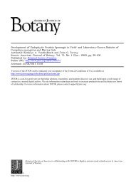
Fig. 12-17.-Fig. 12. Transection near the distal end <strong>of</strong> a root segment showing a lateral root primordium 72 hours<br />
after excision <strong>of</strong> the segment. Wall material remaining from collapsed cortical cells is visible around the tip <strong>of</strong> the primordium,<br />
X 170.-Fig. 13. Transection near the distal end <strong>of</strong> a root segment showing a lateral root 96 hours after excision<br />
<strong>of</strong> the segment. <strong>Root</strong> apex is just penetrating the outer cortex <strong>and</strong> epidermis, X 11O.-Fig. 14. A more highly magnified<br />
view <strong>of</strong> the apex <strong>of</strong> the lateral root shown in Fig. 13. <strong>Root</strong> cap area shows only a limited columnar structure, X280.-<br />
Fig. 15. Longitudinal section <strong>of</strong> a portion <strong>of</strong> the root apex <strong>of</strong> a lateral root after it has reached full diameter <strong>and</strong> growth<br />
rate. The root cap (rc) shows a columnar structure, X305.-Fig. 16. Transection near the proximal end <strong>of</strong> a root segment<br />
44 hours after excision <strong>of</strong> the segment. Pericycle cells (p) have undergone tangential division. e, endodermis, X590.-<br />
Fig. 17. Transection near the proximal end <strong>of</strong> a root segment 72 hours after excision <strong>of</strong> the segment. Divisions <strong>of</strong> the<br />
pericycle cells have resulted in radial files <strong>of</strong> three cells. Enlargement <strong>and</strong> division <strong>of</strong> cells lateral to the protoxylem have<br />
crushed the terminal protoxylem elements, X 340.