Comparative Anatomy of Endogenous Bud and Lateral Root ...
Comparative Anatomy of Endogenous Bud and Lateral Root ...
Comparative Anatomy of Endogenous Bud and Lateral Root ...

Create successful ePaper yourself
Turn your PDF publications into a flip-book with our unique Google optimized e-Paper software.
<strong>Comparative</strong> <strong>Anatomy</strong> <strong>of</strong> <strong>Endogenous</strong> <strong>Bud</strong> <strong>and</strong> <strong>Lateral</strong> <strong>Root</strong> Formation in Convolvulus arvensis<br />
<strong>Root</strong>s Cultured In vitro<br />
Author(s): Howard T. Bonnett, Jr. <strong>and</strong> John G. Torrey<br />
Source: American Journal <strong>of</strong> Botany, Vol. 53, No. 5 (May - Jun., 1966), pp. 496-507<br />
Published by: Botanical Society <strong>of</strong> America<br />
Stable URL: http://www.jstor.org/stable/2440349 .<br />
Accessed: 19/08/2011 13:18<br />
Your use <strong>of</strong> the JSTOR archive indicates your acceptance <strong>of</strong> the Terms & Conditions <strong>of</strong> Use, available at .<br />
http://www.jstor.org/page/info/about/policies/terms.jsp<br />
JSTOR is a not-for-pr<strong>of</strong>it service that helps scholars, researchers, <strong>and</strong> students discover, use, <strong>and</strong> build upon a wide range <strong>of</strong><br />
content in a trusted digital archive. We use information technology <strong>and</strong> tools to increase productivity <strong>and</strong> facilitate new forms<br />
<strong>of</strong> scholarship. For more information about JSTOR, please contact support@jstor.org.<br />
http://www.jstor.org<br />
Botanical Society <strong>of</strong> America is collaborating with JSTOR to digitize, preserve <strong>and</strong> extend access to American<br />
Journal <strong>of</strong> Botany.
Amer. J. Bot. 53(5): 496-507. 1966.<br />
COMPARATIVE ANATOMY OF ENDOGENOUS BUD AND LATERAL<br />
ROOT FORMATION IN CONVOLVULUS ARVENSIS ROOTS<br />
CULTURED IN VITRO1<br />
HOWARD T. BONNETT, JR.2 AND JOHN G. TORREY<br />
The Biological Laboratories, Harvard University, Cambridge, Massachusetts<br />
A B S T R A C T<br />
The anatomy <strong>of</strong> endogenous bud <strong>and</strong> lateral root formation was studied in Convolvulus arvensis<br />
roots cultured in vitro. Although early stages in the initiation <strong>of</strong> buds <strong>and</strong> roots were found to be<br />
identical, continued development <strong>of</strong> primordia into roots or buds was accompanied by differences<br />
in participating primary root tissues, in rates <strong>of</strong> development, <strong>and</strong> in orientation <strong>of</strong> cell divisions.<br />
Evidence for the existence <strong>of</strong> a primordium capable <strong>of</strong> developing into either a bud or root is<br />
discussed with reference to Convolvulus roots <strong>and</strong> other plant parts shown to have similar morphogenetic<br />
potentials.<br />
THE FORMATION <strong>of</strong> buds from roots has been all directions <strong>of</strong> an arc <strong>of</strong> pericycle cells, which<br />
discussed extensively by Beijerinck (1887), van produced a hemispherical protuberance. They be-<br />
Tieghem <strong>and</strong> Duliot (1888), <strong>and</strong> Priestley <strong>and</strong> lieved that as a bud primordium developed it<br />
Swingle (1929). Prior to the monograph <strong>of</strong> Bei- sequentially digested the endodermis <strong>and</strong> then all<br />
jerinck, numerous investigators had contributed the cortical layers in its path to the periphery <strong>of</strong><br />
to a recognition <strong>of</strong> the variety <strong>of</strong> plants which the root. <strong>Lateral</strong> root formation in Convolvulus<br />
form buds from their roots. Irmisch (1857) com- siculus <strong>and</strong> Convolvulus tricolor also proceeded by<br />
piled a list <strong>of</strong> these plants comprising 42 species, tangential divisions <strong>of</strong> six to eight pericycle cells<br />
38 <strong>of</strong> them dicots; this list later grew to 132 species, surrounding a protoxylem pole. As the pericycle<br />
124 <strong>of</strong> them dicots (Wittrock, 1884). Among this cells continued to divide, the endodermis divided<br />
large number <strong>of</strong> plants which develop buds from radially <strong>and</strong> then tangentially; consequently the<br />
their roots, Convolvulus arvensis, the field bind- endodermis remained single-layered at the flanks<br />
weed, was recognized as one <strong>of</strong> a much smaller <strong>of</strong> the lateral root, whereas at the tip it became<br />
group <strong>of</strong> plants, representing several families, in several layers thick. Developmental stages <strong>of</strong><br />
which buds develop from the pericycle in a loca- lateral root formation in Convolvulus arvensis were<br />
tion identical to that <strong>of</strong> lateral root formation. not illustrated, although one drawing <strong>of</strong> a lateral<br />
The anatomy <strong>of</strong> bud formation from Convol- root before emergence showed a single-layered<br />
vulus roots was first studied by Irmisch (1857) endodermis which had undergone only radial<br />
<strong>and</strong> Vogl (1863). They recognized that bud forma- divisions.<br />
tion was endogenous <strong>and</strong> occurred on the pro- These investigators outlined the major steps in<br />
toxylem radii, but because <strong>of</strong> the occurrence <strong>of</strong> the anatomy <strong>of</strong> bud formation from Convolvulus<br />
extensive secondary growth in roots they were roots. As part <strong>of</strong> a study <strong>of</strong> the physiology <strong>of</strong><br />
studying, they were unable to determine the organ formation in cultured Convolvulus roots, a<br />
exact location <strong>of</strong> origin.<br />
reinvestigation <strong>of</strong> the anatomy <strong>of</strong> endogenous<br />
Beijerinck studied bud formation on the hypo- bud <strong>and</strong> root formation was undertaken. Culturcotyl<br />
<strong>and</strong> root <strong>of</strong> Convolvulus seedlings. He found ing these roots has enabled such a study to be made<br />
that buds arose from the pericycle at sites opposite in the absence <strong>of</strong> secondary growth <strong>and</strong> has<br />
the protoxylem poles <strong>and</strong> showed no evidence <strong>of</strong> allowed stages <strong>of</strong> organ formation to be placed in<br />
endodermal participation during their develop- rather precise sequence by their time <strong>of</strong> development.<br />
His attempts to obtain a series <strong>of</strong> stages in ment in organ culture.<br />
the formation <strong>of</strong> lateral roots were unsuccessful MATERIALS AND METHODS-Convolvulus arvensis<br />
because <strong>of</strong> the small number which were present. root segments were cultured in 10-cm petri plates<br />
Beijerinck suggested that endogenous buds de- on a modified Bonner pea root medium (Torrey,<br />
velop in place <strong>of</strong> lateral roots.<br />
1958) solidified with agar <strong>and</strong> adjusted to a post-<br />
Van Tieghem <strong>and</strong> Duliot described the formaautoclave<br />
pH <strong>of</strong> 4.5.<br />
tion <strong>of</strong> bud primordia by growth <strong>and</strong> division in<br />
Prior to a study <strong>of</strong> the anatomy <strong>of</strong> bud <strong>and</strong> root<br />
1 Received for publication June 28, 1965.<br />
formation the distribution <strong>of</strong> buds <strong>and</strong> roots in<br />
This investigation was supported in part by grants from cleared root segments was determined. Intact<br />
the National Institutes <strong>of</strong> Health, U. S. Public Health roots or isolated segments in culture characteris-<br />
Service, GM-10493 to HTB <strong>and</strong> GM-08145 to JGT.<br />
2Present address: Department <strong>of</strong> Biology, University tically developed protuberances which could not<br />
<strong>of</strong> Oregon, Eugene.<br />
he classified reliably as buds or roots. However,<br />
496
May-June, 1966] BONNETT AND TORREY-ORGAN FORMATION IN CONVOLVULUS 497<br />
..<br />
Q~~~~~~~~~~~~~~~~~~~~<br />
498 ti<br />
See page 49 or caption.<br />
... _= toX . ,~~~~~~~~~~~~~~~~~~~<br />
.; 1r- ' _w..! . _~~~~~~~~~~~~~~~~~~~~~~~~~~~~~~~~~~~~<br />
... .<br />
j? F t . | ,> ^@'- iI. _<br />
t.<br />
-.4
498 AMERICAN JOURNAL OF BOTANY [ Vol. 53<br />
in segments cleared by the method <strong>of</strong> Jacobs<br />
(1952) organs could be distinguished before they do<br />
protruded from the root epidermis. It was found<br />
that buds could be distinguished from roots by<br />
the following features: (1) buds are broader<br />
organs than roots; (2) the terminal protoxylem<br />
elements <strong>of</strong> one primary root xylem pole become<br />
separated from the rest <strong>of</strong> the xylem elements in<br />
that pole during bud formation, <strong>and</strong> in a longitudinal<br />
view these protoxylem elements appear<br />
to form arcs which run through portions <strong>of</strong> the bud<br />
primordium; (3) the first xylem <strong>of</strong> the lateral root<br />
differentiates at right angles to the xylem <strong>of</strong> the<br />
parent root segment, whereas the first xylem <strong>of</strong><br />
the bud develops obliquely with respect to the<br />
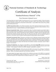
parent xylem. These characteristics are illustrated<br />
in Fig. 1-4. Figure I shows a longitudinal<br />
view <strong>of</strong> a root segment with a cleared bud illustrating<br />
the raised terminal protoxylem elements<br />
running through the middle <strong>of</strong> the bud. Figure 2<br />
shows a bud in which some new xylem has differentiated.<br />
Figure 3 shows a cleared lateral root;<br />
Fig. 4 shows a more highly magnified view <strong>of</strong> the<br />
first mature xylem elements <strong>of</strong> the' lateral root.<br />
The pattern <strong>of</strong> xylem differentiation was adopted<br />
as the most important <strong>of</strong> these criteria, <strong>and</strong><br />
unless otherwise noted, primordia were not<br />
recorded unless they could be positively identified<br />
on this basis.<br />
Figure 5 shows the composite distribution <strong>of</strong><br />
organs formed in 30 segments 15 mm long, excised<br />
from the region <strong>of</strong> the root axis 15-165 mm from<br />
the root apex <strong>and</strong> cleared after 6 weeks in culture.<br />
The distribution <strong>of</strong> organs indicates that<br />
proximal3 regions <strong>of</strong> the segment, which do not<br />
I The terms "proximal" <strong>and</strong> "distal" refer to the segment<br />
orientation with respect to the root from which it was<br />
excised. The distal end <strong>of</strong> a segment is the end which was<br />
closest to the root tip, <strong>and</strong> the proximal end is the end<br />
which was farthest from the root tip.<br />
20<br />
AVE. NO. BUDS/SEGMENT: 2.97<br />
015-<br />
IL<br />
0<br />
w<br />
o-<br />
0<br />
w tO<br />
0-<br />
Z 15.<br />
20<br />
AVE. NOL ROOTS/SEGMENT: 1.23<br />
Fig. 5. The distribution <strong>of</strong> buds <strong>and</strong> roots in 30 segments,<br />
15 mm long, cultured for six weeks, cleared, <strong>and</strong><br />
counted. Each bar represents the total number <strong>of</strong> buds or<br />
roots recorded in each 1-mm interval.<br />
form roots, but which do have a high probability<br />
<strong>of</strong> forming buds can be excised. Distal regions, on<br />
the other h<strong>and</strong>, have a high probability <strong>of</strong> forming<br />
roots <strong>and</strong> also form a few buds.<br />
This polarity <strong>of</strong> organ formation, observed in<br />
cleared root segments, aided in the anatomical<br />
investigation <strong>of</strong> bud <strong>and</strong> root formation. Six root<br />
segments, 15 mm long, were excised from isolated<br />
roots grown in culture for about 6 weeks. The portion<br />
<strong>of</strong> the root axis used was the region 15-105<br />
mm from the root apex. The segments were placed<br />
in a petri plate, each with its distal end to the<br />
periphery <strong>of</strong> the plate. At varying time intervals<br />
after excision, 3-mm segments were excised from<br />
the proximal <strong>and</strong> the distal end <strong>of</strong> a segment <strong>and</strong><br />
examined for stages in the formation <strong>of</strong> buds <strong>and</strong><br />
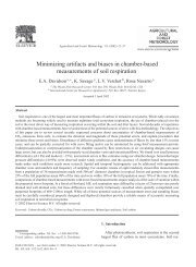
Fig. 1-4.-Fig. 1. <strong>Lateral</strong> view <strong>of</strong> a cleared bud not yet protruding from the root segment. The overlying epidermis<br />
<strong>and</strong> cortex <strong>of</strong> the root have been removed. Terminal protoxylem elements <strong>of</strong> one xylem arm have been separated <strong>and</strong><br />
raised from the rest <strong>of</strong> the xylem elements, traversing the middle <strong>of</strong> the bud (unlabeled arrow). Photographed in polarized<br />
light, X95.-Fig. 2. <strong>Lateral</strong> view <strong>of</strong> a cleared bud not yet protruding from the root segment. The overlving epidermis<br />
<strong>of</strong> the root has been removed. The first xylem to differentiate in association with the bud is marked by arrows. The<br />
raised protoxylem elements are in another focal plane, X 125.-Fig. 3. Cleared lateral root protruding from the cortex<br />
<strong>of</strong> the parent root showing the first mature xylem <strong>of</strong> the lateral root. The overlying epidermis <strong>of</strong> the parent root has been<br />
removed. Photographed in dark field illumination, X90.-Fig. 4. Higher magnification <strong>of</strong> the lateral root-parent root<br />
junction showing the first mature xylem elements <strong>of</strong> two poles <strong>of</strong> the lateral root (unlabeled arrow). Terminal protoxylem<br />
elements <strong>of</strong> the xylem arm <strong>of</strong> the parent root remain adjacent to the rest <strong>of</strong> the xylem elements in that pole. Photographed<br />
in phase contrast, X240.<br />
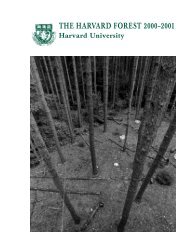
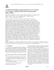
Fig. 6-11.-Fig. 6. Transection <strong>of</strong> a root showing the primary tissues in the region <strong>of</strong> a xylem pole. px, protoxylem;<br />
p, pericycle; e, endodermis; c, cortex, X520.-Fig. 7-11. Transections near the distal end <strong>of</strong> root segments. e, endodermis.<br />
-Fig. 7. The same region shown in Fig. 6-24 hours after excision <strong>of</strong> the segment. Pericycle cells have undergone<br />
tangential division. Photographed in phase contrast, X505.-Fig. 8. A primordium 36 hours after excision. Outer<br />
daughter cells resulting from the first pericycle division have divided tangentially a second time. Photographed in phase<br />
contrast, X360.-Fig. 9. <strong>Root</strong> primordium 44 hours after excision <strong>of</strong> the segment. Radial files <strong>of</strong> four cells derived from<br />
the pericycle have formed. The outermost cells have undergone tangential enlargement <strong>and</strong> divided radially. Some<br />
endodermal cells have divided radially. Photographed in phase contrast, X385.-Fig. 10. <strong>Root</strong> primordium 60 hours<br />
after excision <strong>of</strong> the segment. Divisions <strong>of</strong> the endodermal cells have continued, resulting in two layers <strong>of</strong> cells. Cortical<br />
cells contiguous with the primordium have collapsed, X290.-Fig. 11. <strong>Root</strong> primordium 60 hours after excision <strong>of</strong> the<br />
segment. Cells derived from the endodermis have formed a triple layer at one point (unlabeled arrow). Cells contiguous<br />
with the primordium have collapsed, X260.
May-June, 1966] BONNETT AND TORREY-ORGAN FORMATION IN CONVOLVULUS 499<br />
... . . .....<br />
At<br />
w<br />
AF<br />
41*<br />
.......... .. .<br />
AF<br />
. ... .. .. .. ... .<br />
. ... ... ... .. . .... .. .<br />
.. ....<br />
....<br />
.. ..... . ...... ...... .<br />
VH:,<br />
... .... ... ...
500 AMERICAN JOURNAL OF BOTANY [ Vol. 53<br />
roots, respectively. The 3-mm segments were cells <strong>of</strong> radial growth by the root primordium is<br />
frozen in 10% methyl cyclohexane in isopentane shown in this figure by the collapse <strong>of</strong> cells con-<br />
(v/v) as described by Branton <strong>and</strong> Jacobson tiguous with the primordium.<br />
(1962), fixed <strong>and</strong> dehydrated in 0.1%/ HgCl2 in Figure 11 shows at 60 hours a larger primordiumr<br />
methyl alcohol (Bell, 1959) at -70 C, trans- in which cells derived from the endodermis form a<br />
ferred to n-propyl alcohol, gradually infiltrated triple layer at one point. Several collapsed cells<br />
with polyethylene glycol 400 distearate (Sidman, <strong>of</strong> the innier cortex are still evident.<br />
Mottla, <strong>and</strong> Feder, 1961), <strong>and</strong> embedded in Tissue- By 72 hours (3 days) the young lateral root<br />
mat. Sections were stained with safranin, hema- (Fig. 12) is more than halfway through the cortex.<br />
toxylin, <strong>and</strong> fast green.<br />
Wall material remaining from collapsed cortical<br />
RESULTs-The following series <strong>of</strong> figures for cells is visible around the tip <strong>of</strong> the root. Flattenbud<br />
<strong>and</strong> for root formation are each assigned a ing <strong>of</strong> cells not contiguous with the root, tip, but<br />
time in hours or days. This time refers to the positioned between it <strong>and</strong> the epidermis, is still<br />
length <strong>of</strong> time from excision <strong>of</strong> the 15-mm segment absent.<br />
to fixation <strong>of</strong> the terminal 3-mm portion <strong>of</strong> the Figure 13 shows a lateral root just completing<br />
segment. The stage depicted in most cases cor- penetration <strong>of</strong> the cortex <strong>and</strong> epidermis after 96<br />
responds to the largest primordia which have hours (4 days).<br />
formed within the given time period. The termin- A thorough study <strong>of</strong> the fate <strong>of</strong> the layers <strong>of</strong><br />
ology adopted for planes <strong>of</strong> division is described cells derived from the endodermis has not been<br />
by Esau (1965).<br />
made. Figure 14 is a higher magnification <strong>of</strong> Fig.<br />
Figure 6 is <strong>of</strong> a section <strong>of</strong> the mature primary 13, showing the "root cap" <strong>and</strong> apical initials.<br />
root structure, showing a region where primordia Figure 15 shows the same region <strong>of</strong> such a lateral<br />
have not been initiated. Proceeding from the pro- root after it has reached full diameter <strong>and</strong> growth<br />
toxylem pole (labeled "px" in Fig. 6) towards rate. This root cap shows a columnar structure<br />
the outside <strong>of</strong> the root, the first cell layer is the resulting from transverse divisions in root cap<br />
pericycle (p), followed by the endodermis (e) initial cells. There is a discontinuity in the files <strong>of</strong><br />
with Casparian strips in cross section, <strong>and</strong> then cells <strong>of</strong> the columella <strong>and</strong> the central cylinder.<br />
three cell layers <strong>of</strong> the cortex (c).<br />
Though files <strong>of</strong> cells are also evident in the root<br />
<strong>Lateral</strong> root formation-Figures 7-13 illustrate tip <strong>of</strong> the young lateral root (Fig. 14), the root cap<br />
stages <strong>of</strong> root formation, obtained from transec- area shows only the beginnings <strong>of</strong> a columnar<br />
tions <strong>of</strong> distal ends <strong>of</strong> cultured root segments structure. It is probable that the "root cap" <strong>of</strong><br />
fixed at successive time intervals. Figure 7, at 24 the lateral root at the stage shown in Fig. 13 is<br />
hours, shows a young primordium. Five or six largely derived from the endodermis <strong>and</strong> later<br />
pericycle cells, forming an arc which surrounds sloughed as root cap initials begin transverse<br />
the protoxylem pole, have each undergone radial divisions to form files characteristic <strong>of</strong> the larger<br />
enlargement <strong>and</strong> one tangential division. lateral root.<br />
By 36 hours (Fig. 8) further radial enlargement <strong>Endogenous</strong> bud formation-Figures 16-21 illushas<br />
occurred <strong>and</strong> outer daughter cells have also trate stages in bud development seen in transecdivided<br />
tangentially. Some <strong>of</strong> the endodermal cells tions <strong>of</strong> the proximal ends <strong>of</strong> cultured root segshow<br />
an increase in protoplasmic content. ments. Figure 16 shows a young primordium after<br />
Tangential divisions continue in both the outer 44 hours. As in the root primordium, six or seven<br />
<strong>and</strong> inner rows <strong>of</strong> daughter cells <strong>of</strong> the pericycle pericycle cells around the protoxylem pole have<br />
until radial files <strong>of</strong> four cells are formed. At 44 undergone radial enlargement <strong>and</strong> one tangential<br />
hours (Fig. 9) the outermost cell in this radial division (cf. Fig. 7).<br />
file has undergone tangential enlargement followed By 72 hours (Fig. 17), or 3 days, radial files <strong>of</strong><br />
by radial division. Endodermal cells at this stage three to four cells have formed. Although only<br />
also show radial divisions.<br />
tangential divisions have occurred, alignment<br />
The activation <strong>of</strong> divisions in the endodermis <strong>of</strong> the files <strong>of</strong> daughter cells is less precise than was<br />
continues <strong>and</strong> by 60 hours (Fig. 10) tangential observed in the young root primordium. Enlargedivisions<br />
have produced two cell layers derived ment <strong>and</strong> division <strong>of</strong> the cells lateral to the pr<strong>of</strong>rom<br />
the endodermis. The effect on the cortical toxylem also occur, resulting in the compression<br />
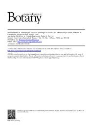
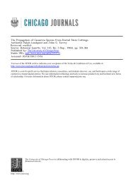
Fig. 12-17.-Fig. 12. Transection near the distal end <strong>of</strong> a root segment showing a lateral root primordium 72 hours<br />
after excision <strong>of</strong> the segment. Wall material remaining from collapsed cortical cells is visible around the tip <strong>of</strong> the primordium,<br />
X 170.-Fig. 13. Transection near the distal end <strong>of</strong> a root segment showing a lateral root 96 hours after excision<br />
<strong>of</strong> the segment. <strong>Root</strong> apex is just penetrating the outer cortex <strong>and</strong> epidermis, X 11O.-Fig. 14. A more highly magnified<br />
view <strong>of</strong> the apex <strong>of</strong> the lateral root shown in Fig. 13. <strong>Root</strong> cap area shows only a limited columnar structure, X280.-<br />
Fig. 15. Longitudinal section <strong>of</strong> a portion <strong>of</strong> the root apex <strong>of</strong> a lateral root after it has reached full diameter <strong>and</strong> growth<br />
rate. The root cap (rc) shows a columnar structure, X305.-Fig. 16. Transection near the proximal end <strong>of</strong> a root segment<br />
44 hours after excision <strong>of</strong> the segment. Pericycle cells (p) have undergone tangential division. e, endodermis, X590.-<br />
Fig. 17. Transection near the proximal end <strong>of</strong> a root segment 72 hours after excision <strong>of</strong> the segment. Divisions <strong>of</strong> the<br />
pericycle cells have resulted in radial files <strong>of</strong> three cells. Enlargement <strong>and</strong> division <strong>of</strong> cells lateral to the protoxylem have<br />
crushed the terminal protoxylem elements, X 340.
May-June, 1966] BONNETT AND TORREY-ORGAN FORMATION IN CONVOLVULUS 501<br />
(I<br />
...U......<br />
. ........ .<br />
_ _ n!|L, s--- s<br />
_<br />
'<br />
~~~~~~~~~~~~~~~~~~~~~~~~~~~~~~~~~~~~~~~~~~.<br />
......i..i .:....
502 AMERICAN JOURNAL OF BOTANY [Vol. 53
lI\ay-June, 1966] BONNETT AND TORREY-ORGAN FORMATION IN CONVOLVULUS 503<br />
<strong>of</strong> terminal protoxylem elements. The rate <strong>of</strong> elusion <strong>of</strong> separate figures for these stages is based<br />
development <strong>of</strong> buds can be compared to that <strong>of</strong> only on the procedure <strong>of</strong> describing root <strong>and</strong> bud<br />
roots by consulting Fig. 12, also at 72 hours, in formation at opposite ends <strong>of</strong> the 15-mm segments.<br />
which the root is more than halfway through the In fact, from a consideration <strong>of</strong> the data in the<br />
cortex.<br />
next section, one may not exclude the possibility<br />
By 5 days (Fig. 18) cells <strong>of</strong> the endodermis <strong>of</strong> that the primordia at these earliest stages were<br />
the bud primordium show only enlargement <strong>and</strong> present prior to excision <strong>of</strong> the segments.<br />
the beginning <strong>of</strong> radial divisions, illustrated by Organ formation in intact cultured roots-Althe<br />
metaphase plate in one <strong>of</strong> the endodermal though there were no externally visible primordia<br />
cells. The root primordium at a comparable size for at least several hundred millimeters behind<br />
already has a two-layered endodermis (Fig. 10). the tips <strong>of</strong> Convolvutus roots grown in culture,<br />
Figure 19 shows a bud primordium at about histological sections <strong>of</strong> the region <strong>of</strong> the root from<br />
8 days. Endodermal cells have undergone ex-. which the 15-mm segments were routiinely extensive<br />
enlargement <strong>and</strong> have divided radially cised revealed that small primordia were present<br />
By the time bud primordia have reached this at the time <strong>of</strong> segment excision. Therefore, it was<br />
size, a distinction among cells <strong>of</strong> the bud primor- necessary to determine the nature <strong>and</strong> stage <strong>of</strong> dedium,<br />
endodermal cells, <strong>and</strong> adjacent cortical cells velopment <strong>of</strong> these primordia hich, because <strong>of</strong><br />
cannot always be made. As yet, there is no histogen their small size, could be done o'ly by a histologiorganization<br />
typical <strong>of</strong> the bud apex.<br />
cal study. Rather than section the entire lengths<br />
At 11 days (Fig. 20) a meristematic dome <strong>of</strong> <strong>of</strong> roots, representative 5-mm lengths along each<br />
cells has developed at the outer periphery <strong>of</strong> the root were fixed <strong>and</strong> sectioned. Seven roAt cultures,<br />
primordium. Cortical cells around the flanks <strong>and</strong> grown in the dark for about 6 weeks, were<br />
at the tip <strong>of</strong> the primordium show flattening in observed for the 24-hour period before fixation to<br />
the directions which would be predicted as a re- confirm that they were growing at a normal rate<br />
sult <strong>of</strong> pressure exerted by the developing bud. (greater than 20 mm/day). From these roots 5-<br />
Cells betweeln the epidermis <strong>and</strong> the bud apex are mm segments were excised <strong>and</strong> immediately preflattened<br />
so that their longest axis, seen in cross pared for histological study. The first column in<br />
section, is parallel to the epidermis. Cells along the Table 1 shows the distance from the root tip to the<br />
flank <strong>of</strong> the bud primordium are obliquely flat- 5-mm segments studied. Data from the seven 5-<br />
tened from the radial <strong>and</strong> tangential growth <strong>of</strong> the mm segments, each the same distance from the<br />
bud primordium. Continued divisions <strong>and</strong> en- root tip, were pooled so that the number <strong>of</strong> prilargement<br />
<strong>of</strong> the cells lateral to the terminal mordia reported in the second column <strong>of</strong> Table 1<br />
xylem elements have completely crushed the represents the total number <strong>of</strong> primordia obterminal<br />
protoxylem element <strong>and</strong> separated it served for that interval in all seven roots.<br />
from the rest <strong>of</strong> the xylem arm (see arrow in Fig. Primordia were rated by class. Primordia<br />
20). This process confirms one <strong>of</strong> the diagnostic -vhich showed one or a few tangential divisions<br />
features <strong>of</strong> cleared buds illustrated in Fig. 1. Such in the pericycle, but not a complete arc <strong>of</strong> divided<br />
an effect has been observed in only one root but cells, were assigned to Class 1. Class 2 corresin<br />
nearly all cleared or sectioned buds.<br />
ponds to primordia such as those illustrated in<br />
By about 2 weeks (Fig. 21) a bud apex has Fig. 7, 16; Class 3 to those shown in Fig. 8, 17;<br />
developed which has penetrated the outer cortex Class 4 to that shown in Fig. 18; Class 5 to that<br />
<strong>and</strong> epidermis <strong>of</strong> the root <strong>and</strong> which shows a clear shown in Fig. 19; <strong>and</strong> Class 6 to that shown in<br />
single-layered tunica <strong>and</strong> two leaf primordia. Fig. 20. No primordia were found which could be<br />
Although the series <strong>of</strong> stages from a single- identified as root primordia (this would require<br />
layered pericycle to a clearly recognizable organ that they be at least as large as the primordium<br />
are figured separately for the root (Fig. 7-13) <strong>and</strong> illustrated in Fig. 9), so all primordia have been<br />
the bud (Fig. 16-21), there are no entirely con- assigned to classes corresponding to the developsistent<br />
anatomical features which allow the mental stages <strong>of</strong> bud formation.<br />
earliest stages <strong>of</strong> organ formation at the distal end From Table 1 it is clear that primordia are<br />
(Fig. 7, 8) to be distinguished from the earliest initiated at a distance <strong>of</strong> only 5-10 mm from the<br />
stages at the proximal end (Fig. 16-17). The in- actively growing root apex <strong>and</strong> that the first<br />
Fig. 18-21. Transections near the proximal end <strong>of</strong> root segments.-Fig. 18. <strong>Bud</strong> primordium five days after excision<br />
<strong>of</strong> the segment. Division <strong>of</strong> endodermal cells has not occurred with the exception <strong>of</strong> one cell in metaphase (unlabeled<br />
arrow), X320.-Fig. 19. <strong>Bud</strong> primordium about eight days after excision <strong>of</strong> the segment. Endodermal cells have under-<br />
gone enlargement <strong>and</strong> divided radially. e, endodermal cell at the flank <strong>of</strong> the primordium, which has not enlarged; E,<br />
endodermal cell which has enlarged <strong>and</strong> divided radially, X270.-Fig. 20. <strong>Bud</strong> primordium about 11 days after excision<br />
<strong>of</strong> the segment. A meristematic dome <strong>of</strong> cells has developed at the periphery <strong>of</strong> the primordium. The terminal protoxylem<br />
element (unlabeled arrow) has been crushed <strong>and</strong> separated from the rest <strong>of</strong> the xylem arm, X 185.-Fig. 21. <strong>Bud</strong> about<br />
14 days after excisioin <strong>of</strong> the segment. The bud apex, which has penetrated the outer cortex <strong>and</strong> epidermis <strong>of</strong> the root, has<br />
a single-layered tunica <strong>and</strong> two leaf primordia, X 135.
504 AMERICAN JOURNAL OF BOTANY [Vol. 53<br />
TABLE 1. Primordia present in the terminal 135 mm <strong>of</strong> Convolvulus roots based on a histological analysis <strong>of</strong> segments from<br />
seven roots cultured for about six weeks<br />
Class: 1 2 3 4 5 6<br />
Corresponding bud primor- - 16 17 18 19 20<br />
dium stage shown by Fig.:<br />
<strong>Bud</strong> primordium age: - 44 hr 72 hr 5 da 8 da 11 da<br />
Distance <strong>of</strong> Total num- Corresponding root primor- 7 8<br />
5-mm segment ber <strong>of</strong> pri- dium stage shown by Fig.:<br />
from root mordia/<br />
tip (mm) 7 segments <strong>Root</strong> primordium age: 24 hr 36 hr<br />
5-10 2 2<br />
15-20 11 11<br />
30-35 14 6 7 1<br />
50-55 12 2 9 1<br />
70-75 14 1 11 2<br />
90-95 16 4 12 -<br />
110-115 13 1 11 1<br />
130-135 10 9 1<br />
150-155 17 2 12 2 1<br />
170-175 8 1 4 3<br />
190-195 11 1 6 2 1 1<br />
210-215 14 2 7 3 2<br />
230-235 8 - 8<br />
250-255 8 5 2 1<br />
stages in bud <strong>and</strong> root formation (Fig. 7, 8, 16, development in excised segments to the rate <strong>of</strong><br />
17) could be accounted for by primordia found to bud development in the intact root.<br />
be present at the time <strong>of</strong> excision in the region <strong>of</strong> A bud at the 5-day stage (Class 4; Fig. 18) <strong>of</strong><br />
the root axis from which the 15-mm segments development is no larger than a bud which might<br />
were excised (15-105 mm from the root apex). be seen in the same region <strong>of</strong> the root axis after<br />
However, later stages in the formation <strong>of</strong> buds the root had grown an additional 23 mm/day for<br />
(Fig. 18-21) or roots (Fig. 9-13) cannot be ex- 5 days. The region <strong>of</strong> the root axis from which<br />
plained by pre-existing primordia at the same the segments were excised (15-105 mm from the<br />
stage <strong>and</strong> must represent organ development after root apex) would be 130-220 mm from the apex<br />
segment excision. Although primordia represent- after 5 days <strong>of</strong> growth (Table 1). Similarly,<br />
ing more advanced stages in bud development a bud at the 8-day stage (Class 5; Fig. 19) is no<br />
were observed at increasing distances from the larger than a bud that might be found 199-289<br />
root apex, they never occurred in greater numbers mm from the root apex. Consequently, some bud<br />
than could be accounted for by initiation <strong>of</strong> all primordia may develop in the intact cultured<br />
primordia within the first 20 mm <strong>of</strong> the root. roots at a rate comparable to that observed in<br />
Since few, if any, new primordia appeared to be excised segments. No evidence was obtained for<br />
initiated after the first 20 mm, <strong>and</strong> since the lateral root development in this region <strong>of</strong> the<br />
largest proportion <strong>of</strong> the primordia was always in intact root except for stages too small to disthe<br />
first two classes, most primordia must have tinguish as bud or root primordia.<br />
been inhibited from further development. Those To extend the analysis <strong>of</strong> organ production by<br />
primordia which did develop became recognizable root cultures, entire roots were cleared <strong>and</strong> the<br />
bud primordia.<br />
distribution <strong>of</strong> organs along their length was<br />
The age <strong>of</strong> a 15-mm region <strong>of</strong> the root axis may determined. <strong>Root</strong>s used in this study were checked<br />
be figured either by the number <strong>of</strong> days the region for a 24-hour period to confirm that they were<br />
has been cultured as an excised segment or, in the growing at a rate <strong>of</strong> at least 20 mm/day. Primorintact<br />
root, by the increase in distance <strong>of</strong> the region dia were assigned to one <strong>of</strong> the following four<br />
from the root tip. Since the mean growth rate <strong>of</strong> groups: (A) primordia which could not be identi-<br />
Convolvulus roots is about 23 mm/day, each 23- fied as root or bud; (B) larger primordia in which<br />
mm interval between the 15-mm region <strong>and</strong> the the terminal xylem element was separated, but<br />
root tip is equivalent to one day in culture for the no new xylem had differentiated in association<br />
excised segment. This relationship forms the basis with the primordium; (C) buds; <strong>and</strong> (D) roots.<br />
for the following comparisons <strong>of</strong> the rate <strong>of</strong> bud The identification <strong>of</strong> primordia in cleared whole
May-June, 1966] BONNETT AND TORREY-ORGAN FORMATION IN CONVOLVULUS i&5-<br />
TABLE 2. Organ production in whole root cultures<br />
Groupb<br />
Distance<br />
from tip<br />
(mm)<br />
<strong>Root</strong> la <strong>Root</strong> 2 <strong>Root</strong> 3<br />
A B C D A B C D A B C D<br />
0-100 - - 1<br />
100-200 - 4 - 1<br />
200-300 1 2 - 4 -<br />
300-400 2 1 2 2 -<br />
400-500 7 5 - - 4 -<br />
500-600 8 1 1 8 7 2 5 1 1<br />
600-700 8 1 6 6 1 7 --<br />
700-800 7 - 1 11 1 3 8 2 1 -<br />
800-900 12 1 23 11 1 9 1 1 -<br />
900-1000 9 2 14 5 1 15 1 5<br />
1000-1100 18 1 6 1 6 3 7 4 - 2 -<br />
1100-1200 2 1 6 14 2 7 2<br />
1200-1300 3 4 3 5 2 5 1 3 2 1<br />
1300-1400 2 5 1 3<br />
1400-1500 2 2 1<br />
a <strong>Root</strong> 1 was 1270 mm long; root 2 was 1450 mm long; root 3 was 1330 mm long.<br />
b A, indeterminate primordium; B, primordium with separated protoxylem; C, bud primordium;j), root primordium.<br />
roots is more difficult <strong>and</strong> less certain at the the formation <strong>of</strong> roots <strong>and</strong> buds. Similarly, under<br />
early stages than in sectioned roots. Even so, root the conditions <strong>of</strong> root culture used in this investiprimoridia<br />
can be classified definitely between gation, there was no evidence <strong>of</strong> vascular cambium<br />
the 72- <strong>and</strong> 96-hour stage (Fig. 12, 13). <strong>Bud</strong> pri- or phellogen activity accompanying or preceding<br />
mordia can be recognized by the 11-day stage (Fig. organ formation.<br />
20). Results from the three roots studied in this In the earliest stages there were no differences<br />
way are presented in Table 2. All organs within observed in cellular patterns <strong>of</strong> bud <strong>and</strong> root<br />
each 100-mm interval <strong>of</strong> the root axis have been formation. Both organs originate from an arc <strong>of</strong><br />
grouped.<br />
pericycle cells by tangential divisions <strong>of</strong> these<br />
Although a few buds may develop in the cells. Later stages differ not only in their rates <strong>of</strong><br />
presence <strong>of</strong> the root tip at the same rate as they development, but also in the behavior <strong>of</strong> the endodevelop<br />
in excised segments, the data presented dermis, in cellular patterns <strong>of</strong> organization, <strong>and</strong><br />
in Table 2 show that excision <strong>of</strong> segments from the in participation <strong>of</strong> central cylinder cells around<br />
-root axis greatly promotes the number <strong>of</strong> buds the protoxylem pole. Van Tieghem <strong>and</strong> Duliot<br />
which develop. Ten 15-mm segments were found believed that the bud digests first the endodermis<br />
to produce 30 buds after 4 weeks in culture. No <strong>and</strong>, subsequently, all the cortical layers until<br />
150-mm region <strong>of</strong> these roots <strong>of</strong> a comparable age it reaches the outside <strong>of</strong> the root. In this study<br />
or older produced nearly as many buds.<br />
there was no indication <strong>of</strong> digestion <strong>of</strong> any cells<br />
Excision <strong>of</strong> segments from the root axis also during bud development, but there was some<br />
greatly affects lateral root formation. Segments indication <strong>of</strong> mechanical distortion. Rather than<br />
15 mm in length develop lateral roots large enough being digested, the endodermis underwent radial<br />
-to recognize in cleared root segments in only 3-4 divisions <strong>and</strong> great cell enlargement. Because <strong>of</strong><br />
Kdays (equivalent to less than 100 mm growth <strong>of</strong> this enlargement in bud primordia, it became<br />
-the main axis). In intact roots (Table 2) no lateral difficult to distinguish the endodermis from other<br />
roots were recorded closer to the tip than about inner cortical layers or from the apical primordial<br />
1,000 mm (equivalent to about 6 weeks' growth). cells prior to organization <strong>of</strong> the bud apex. A<br />
DISCUSSION-Although the methods used in<br />
study <strong>of</strong> longitudinal sections might have clarified<br />
this anatomical investigation <strong>of</strong> Convolvulus root<br />
<strong>and</strong> bud formation involved excision <strong>of</strong><br />
the fate <strong>of</strong> the endodermis, but no study <strong>of</strong> this<br />
segments,<br />
it is clear that excision is not required to set <strong>of</strong>f type was made. Therefore, the possible participaprimordium<br />
initiation. As a result <strong>of</strong> cutting no tion <strong>of</strong> the endodermis <strong>and</strong> other cortical cells<br />
callus was formed, <strong>and</strong> although occasional cell in the organization <strong>of</strong> the bud apex cannot be<br />
.divisions may have occurred at the cut surface, ruled out.<br />
most meristematic activity was associated with In root development the activation <strong>of</strong> the endo-
506 AMERICAN JOURNAL OF BOTANY [Vol. 53<br />
dermis to repeated divisions culminated in a system with a limited number <strong>and</strong> capacity <strong>of</strong><br />
series <strong>of</strong> layers <strong>of</strong> cells. Cortical collapse at the sites for organ formation. In a later paper, Dore<br />
advancing margins <strong>of</strong> these endodermal deriva- <strong>and</strong> Williams (1956) found that five organs/<br />
tives suggested at least partial enzymatic penetra- lateral root trace were formed in 1-mm cuttings.<br />
tion. It is possible that the activation <strong>of</strong> the The average value <strong>of</strong> one or two organs/site was<br />
endodermis by the developing root may aid in observed in longer segments. It is attractive to<br />
its penetration <strong>of</strong> the cortex, but such an interpre- suggest that similarity in young stages <strong>of</strong> primortation<br />
is based only on the appearance <strong>of</strong> the dium formation indicates an undetermined primorcortex<br />
during the radial growth <strong>of</strong> bud <strong>and</strong> root dium, but such similarity cannot be accepted as<br />
primordia.<br />
pro<strong>of</strong>.<br />
Though the earliest stages <strong>of</strong> root <strong>and</strong> bud In root <strong>and</strong> bud formation in Convolvulus,<br />
initiation appeared identical, conclusive evidence although only pericycle cells around protoxylem<br />
for an undetermined primordium, or an unde- radii participate in primordial initiation, restrictermined<br />
primordium site, has not been obtained. tions on the number <strong>of</strong> primordial sites in the<br />
There is ample evidence in favor <strong>of</strong> the existence <strong>of</strong> longitudinal direction are little understood. Horan<br />
undetermined primordium which may develop monal treatment <strong>of</strong> Convolvulus roots can greatly<br />
into a leaf or a bud. Wardlaw (1949) showed by increase the number <strong>of</strong> organs (Bonnett anid<br />
surgical isolation that an incipient leaf site on the Torrey, 1965), making any statistical pro<strong>of</strong> <strong>of</strong><br />
apex <strong>of</strong> Dryopteris aristata could instead develop different developmental potentialities <strong>of</strong> early<br />
into a bud. In an accompanying histological primordia based on a limited number <strong>of</strong> available<br />
study, he concluded that small leaf <strong>and</strong> bud pri- sites untenable<br />
mordia were indistinguishable. Cutter (1956), using<br />
apices <strong>of</strong> the same fern, showed that the three<br />
youngest leaf primordia could be induced to<br />
LITERATURE CITED<br />
develop as buds by surgical isolation. By excising<br />
BEIJERINCK, M. W. 1887.<br />
<strong>and</strong> culturing young fern leaf primordia <strong>of</strong> Os-<br />
Beobachtungen und Betrachtungen<br />
fuber Wurzelknospen und Nebenwurzeln.<br />
munda, Steeves (1961) was able to obtain de-<br />
Natuurk. Verh<strong>and</strong>l. der kon. Akademie der Wetensch.<br />
velopment <strong>of</strong> the first nine leaf primordia into Amsterdam 25: 1-146.<br />
buds.<br />
BELL, L. G. E. 1959. The combination <strong>of</strong> a portion <strong>of</strong> the<br />
The case regarding the existence <strong>of</strong> primordia cytoplasmie ribonucleic acid compounds with mercury.<br />
which may develop into either a bud or a root is Exptl. Cell Res. 16: 615-623.<br />
much less secure. Beijerinck described the con- BONNETT, H. T., JR., AND J. G. TORREY. 1965. Chemversion<br />
<strong>of</strong> a bud developing on the roots <strong>of</strong> Rumex ical control <strong>of</strong> organ formation in root segments <strong>of</strong><br />
acetosella into a root. The change from bud-like Convolvulus cultured in vitro. Plant Physiol. 40:<br />
1228-1236.<br />
vascular organization to root-like vascular organi-<br />
BRANTON, D., AND L. JACOBSON. 1962. Iron localization<br />
zation also corresponded to the change in mor- in pea plants. Plant Physiol. 37: 546-551.<br />
phology <strong>of</strong> the organ. Such an organ thereby CUTTER, E. G. 1956. Experimental <strong>and</strong> analytical studies<br />
possessed one or two leaf primordia at its base, <strong>of</strong> pteridophytes. XXXIII. The experimental inducbut<br />
terminated in a root apex. This observation tion <strong>of</strong> buds from leaf primordia in Dryopteris aristata.<br />
was experimentally confirmed by Edmonson in Ann. Bot. n. s. 20: 143-165.<br />
1925 (cited by Priestley <strong>and</strong> Swingle, 1929). DORER J. 1955. Studies in the regeneration <strong>of</strong> horseradish.<br />
In Rumex acetosella buds are formed at intervals I. A re-examination <strong>of</strong> the morphology <strong>and</strong> anatomy<br />
<strong>of</strong><br />
along the length <strong>of</strong> the root. If a segment <strong>of</strong> the regeneration. Ann. Bot. n. s. 19: 127-137.<br />
- AND W. T. WILLIAMS. 1956. Studies on the<br />
root is excised <strong>and</strong> placed upright in damp soil,<br />
regeneration <strong>of</strong> horseradish. II. Correlation phenombuds<br />
near the lower end sometimes develop into ena. Ann. Bot. n. s. 20:231-250.<br />
roots.<br />
ESAU, K. 1965. Plant anatomy. John Wiley <strong>and</strong> Sons,<br />
Dore (1955) studied the formation <strong>of</strong> roots <strong>and</strong> New York.<br />
buds from thickened root cuttings <strong>of</strong> horseradish IRMISCH, T. 1857. fiber die Keimung und die Erneu-<br />
(Armoracia rusticana). These organs arose only erungsweise von Convolvulus sepium und Convolvulus<br />
after the cortex had been sloughed; they were arvensis, so wie fiber hypokotylische Adventivknospen<br />
restricted to sites in association with old lateral bei krautartigen phanerogamen Pflanzen. Bot. Zeit.<br />
15: 433-443, 449-472, 481-484, 489-497.<br />
root traces. Usually only one, or occasionally<br />
JACOBS, W. P. 1952. The role <strong>of</strong> auxin in differentiation<br />
two, primordia developed at each site: either <strong>of</strong> xylem around a wound. Amer. J. Bot. 39: 301-309.<br />
two roots, two buds, or one <strong>of</strong> each.<br />
PRIESTLEY, J. H., AND C. F. SWINGLE. 1929. Vegetative<br />
Evidence that an undetermined primordium propagation from the st<strong>and</strong>point <strong>of</strong> plant anatomy.<br />
existed in these cuttings was based on the early U. S. Dept. Agr. Tech. Bul. 151, Washington, D. C.<br />
SIDMAN, histological identity <strong>of</strong> all primordia <strong>and</strong> on the<br />
R. L., P. A. MOTTLA, AND N. FEDER. 1961.<br />
Improved polyester wax embedding for histology.<br />
fact that submergence <strong>of</strong> the cuttings in water<br />
Stain Technol. 36: 279-284.<br />
caused the proportion <strong>of</strong> buds to roots to decrease STEEVES, T. A. 1961. A study <strong>of</strong> the developmental<br />
while the total number <strong>of</strong> organs remained con- potentialities <strong>of</strong> excised leaf primordia in sterile<br />
stant. Such evidence may be obta ned oni v in a culture. Phytomorphology 11: 346-359.
May-June, 1966] CONSTANTIN AND MULLENAX-SHOOT APEX IN LACTUCA 507<br />
TIEGHEM, P. VAN, AND H. DULIOT. 1888. Recherches<br />
comparatives sur l'origine des membres endogenes<br />
dans les plantes vasculaires. Ann. Sci. Nat., Bot. 8:<br />
1-660.<br />
TORREY, J. G. 1958. <strong>Endogenous</strong> bud <strong>and</strong> root formation<br />
by isolated roots <strong>of</strong> Convolvulus grown in vitro. Plant<br />
Physiol. 33: 258-263.<br />
VOGL, A. 1863. Beitrage zur Anatomie und Histologie<br />
Amer. J. Bot. 53(5): 507-511. 1966.<br />
der unterirdischen Theile von Convolvulus arvensis L.<br />
Verh<strong>and</strong>l. Zool.-Bot. Ges. Wien. 13: 257-300.<br />
WARDLAW, C. W. 1949. Experiments on organogenesis in<br />
ferns. Growth (suppl.) 9: 93-131.<br />
WITTROCK, V. B. 1884. Ueber Wurzelsprossen bei<br />
krautartigen Gewachsen, mit besonderer Rulcksicht<br />
auf ihre verschiedene biologische Bedeutung. Bot.<br />
Centralbl. 17: 227-232, 258-264.<br />
STRUCTURE OF THE SHOOT APEX IN LACTUCA SATIVAl<br />
M. J. CONSTANTIN AND R. H. MULLENAX<br />
Agricultural Research Laboratory <strong>of</strong> the University <strong>of</strong> Tennessee, Oak Ridge, Tennessee2<br />
AB STRACT<br />
Histological observations <strong>of</strong> the leaf lettuce 'Black Seeded Simpson' showed the dormant embryo<br />
to possess two visible leaves <strong>and</strong> a flat to slightly depressed plumular apex. Observations conducted<br />
over a 12-day period <strong>of</strong> germination <strong>and</strong> growth showed the development <strong>of</strong> L1 <strong>and</strong> L2, emergence<br />
<strong>of</strong> L3 <strong>and</strong> L4, <strong>and</strong> periodic changes in size <strong>of</strong> the apex which were associated with leaf emergence.<br />
Thus the dormant embryo <strong>of</strong> Lactuca appears to be considerably rrore advanced in development<br />
than was previously believed. The shoot apex appeared flat to slightly depressed at all develop-<br />
mental stages studied.<br />
THE LACTUCA embryo is used frequently to Additional information pertinent to Lactuca: (a)<br />
exemplify embryonic simplicity among dicotyle- the young seedling stems do not undergo interdonous<br />
plants. The mature Lactuca embryo was nodal elongation <strong>and</strong> form either a head or a basal<br />
shown by Esau (1953) to possess a radicle <strong>and</strong> a rosette <strong>of</strong> leaves, <strong>and</strong> (b) the leaves are crowded on<br />
hypocotyl with two cotyledons subtending an the axis in a 2/5 spiral phyllotaxy (Hayward,<br />
epicotyl having a convex apex without differenti- 1938).<br />
ated leaf primordia. Jones (1927) did not describe Generally, apical meristems consist <strong>of</strong> meristethe<br />
Lactuca epicotyl but sketched transverse matic initials <strong>and</strong> their immediate derivations <strong>of</strong> a<br />
sections <strong>of</strong> developing achenes showing the embryo shoot <strong>and</strong> occur as dome shaped structures that<br />
with cotyledons <strong>and</strong> a plumule that consisted <strong>of</strong> extend above the base <strong>of</strong> the last formed leaf or<br />
two unidentified structures. Hayward (1938) pro- leaf pair. However, shoot apices <strong>of</strong> seed plants vary<br />
vided the only information about the shoot apex anatomically from a shape which is convex to one<br />
in developing seedlings. The shoot apex <strong>of</strong> a that is flat <strong>and</strong> even partially depressed, as for<br />
C-day-old seedling <strong>of</strong> 'New York Regular' was example in Drimys (Gifford, 1950), Helianthus<br />
shown to be convex <strong>and</strong> subtended by a single, well- (Esau, 1945), <strong>and</strong> Rauwolfia (Mia, 1960). There is<br />
developed leaf. He also mentioned that after evidence that apices change both in size <strong>and</strong> shape<br />
emergence the cotyledons become oriented almost during plant growth <strong>and</strong> development; therefore,<br />
vertically; then by expansion they expose a the plumular or embryonic apex may be considerminute<br />
epicotyl, <strong>and</strong> within a few days the first ably different from the seedling shoot apex.<br />
leaf appears. Thus it may be inferred that the shoot Seasonal changes also appear to play an important<br />
apex <strong>of</strong> the dormant embryo is convex <strong>and</strong> role in determining apical size <strong>and</strong> shape. There<br />
possesses at best only a single, poorly developed are many excellent reviews <strong>of</strong> plant apices <strong>and</strong><br />
leaf primordium.<br />
related subjects; therefore, no effort will be made<br />
1 Received for publication July 5, 1965.<br />
to provide a complete literature review. Instead,<br />
The authors thank Dr. E. A. Ball, Departm ent <strong>of</strong> Botany, readers are asked to consult Foster (1939, 1949),<br />
North Carolina State University, Drs. J. D. Caponetti <strong>and</strong> Philipson (1949, 1954), Popham (1951), Buvat<br />
A. S. Heilman, Department <strong>of</strong> Botany, University <strong>of</strong> (1952), Esau (1953), Gifford<br />
Tennessee, for comments <strong>and</strong> (1954), Sinnott<br />
suggestions (1960),<br />
concerning the<br />
manuscript.<br />
or Clowes (1961) among many others. A more<br />
2 Operated by the Tennessee Agricultural Experirrent recent collection <strong>of</strong> pertinent data appears in the<br />
Station for the U.S. Atomic Energy Commission under Brookhaven Symposium in Biology (1964) con-<br />
Contract No. AT-40-1-GEN-242. This manuscript is<br />
published with the cerning meristems <strong>and</strong><br />
permission <strong>of</strong> the differentiation.<br />
Director <strong>of</strong> the<br />
University <strong>of</strong> Tennessee Agricultural Experiment Station, The present study characterizes the plumular or<br />
Knoxville.<br />
embryonic shoot apex <strong>and</strong> also apical changes