The C. elegans Hand gene controls embryogenesis and early ...
The C. elegans Hand gene controls embryogenesis and early ...
The C. elegans Hand gene controls embryogenesis and early ...

You also want an ePaper? Increase the reach of your titles
YUMPU automatically turns print PDFs into web optimized ePapers that Google loves.
Development 130, 2881-2892<br />
© 2003 <strong>The</strong> Company of Biologists Ltd<br />
doi:10.1242/dev.00483<br />
<strong>The</strong> C. <strong>elegans</strong> <strong>H<strong>and</strong></strong> <strong>gene</strong> <strong>controls</strong> embryo<strong>gene</strong>sis <strong>and</strong> <strong>early</strong> gonado<strong>gene</strong>sis<br />
Laura D. Mathies 1 , Samuel T. Henderson 2 <strong>and</strong> Judith Kimble 1, *<br />
1Howard Hughes Medical Institute <strong>and</strong> Department of Biochemistry, University of Wisconsin-Madison, Madison, WI 53706-1544,<br />
USA<br />
2Institute for Behavioral Genetics, University of Colorado-Boulder, Boulder, CO 80309-0447, USA<br />
*Author for correspondence (e-mail: jekimble@facstaff.wisc.edu)<br />
Accepted 11 March 2003<br />
SUMMARY<br />
<strong>The</strong> C. <strong>elegans</strong> genome encodes a single <strong>H<strong>and</strong></strong> bHLH<br />
transcription factor. Either hnd-1(RNAi) or a hnd-1<br />
deletion causes partially penetrant defects in viability <strong>and</strong><br />
gonado<strong>gene</strong>sis. Dead embryos <strong>and</strong> young larvae are often<br />
misshapen at the posterior end. Our primary focus has<br />
been the role of hnd-1 in gonado<strong>gene</strong>sis. Wild-type C.<br />
<strong>elegans</strong> has two somatic gonadal precursors <strong>and</strong> two<br />
primordial germ cells in stereotyped positions within its<br />
four-celled gonadal primordium. <strong>The</strong> hnd-1 <strong>gene</strong> affects<br />
the presence <strong>and</strong> position of both the somatic gonadal<br />
precursors <strong>and</strong> primordial germ cells within the<br />
primordium, but does not appear to have any role in later<br />
gonado<strong>gene</strong>sis. hnd-1 probably acts within the somatic<br />
INTRODUCTION<br />
An organ primordium consists of precursor cells that <strong>gene</strong>rate<br />
all the diverse cell types of the mature organ. For some organs,<br />
‘organ selector’ <strong>gene</strong>s control all precursor cells within the<br />
organ primordium, regardless of cell type (Gaudet <strong>and</strong> Mango,<br />
2002; Gehring <strong>and</strong> Ikeo, 1999). By contrast, precursor cells in<br />
other organ primordia are specified by the intersection of<br />
global patterning <strong>gene</strong>s <strong>and</strong> may not rely on a single organ<br />
selector <strong>gene</strong> (Bradley et al., 2001; Lockwood <strong>and</strong> Bodmer,<br />
2002). However, the <strong>gene</strong>rality of these mechanisms remains<br />
unknown.<br />
We have focused on developmental <strong>controls</strong> of the gonadal<br />
primordium in the nematode Caenorhabditis <strong>elegans</strong>. This organ<br />
primordium is unusually simple: it is composed of two somatic<br />
gonadal precursor cells (SGPs) <strong>and</strong> two primordial germ cells<br />
(PGCs). <strong>The</strong> SGPs <strong>gene</strong>rate all somatic tissues of the gonad<br />
proper (i.e. ovary or testis), as well as genital ducts (e.g. uterus,<br />
vas deferens), whereas the PGCs give rise to all germ cells,<br />
including gametes. <strong>The</strong> SGPs <strong>and</strong> PGCs arise from distinct<br />
embryonic blastomeres <strong>and</strong> assemble into the gonadal<br />
primordium midway through embryo<strong>gene</strong>sis (Fig. 1A,B)<br />
(Sulston et al., 1983). Within the mature primordium, the SGPs<br />
(Z1 <strong>and</strong> Z4) reside at the distal poles <strong>and</strong> the PGCs (Z2 <strong>and</strong> Z3)<br />
are situated proximally (Fig. 1C) (Kimble <strong>and</strong> Hirsh, 1979). In<br />
addition to this proximal-distal polarity, the primordium displays<br />
left-right <strong>and</strong> dorsal-ventral polarity (Fig. 1C). <strong>The</strong>refore, the<br />
four-celled gonadal primordium is patterned in three axes.<br />
2881<br />
gonadal precursors or their mesodermal predecessors;<br />
defects in primordial germ cells <strong>and</strong> germ line appear to be<br />
secondary. In hnd-1 mutants, somatic gonadal precursors<br />
are <strong>gene</strong>rated normally, but are not maintained properly<br />
<strong>and</strong> sometimes die. A similar role in controlling the<br />
maintenance of precursor fates has been described for<br />
other <strong>gene</strong>s governing <strong>early</strong> organo<strong>gene</strong>sis, including the<br />
zebrafish <strong>H<strong>and</strong></strong> <strong>gene</strong> h<strong>and</strong>s off. We also report the discovery<br />
of two <strong>gene</strong>s, ehn-1 <strong>and</strong> ehn-3, that have overlapping<br />
functions with hnd-1 in embryo<strong>gene</strong>sis <strong>and</strong> gonado<strong>gene</strong>sis.<br />
Key words: C. <strong>elegans</strong>, Gonado<strong>gene</strong>sis, HAND, Organ primordium,<br />
hnd-1, ehn<br />
Somatic <strong>and</strong> germline precursors originate from distinct<br />
embryonic lineages during gonado<strong>gene</strong>sis in other organisms<br />
as well. In Drosophila, SGPs are specified by global patterning<br />
<strong>gene</strong>s that subdivide the mesoderm into discreet regions (Boyle<br />
et al., 1997; Boyle <strong>and</strong> DiNardo, 1995). Once specified, the<br />
clift (eyes absent – FlyBase) <strong>gene</strong>, which encodes a novel<br />
nuclear protein, maintains the SGP fate (Boyle et al., 1997).<br />
<strong>The</strong> mammalian gonadal mesoderm may similarly rely on<br />
<strong>gene</strong>s that pattern the embryo as a whole (Capel, 2000). In<br />
addition, several transcription factors affect development of the<br />
gonadal mesoderm in mice: Wt1, Sf1, Lim1 (Lhx1 – Mouse<br />
Genome Informatics) <strong>and</strong> Emx2 control both gonadal <strong>and</strong> nongonadal<br />
development (Kreidberg et al., 1993; Miyamoto et al.,<br />
1997), whereas Lhx9 appears specific for gonado<strong>gene</strong>sis (Birk<br />
et al., 2000). PGCs, on the other h<strong>and</strong>, are often formed outside<br />
the gonad <strong>and</strong> later migrate to the developing gonad (reviewed<br />
by Starz-Gaiano <strong>and</strong> Lehmann, 2001; Wylie, 2000). Once<br />
there, the PGCs depend on the somatic gonad for survival <strong>and</strong><br />
for cell fate decisions within the germ line (Kimble <strong>and</strong> White,<br />
1981; McCarter et al., 1997; Starz-Gaiano <strong>and</strong> Lehmann,<br />
2001).<br />
Several <strong>gene</strong>s have been identified that control <strong>early</strong><br />
gonado<strong>gene</strong>sis in C. <strong>elegans</strong> (Fig. 1C) (Hubbard <strong>and</strong><br />
Greenstein, 2000). For example, gon-2 <strong>and</strong> gon-4 control the<br />
onset <strong>and</strong> timing of gonadal cell divisions (Friedman et al.,<br />
2000; Sun <strong>and</strong> Lambie, 1997), <strong>and</strong> lin-17, sys-1, wrm-1, lit-1<br />
<strong>and</strong> pop-1 govern the asymmetric division of the SGPs<br />
(Siegfried <strong>and</strong> Kimble, 2002; Sternberg <strong>and</strong> Horvitz, 1988).
2882<br />
L. D. Mathies, S. T. Henderson <strong>and</strong> J. Kimble<br />
A 260 minutes<br />
ventral view<br />
B 320 minutes<br />
ventral view<br />
C L1 larva<br />
ventral view<br />
lateral view<br />
Z1 Z2 Z3 Z4<br />
DTC AC DTC<br />
For germline development, pie-1 <strong>and</strong> nos-2 control PGC fate <strong>and</strong><br />
influence their incorporation into the gonadal primordium<br />
(Seydoux et al., 1996; Subramaniam <strong>and</strong> Seydoux, 1999;<br />
Tenenhaus et al., 2001). In this paper, we report that the C.<br />
<strong>elegans</strong> <strong>H<strong>and</strong></strong> bHLH transcription factor hnd-1 is important for<br />
<strong>early</strong> gonado<strong>gene</strong>sis as well as for embryo<strong>gene</strong>sis. Specifically,<br />
hnd-1 influences the number <strong>and</strong> position of SGPs in the gonadal<br />
primordium, <strong>and</strong> affects body shape in the embryo. <strong>The</strong> hnd-1<br />
<strong>gene</strong> is expressed broadly in the embryonic mesoderm <strong>and</strong> then<br />
more specifically in the SGPs. Our results suggest that hnd-1<br />
governs maintenance of SGP fate <strong>and</strong> SGP survival. We also<br />
report the discovery of two <strong>gene</strong>tic enhancers of hnd-1, named<br />
ehn-1 <strong>and</strong> ehn-3 (for enhancer of <strong>H<strong>and</strong></strong>), that have overlapping<br />
functions with hnd-1 in embryo<strong>gene</strong>sis <strong>and</strong> gonado<strong>gene</strong>sis.<br />
MATERIALS AND METHODS<br />
dorsal<br />
ventral<br />
specification<br />
migration<br />
coalescence<br />
cell division control<br />
gon-2<br />
gon-4<br />
asymmetric division<br />
lin-17 wrm-1<br />
pop-1 lit-1<br />
sys-1<br />
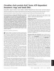
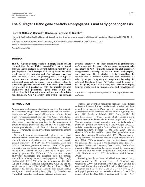
Fig. 1. Early gonado<strong>gene</strong>sis in C. <strong>elegans</strong>. Somatic gonadal<br />
precursors (SGPs: Z1 <strong>and</strong> Z4), dark gray; primordial germ cells<br />
(PGCs: Z2 <strong>and</strong> Z3), light gray. (A) SGPs are specified within the<br />
mesodermal layer (white circles) <strong>and</strong> then migrate to meet PGCs.<br />
(B) SGPs <strong>and</strong> PGCs coalesce into the gonadal primordium, which at<br />
this stage has a left-right orientation. (C) During embryo<br />
morpho<strong>gene</strong>sis, the gonadal primordium shifts to an anteriorposterior<br />
orientation, <strong>and</strong> acquires left-right <strong>and</strong> dorsal ventral axes.<br />
<strong>The</strong> first SGP division is asymmetric <strong>and</strong> segregates the potential to<br />
make two regulatory cells: anchor cells (AC) <strong>and</strong> distal tip cells<br />
(DTC). Genes crucial for <strong>early</strong> SGP divisions are noted.<br />
Strains<br />
Animals were grown at 20°C unless otherwise noted. All strains were<br />
derivatives of Bristol strain N2 (Sulston <strong>and</strong> Horvitz, 1977). <strong>The</strong><br />
following mutations are described by Hodgkin (Hodgkin, 1997) or<br />
cited references. LGI: gon-2(q388) (Sun <strong>and</strong> Lambie, 1997) <strong>and</strong> sys-<br />
1(q544) (Miskowski et al., 2001). LGII: hlh-1(cc450) (Chen et al.,<br />
1994); unc-104(e1265); rol-6(e187); <strong>and</strong> mnDf93 (Sigurdson et al.,<br />
left<br />
right<br />
1984). LGIV: ced-2(e1752); ced-3(n717); gon-4(q519) (Friedman et<br />
al., 2000); unc-24(e138); unc-5(e53); dpy-13(e184); <strong>and</strong> nDf41. LGX:<br />
unc-9(e101). Dominant GFP balancers: mIn1[mIs14] for LGII<br />
(Edgley <strong>and</strong> Riddle, 2001); hT2[qIs48] for LGI; <strong>and</strong> nT1[qIs50] for<br />
LGIV. qIs48 <strong>and</strong> qIs50 are insertions of ccEx9747 onto hT2 <strong>and</strong> nT1,<br />
respectively. Molecular markers: qIs55 [hnd-1(N)::GFP]; qIs69 [hnd-<br />
1::GFPlacZ]; qIs56 [lag-2::GFP] (Siegfried <strong>and</strong> Kimble, 2002);<br />
leIs129 [pes-1::GFP] (Molin et al., 2000); qIs61 [pes-1::GFP]; ayIs7<br />
[hlh-8::GFP] (Harfe et al., 1998); <strong>and</strong> qIs77 [unc-122::GFP]<br />
(Miyabayashi et al., 1999). qIs56 <strong>and</strong> qIs61 were <strong>gene</strong>rated by<br />
microparticle bombardment (Praitis et al., 2001).<br />
Plasmids <strong>and</strong> trans<strong>gene</strong>s<br />
All cloning was performed by st<strong>and</strong>ard methods (Sambrook et al.,<br />
1989). PCR products were sequenced. Primer sequences are available<br />
upon request. Trans<strong>gene</strong>s were <strong>gene</strong>rated as simple arrays unless<br />
otherwise noted.<br />
hnd-1 cDNA (pJK849 <strong>and</strong> pJK901)<br />
Using a probe from the coding region of C44C10.8, we isolated a<br />
hnd-1 cDNA from an embryonic C. <strong>elegans</strong> cDNA library (a gift from<br />
P. Okkema) <strong>and</strong> subcloned it to make pJK849. <strong>The</strong> hnd-1 5′ end was<br />
cloned by RT-PCR using embryonic total RNA, a primer to the SL1<br />
trans-spliced leader <strong>and</strong> internal hnd-1-specific primers. A full-length<br />
hnd-1 cDNA (pJK901) was assembled from the SL1 RT-PCR product<br />
<strong>and</strong> pJK849.<br />
hnd-1(FL)::GFP (pJK850)<br />
GFP coding sequences were amplified by PCR from pPD95.81 (a gift<br />
from A. Fire) <strong>and</strong> subcloned into a hnd-1 genomic fragment (pJK906).<br />
pJK850 includes 1568 bp of the hnd-1 sequence upstream of the<br />
5′UTR <strong>and</strong> 182 bp downstream of the 3′UTR. pJK850 was injected<br />
with pRF4[Rol] (Mello et al., 1991) into hnd-1 to <strong>gene</strong>rate qEx486;<br />
this array rescued hnd-1 gonadal defects completely (n=136) <strong>and</strong><br />
reduced lethality from 28% to 7% (n=190).<br />
hnd-1(N)::GFP (pJK848)<br />
<strong>The</strong> first two exons <strong>and</strong> 1540 bp upstream of the hnd-1 5′UTR were<br />
PCR amplified <strong>and</strong> cloned into pPD95.81 (a gift from A. Fire).<br />
pJK848 was injected into unc-4(e120) with the co-injection marker<br />
pNC4-21[unc-4+] (Miller <strong>and</strong> Niemeyer, 1995) <strong>and</strong> N2 DNA to<br />
create qEx447 <strong>and</strong>, subsequently, qIs55. With the exception of SGPs,<br />
hnd-1(N)::GFP was detected in cells that also express hlh-1, a marker<br />
for body muscle (Krause et al., 1990).<br />
hnd-1::GFPlacZ (pJK900)<br />
<strong>The</strong> hnd-1 promoter (plus 11 N-terminal codons) was PCR amplified<br />
<strong>and</strong> cloned into pPD96.04 (a gift from A. Fire). pJK900 was injected<br />
with pRF4[Rol+] to create qEx492 <strong>and</strong>, subsequently, qIs69. pJK850<br />
<strong>and</strong> pJK900, but not pJK848, express GFP in several head cells that<br />
we have not identified.<br />
HS-hnd-1 (pJK902)<br />
<strong>The</strong> hnd-1 cDNA from pJK901 was cloned into pPD49.78 (a gift from<br />
A. Fire) to <strong>gene</strong>rate pJK902, which was injected into qIs61 with the<br />
co-injection marker pRF4[Rol+] to make qEx493. Embryos were<br />
subjected to two 30-minute heat pulses at 33°C, with a one hour<br />
recovery interval. Resulting L1 larvae were scored for extra SGPs<br />
using pes-1::GFP.<br />
hlh-1::hnd-1GFP (pJK904)<br />
A hnd-1::GFP fusion was <strong>gene</strong>rated by inserting GFP into the RsrII<br />
site of the full-length hnd-1 cDNA (pJK901). hnd-1::GFP was then<br />
cloned into pPD51.45 (Krause et al., 1990) to <strong>gene</strong>rate pJK904, which<br />
was injected into hnd-1 with the co-injection marker pRF4[Rol+] to<br />
make qEx496; this array rescued hnd-1 gonadal defects <strong>and</strong><br />
marginally rescued lethality (20%, n=372).
lag-2::hnd-1GFP (pJK905)<br />
A hnd-1::GFP fusion (see above) was cloned into pJK590 (Blelloch<br />
et al., 1999) to <strong>gene</strong>rate pJK905, which was injected into hnd-1 with<br />
the co-injection marker pRF4[Rol+] to make qEx497; this array<br />
partially rescued hnd-1 gonado<strong>gene</strong>sis defects <strong>and</strong> did not rescue<br />
lethality (24%, n=192).<br />
hnd-1 genomic DNA (pJK906)<br />
A plasmid carrying hnd-1 genomic DNA was amplified by PCR; it<br />
contained the same upstream <strong>and</strong> downstream sequences as in hnd-<br />
1(FL)::GFP. pJK906 was injected with pPD136.64 [myo-3::YFP] (a<br />
gift from A. Fire) <strong>and</strong> pJK907 [pes-1::CFP] into hnd-1 to make<br />
qEx495; this array was used for mosaic analysis.<br />
pes-1::CFP (pJK907)<br />
<strong>The</strong> pes-1 promoter from pUL#MJA1 (Molin et al., 2000) was cloned<br />
into pPD136.64 (a gift from A. Fire).<br />
hnd-1 RNA interference <strong>and</strong> deletion<br />
Double-str<strong>and</strong>ed hnd-1 RNA was <strong>gene</strong>rated, using pJK849 as<br />
template, <strong>and</strong> injected at 1 mg/ml. <strong>The</strong> hnd-1(q740) deletion was<br />
isolated essentially as described by Kraemer et al. (Kraemer et al.,<br />
1999), <strong>and</strong> backcrossed eight times. To test for maternal effects,<br />
hnd-1 females, <strong>gene</strong>rated by fog-1 RNAi (Jin et al., 2001), were<br />
crossed with N2 males [17% of the cross-progeny died as embryos or<br />
young larvae (n=313), <strong>and</strong> all adult progeny had normal gonads<br />
(n=260)]. To test for zygotic lethality, we scored progeny of unc-9<br />
hnd-1/++ mothers [6% died as embryos or larvae (n=235)]. To<br />
investigate whether hnd-1(q740) was a null allele, RT-PCR was<br />
performed on mutant <strong>and</strong> wild-type worms, using primers to a region<br />
retained in the hnd-1 deletion. Template RNA was prepared from 20<br />
gravid adults using TRI reagent (Molecular Research Center). A PCR<br />
product was obtained only from wild-type worms.<br />
Tests for hnd-1 <strong>gene</strong>tic interactions<br />
hlh-1<br />
Progeny of hlh-1/+; hnd-1 mothers had 55% embryonic <strong>and</strong> larval<br />
lethality, compared with 25% defects for hlh-1/+ (Chen et al., 1994)<br />
<strong>and</strong> 28% defects for hnd-1 (this work).<br />
<strong>The</strong> following were evaluated using number of gonadal arms as a<br />
measure:<br />
sys-1<br />
100% of sys-1/+; hnd-1/+ worms had two arms (n=89). sys-1/+;<br />
hnd-1 had 68% gonadal arms, compared with 70% for hnd-1 alone<br />
<strong>and</strong>
2884<br />
L. D. Mathies, S. T. Henderson <strong>and</strong> J. Kimble<br />
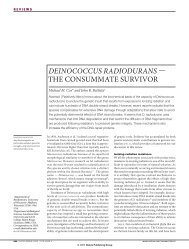
Fig. 2. <strong>The</strong> hnd-1 <strong>gene</strong> encodes the C.<br />
<strong>elegans</strong> <strong>H<strong>and</strong></strong> transcription factor.<br />
(A) hnd-1 (formerly C44C10.8) genomic<br />
organization. White, untranslated regions;<br />
gray <strong>and</strong> black, coding region; black, basic<br />
helix-loop-helix (bHLH) domain. Brackets<br />
below mark end points of hnd-1 deletion.<br />
(B) Amino acid sequence alignment of<br />
bHLH domains of human (h), Xenopus (x),<br />
zebrafish (z), Drosophila (Dm) <strong>and</strong><br />
C. <strong>elegans</strong> (Ce) HAND proteins. (C) hnd-1<br />
reporters. Top, hnd-1(FL)::GFP inserts<br />
GFP after amino acid 163. Middle, hnd-<br />
1(N)::GFP fuses GFP to the N terminus of<br />
HND-1 <strong>and</strong> uses the unc-54 3′UTR (light<br />
gray). Bottom, hnd-1::GFPlacZ replaces<br />
most of the hnd-1 coding region with a<br />
GFPlacZ fusion <strong>and</strong> the unc-54 3′UTR<br />
(light gray). GFP <strong>and</strong> GFPlacZ are not to<br />
scale.<br />
surviving adults (52%, n=656). Aside from gonadal defects<br />
<strong>and</strong> minor body wall abnormalities, hnd-1 survivors appeared<br />
normal. <strong>The</strong> hnd-1 mutant was recessive: the gonadal defects<br />
had no maternal effect, but both maternal <strong>and</strong> zygotic hnd-1<br />
activities were important for viability.<br />
hnd-1 affects embryo morpho<strong>gene</strong>sis<br />
hnd-1 mutants can die as embryos or young larvae with<br />
A<br />
B<br />
hnd-1(q740): 1291bp deletion<br />
basic helix loop helix<br />
hh<strong>and</strong>1 R K G S G P K K E R R R T E S I N S A F A E L R E C I P N V P A D T K . . L S K I K T L R L A T S Y I A Y L M D V L<br />
xh<strong>and</strong>1 R K G A P P K K E R R R T E S I N S A F A E L R E C I P N V P A D T K . . L S K I K T L R L A T S Y I G Y L M D V L<br />
hh<strong>and</strong>2 R R G T A N R K E R R R T Q S I N S A F A E L R E C I P N V P A D T K . . L S K I K T L R L A T S Y I A Y L M D L L<br />
xh<strong>and</strong>2 R R G T A N R K E R R R T I S I N S A F A E L R E C I P N V P A D T K . . L S K I K T L R L A T S Y I A Y L M D L L<br />
zh<strong>and</strong>2 R R P T A N R K E R R R T Q S I N S A F A E L R E C I P N V P A D T K . . L S K I K T L R L A T S Y I A Y L M D I L<br />
Dmh<strong>and</strong> K R N T A N K K E R R R T Q S I N N A F S Y L R E K I P N V P T D T K . . L S K I K T L K L A I L Y I N Y L V N V L<br />
Ceh<strong>and</strong> R K E K S R E K E H R R A Q C I N S A F E I L Q Q H I P Y L K S E E R K S L P K I K T L R L A M Q Y I D H L K K L L<br />
C<br />
hnd-1 (C44C10.8)<br />
hnd-1(FL)::GFP<br />
hnd-1(N)::GFP<br />
hnd-1::GFPlacZ<br />
SL1<br />
bHLH domain<br />
GFP<br />
GFP<br />
GFPlacZ<br />
100 bp<br />
100 bp<br />
3'UTR<br />
3'UTR<br />
variable body shape defects, typically in the posterior (Fig.<br />
3A,B). Most hnd-1 embryos contained pharynx <strong>and</strong> gut (Fig.<br />
3C,D), as well as muscle, as evidenced by twitching. Because<br />
hnd-1::GFP is expressed in mesodermal precursors (see<br />
below), we compared body wall muscles in wild-type <strong>and</strong><br />
hnd-1 embryos using an α-myosin antibody (Miller et al.,<br />
1983). Both wild-type <strong>and</strong> hnd-1 late-stage embryos have four<br />
quadrants of body muscle (Fig. 3E,F) (Miller et al., 1983),<br />
although muscle fibers were sometimes disorganized in<br />
mutants (Fig. 3F).<br />
hlh-1 has striking similarities to hnd-1. <strong>The</strong> hlh-1 <strong>gene</strong><br />
encodes a MYOD-like bHLH protein, <strong>and</strong> hlh-1 mutants have<br />
severe defects in embryo morpho<strong>gene</strong>sis but <strong>gene</strong>rate body<br />
muscle normally (Chen et al., 1994). We examined hnd-1;<br />
hlh-1 double mutants to determine whether these two bHLH<br />
proteins might have overlapping functions, but double mutants<br />
made body muscle (not shown). Furthermore, we found no<br />
significant <strong>gene</strong>tic interaction between hlh-1 <strong>and</strong> hnd-1<br />
(Materials <strong>and</strong> Methods). <strong>The</strong>refore, hnd-1 <strong>and</strong> hlh-1 appear to<br />
function independently.<br />
hnd-1 governs SGP number <strong>and</strong> position<br />
Wild-type hermaphrodites possess two gonadal arms. By<br />
contrast, adult hermaphrodites depleted for hnd-1 displayed a<br />
range of gonadal shapes: two gonadal arms (Fig. 4A); a single<br />
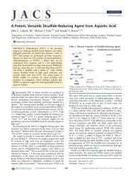
Fig. 3. hnd-1 morpho<strong>gene</strong>sis defect. (A,C,E) wild type, (B,D,F) hnd-<br />
1(q740). (A) Wild-type L1 with normal body morphology. <strong>The</strong><br />
gonadal primordium is bracketed. Three cells are visible: Z1, black<br />
arrow; PGCs, white arrows; Z4 is in a different focal plane. (B) hnd-1<br />
L1 with typical body shape defect (black arrow). Arrested larvae<br />
often have vacuoles in the head (arrowheads). (C) Wild-type pretzelstage<br />
embryo is elongated <strong>and</strong> contains pharynx (ph) <strong>and</strong> gut.<br />
(D) hnd-1 pretzel-stage embryo has not elongated posteriorly<br />
(arrow), but has a fully developed pharynx <strong>and</strong> gut tissue, which can<br />
be disorganized. (E) Wild-type embryos stained with α-myosin<br />
antibodies. Two muscle quadrants are visible in this plane<br />
(arrowheads). (F) hnd-1 embryos <strong>gene</strong>rate four muscle quadrants,<br />
which can be disorganized posteriorly (arrow); three quadrants are<br />
visible in this plane (arrowheads). Scale bar: 10 μm.
Fig. 4. hnd-1 gonadal defects. (A-D) L4 hnd-1(RNAi)<br />
hermaphrodites, DAPI stained to highlight nuclei. Dashed line<br />
delineates the extent of the gonad. Arrowhead, distal end; carat,<br />
center of gonad (vulva). (A,B) Anterior half of animal is on the left,<br />
posterior on the right. (A) Two-armed gonad. (B) One-armed gonad.<br />
(C) No apparent gonad. (D) Abnormal gonad.<br />
gonadal arm (Fig. 4B); no apparent gonad (Fig. 4C); or<br />
abnormal gonads (Fig. 4D). ‘Abnormal gonads’ include a<br />
variety of shapes, most typically an amorphous mass (Fig. 4D).<br />
One-armed <strong>and</strong> two-armed gonads were frequent <strong>and</strong> were<br />
usually fertile, whereas absent <strong>and</strong> abnormal gonads were less<br />
common <strong>and</strong> were always sterile (Table 1). A similar, but less<br />
penetrant effect was seen in males (not shown). We conclude<br />
that hnd-1 is important, but not essential, for gonado<strong>gene</strong>sis.<br />
<strong>The</strong> gonadal morphologies in hnd-1 mutants suggested a<br />
defect <strong>early</strong> in gonado<strong>gene</strong>sis. <strong>The</strong>refore, we examined SGPs<br />
in hnd-1 gonadal primordia, using either nuclear pes-1::GFP<br />
(Molin et al., 2000) (Fig. 5A-C) or cytoplasmic lag-2::GFP,<br />
which reveals cellular processes (Blelloch et al., 1999) (Fig.<br />
5G,J). Whereas all wild-type gonadal primordia had two SGPs<br />
(Table 2A), hnd-1 primordia could have two (Fig. 5A,B), one<br />
(Fig. 5C), zero (Fig. 5D) or even three SGPs (Table 2A).<br />
<strong>The</strong>refore, hnd-1 is important for determining SGP number.<br />
SGP position was also affected in hnd-1 mutants. In wild<br />
type, the two SGPs reside at the distal poles of the primordium,<br />
flanking the PGCs <strong>and</strong> extending cytoplasmic processes to<br />
meet mid-ventrally. In most hnd-1 mutants, SGPs occupied<br />
similar polar positions (Table 2B), <strong>and</strong> extended ventral<br />
processes (Fig. 5E-G). However, in some hnd-1 mutants, one<br />
or both SGPs were not at the pole, but instead were found more<br />
centrally in the primordium (Table 2B). When an SGP was<br />
misplaced dorsally, it extended cytoplasmic processes along<br />
the dorsal surface of the primordium (Fig. 5H-J). Finally, in<br />
some hnd-1 mutants, SGPs were observed ectopically (Table<br />
2B). <strong>The</strong>se ectopic SGPs could be in animals with either two<br />
or three total SGPs; as predicted, ectopic gonadal arms have<br />
been observed in rare hnd-1 mutants. <strong>The</strong>refore, hnd-1 can<br />
hnd-1 <strong>and</strong> gonado<strong>gene</strong>sis<br />
2885<br />
Fig. 5. Gonadal primordia in hnd-1 L1 larvae. All images are hnd-<br />
1(RNAi). Black arrow, SGP with name of cell; asterisk, PGC.<br />
(A-C) pes-1::GFP marks SGPs, GFP overlays DIC image.<br />
(A,B) Primordium with two SGPs. Right focal plane shows Z1<br />
(A). Left focal plane shows Z4 (B). (C) Primordium with one SGP,<br />
left focal plane. (D) Primordium with no SGPs, two PGCs are<br />
present, but are separated. (E-J) lag-2::GFP marks SGPs. (E-G) Two<br />
SGPs in normal positions. Z1 is at anterior pole on right (E), Z4 is at<br />
posterior pole on left (F). Z1 <strong>and</strong> Z4 extend cytoplasmic processes to<br />
meet mid-ventrally (G, white arrow). (H-J) Primordium with one<br />
misplaced SGP. Z1 is displaced dorsally (H), Z4 is located at the<br />
posterior pole (I). Z1 <strong>and</strong> Z4 meet mid-dorsally via a thin<br />
cytoplasmic process (J, white arrow). Scale bar: 5 μm.<br />
affect the position of the SGPs within the primordium <strong>and</strong><br />
within the animal.<br />
SGP number <strong>and</strong> position are crucial for<br />
gonado<strong>gene</strong>sis<br />
We used hnd-1 mutants born with aberrant gonadal primordia<br />
to investigate how organization of that primordium affected<br />
gonado<strong>gene</strong>sis. Specifically, we used pes-1::GFP to score<br />
SGPs in hnd-1 L1 larvae, permitted the animals to develop <strong>and</strong><br />
then examined them again as L4s. Our results (Table 3) led to<br />
three conclusions. First, most hnd-1 primordia with a wild-type<br />
appearance (two SGPs placed at the poles) <strong>gene</strong>rated wild-type<br />
appearing adults with two gonadal arms (93%, n=70).<br />
<strong>The</strong>refore, hnd-1 appears to play little or no role in<br />
gonado<strong>gene</strong>sis after formation of the gonadal primordium.<br />
Second, most primordia containing one SGP <strong>gene</strong>rated adult<br />
gonads with only a single arm (98%, n=53); none made two
2886<br />
L. D. Mathies, S. T. Henderson <strong>and</strong> J. Kimble<br />
Table 2. Effect of hnd-1/EHN mutants on SGP number<br />
<strong>and</strong> position<br />
A SGP number<br />
Percentage of animals<br />
Three Two One No<br />
Genotype* SGPs SGPs SGP SGP n<br />
Wild type 0 100 0 0 64<br />
hnd-1(q740) 2 57 39 3 103<br />
hnd-1(RNAi) 2 61 36 1 87<br />
ehn-1(q638) 0 90 10 0 106<br />
ehn-3(q689) 0 89 8 2 171<br />
ehn-1(q638); ehn-3(q689) 0 5 35 60 43<br />
ehn-3(q689); hnd-1(RNAi) 0 4 29 67 49<br />
*All contain qIs61, which is an integrated pes-1::GFP.<br />
n, number of animals scored.<br />
B SGP position<br />
Percentage of SGPs in each position<br />
Genotype* Pole Position † Central ‡ Ectopic § n<br />
Wild type 100 0 0 128<br />
hnd-1(q740) 87 9 4 162<br />
hnd-1(RNAi) 83 15 3 143<br />
ehn-1(q638) 90 9 1 196<br />
ehn-3(q689) 96 4 0 320<br />
*All contain qIs61, which is an integrated pes-1::GFP.<br />
† SGP at its normal position within the primordium.<br />
‡ SGP misplaced centrally within the primordium.<br />
§ pes-1::GFP-expressing cells outside of the gonadal primordium.<br />
n, number SGP cells scored.<br />
arms. Finally, primordia with wild-type SGP number but<br />
aberrant SGP position often <strong>gene</strong>rated defective gonads (26%,<br />
n=23). <strong>The</strong>refore, SGP position within the primordium may be<br />
important for gonado<strong>gene</strong>sis.<br />
Effects on germline development in hnd-1 mutants<br />
<strong>The</strong> gonadal primordium in hnd-1 mutants sometimes lacked<br />
PGCs. In primordia with two SGPs, 7% lacked one or both<br />
PGCs (n=59), <strong>and</strong> in primordia with one SGP, 40% were<br />
missing at least one PGC (n=47). To investigate whether<br />
the two PGCs were made in hnd-1 mutants, we stained ~100cell<br />
embryos with a germline-specific antibody, α-PGL-1<br />
(Kawasaki et al., 1998): all had two PGL-1-staining cells<br />
(n=237). <strong>The</strong>refore, hnd-1 does not affect the <strong>gene</strong>ration of<br />
PGCs, but instead affects their maintenance within the gonadal<br />
primordium. A simple interpretation is that hnd-1 acts in SGPs,<br />
which in turn are essential for germline survival <strong>and</strong><br />
positioning.<br />
hnd-1 expression during wild-type development<br />
To investigate hnd-1 expression, we constructed three GFP<br />
reporters (Fig. 2C). hnd-1(FL)::GFP inserts GFP coding<br />
sequences into the third exon of the full-length HND-1 protein;<br />
this reporter rescued hnd-1 mutants (Materials <strong>and</strong> Methods).<br />
hnd-1(N)::GFP inserts GFP more N-terminally <strong>and</strong> replaces<br />
the hnd-1 3′UTR with the unc-54 3′UTR. hnd-1::GFPlacZ<br />
replaces most of the hnd-1 coding region with GFP <strong>and</strong> βgalactosidase<br />
coding sequences, <strong>and</strong> the unc-54 3′UTR. All<br />
three hnd-1 reporters expressed GFP in largely the same cells,<br />
but hnd-1(N)::GFP <strong>and</strong> hnd-1::GFPlacZ expressed GFP at a<br />
higher level <strong>and</strong> expression persisted longer. <strong>The</strong> rescue by<br />
hnd-1(FL)::GFP suggests that its expression is relevant to<br />
hnd-1 function.<br />
<strong>The</strong> hnd-1 reporters expressed GFP in the MS, C <strong>and</strong> D<br />
embryonic lineages (Fig. 6A). Expression was first observed in<br />
four MS great-gr<strong>and</strong>daughters, four C great-gr<strong>and</strong>daughters<br />
<strong>and</strong> two D daughters (Fig. 6B). <strong>The</strong>se MS descendants give<br />
rise to the SGPs <strong>and</strong> other mesodermal cells (Fig. 6A) (Sulston<br />
et al., 1983); the C- <strong>and</strong> D-expressing cells all <strong>gene</strong>rate body<br />
wall muscle (Sulston et al., 1983). Expression continued<br />
through one cell division (Fig. 6C) <strong>and</strong> then became difficult<br />
to detect using hnd-1(FL)::GFP. hnd-1(N)::GFP remained<br />
detectable in some cells within these MS <strong>and</strong> C lineages (Fig.<br />
6D), but disappeared from most body muscle cells by the<br />
comma stage of embryo<strong>gene</strong>sis (Fig. 6E). <strong>The</strong>n, the hnd-1<br />
reporters were expressed in the SGPs (Z1 <strong>and</strong> Z4) as they<br />
approached the PGCs to form the gonadal primordium<br />
(Fig. 6E,F). Shortly after the primordium was assembled, hnd-<br />
1(N)::GFP expression was reduced or disappeared (Fig. 6G).<br />
GFP was not detected in the SGPs at hatching or postembryonically<br />
(not shown). <strong>The</strong>refore, hnd-1 appears to be<br />
expressed during embryo<strong>gene</strong>sis in mesodermal precursor cells<br />
that <strong>gene</strong>rate predominantly body wall muscle, <strong>and</strong> then in<br />
SGPs.<br />
<strong>The</strong> hnd-1 <strong>gene</strong> is not required for specification of<br />
the SGP fate<br />
<strong>The</strong> most common gonadal defect in hnd-1 mutants is a<br />
missing SGP (Table 2A). To investigate whether both SGPs are<br />
made in hnd-1-mutant embryos, we used the hnd-1(N)::GFP<br />
reporter, which is an <strong>early</strong> SGP marker. We found both SGPs<br />
Table 3. Correlation of gonadal primordium <strong>and</strong> adult gonad in hnd-1 mutants<br />
Gonadal primordium Adult gonad (%)<br />
Number None<br />
Genotype* of SGPs SGP position ‡ Two arms One arm Abnormal visible n<br />
Wild type 2 Both at poles 100 0 0 0 64<br />
hnd-1 † 2 Both at poles 93 6 1 0 70<br />
hnd-1 † 2 One at pole, one misplaced 74 9 17 0 23<br />
hnd-1 † 1 Pole 0 98 2 0 53<br />
hnd-1 † 1 Misplaced 0 71 29 0 7<br />
hnd-1 † 0 Not applicable 0 0 0 100 4<br />
*All contain qIs61, an integrated pes-1::GFP.<br />
† Includes both hnd-1(RNAi) <strong>and</strong> hnd-1(q740).<br />
‡ Presence <strong>and</strong> position of Z1 <strong>and</strong> Z4 were scored by pes-1::GFP expression.
hnd-1 <strong>and</strong> gonado<strong>gene</strong>sis<br />
Fig. 6. hnd-1::GFP expression.<br />
(A) Lineage diagram depicting<br />
cells that express hnd-1::GFP<br />
(green). Dashed lines indicate<br />
approximate stage of embryos<br />
in panels B-D. (B-H) Confocal<br />
images of embryos expressing<br />
hnd-1::GFP (green).<br />
(B-D) Projections of z-series;<br />
embryos were observed over<br />
time to identify cells. (B) hnd-<br />
1(N)::GFP expression is first<br />
detected in four gr<strong>and</strong>daughters<br />
of MS, descendants of MS.ap<br />
<strong>and</strong> MS.pp, in four<br />
gr<strong>and</strong>daughters of C,<br />
descendants of C.ap <strong>and</strong> C.pp<br />
(C.xp), <strong>and</strong> in two daughters of<br />
D. (C) hnd-1(FL)::GFP<br />
expression in gr<strong>and</strong>daughters of<br />
MS.ap/MS.pp <strong>and</strong> daughters of<br />
C.ap/C.pp. (D) hnd-1(N)::GFP<br />
expression fades after the next<br />
division of most hnd-1expressing<br />
cells but is retained<br />
in daughters of MS.appp <strong>and</strong><br />
MS.pppp, <strong>and</strong> in daughters<br />
of C.ppp <strong>and</strong> C.app.<br />
(E-H) Embryos fixed <strong>and</strong><br />
stained with α-HLH-1 or α-<br />
PGL-1 (red). (E) Unlike HLH-1<br />
(red), which is detected in body<br />
muscle lineages throughout<br />
embryo<strong>gene</strong>sis (Krause et al.,<br />
1990), hnd-1(N)::GFP is absent from body muscles by the comma-stage of embryo<strong>gene</strong>sis; at this time, expression is seen in Z1/Z4. (F) hnd-<br />
1(N)::GFP is detected in Z1 <strong>and</strong> Z4 as they meet the PGCs (Z2, Z3), marked by PGL-1 (red). (G) Shortly after, expression is absent from Z1<br />
<strong>and</strong> Z4. (H) hnd-1 embryos express hnd-1(N)::GFP in Z1/Z4.<br />
present <strong>and</strong> in their normal position near the PGCs (Fig. 6H;<br />
n=29). <strong>The</strong>refore, hnd-1 is not necessary for SGP specification<br />
or SGP migration to the PGCs.<br />
After formation of the gonadal primordium, SGPs remained<br />
associated with PGCs in hnd-1 embryos; none were seen<br />
detaching. Instead, SGP nuclei sometimes became smaller <strong>and</strong><br />
hnd-1 reporter expression faded prematurely (38%, n=13). <strong>The</strong><br />
simplest hypothesis is that hnd-1 is required for maintenance<br />
of SGP fate <strong>and</strong> possibly SGP survival.<br />
We next investigated whether ectopic hnd-1 expression<br />
could transform other cells to the SGP fate. To this end, we<br />
used a heat-inducible promoter to express hnd-1 during<br />
embryo<strong>gene</strong>sis but found no ectopic SGPs, as assayed by pes-<br />
1::GFP (Materials <strong>and</strong> Methods). We also expressed a fulllength<br />
HND-1::GFP fusion protein under control of either of<br />
two mesodermal promoters (see below), but again did not<br />
observe ectopic SGPs. <strong>The</strong>se results are consistent with the<br />
proposed role for hnd-1 in controlling SGP maintenance or<br />
survival.<br />
What becomes of SGPs in hnd-1 mutants?<br />
<strong>The</strong> missing SGPs in hnd-1 mutants might be explained by<br />
transformation to a different cell type, or by cell death. To explore<br />
the first idea, we reasoned that the most likely transformation<br />
would be to a different mesodermal cell type. We tested this using<br />
2887<br />
hlh-8::GFP to mark the M mesoblast (Harfe et al., 1998) <strong>and</strong><br />
unc-122::GFP to mark coelomocytes (Miyabayashi et al., 1999).<br />
All wild-type L1s had a single hlh-8::GFP-expressing M<br />
mesoblast, as expected (n=61). Similarly, most hnd-1 mutants<br />
had a single M cell, but a few had two M cells (5%, n=63) or no<br />
M cell (2%, n=63). Those with an additional M cell had two<br />
SGPs, suggesting that extra M cells were not transformed SGPs.<br />
Likewise, occasional extra coelomocytes were seen, but overall<br />
hnd-1 mutants had marginally fewer coelomocytes than wild type<br />
(5.5 versus 5.9 on average per animal, n>30). Importantly, the<br />
extra coelomocytes could be in worms with two gonadal arms.<br />
<strong>The</strong>refore, hnd-1 appears to have a low-penetrance effect on M<br />
cells <strong>and</strong> coelomocytes, but this is unlikely to account for the<br />
missing SGPs.<br />
To determine whether SGPs are lost as a result of<br />
programmed cell death in hnd-1 mutants, we examined hnd-1;<br />
ced-3 double mutants using the pes-1::GFP marker. <strong>The</strong> ced-3<br />
<strong>gene</strong> is required for all programmed cell deaths (Ellis <strong>and</strong><br />
Horvitz, 1986). In hnd-1 single mutants, 42% were missing at<br />
least one SGP (Table 2A), <strong>and</strong>, in ced-3; hnd-1 double mutants,<br />
45% lacked at least one SGP (n=69). <strong>The</strong>refore, SGP loss does<br />
not appear to rely on ced-3-dependent programmed cell death.<br />
Next, we investigated whether SGPs died in hnd-1 mutants.<br />
In C. <strong>elegans</strong>, cell corpses resulting from either programmed<br />
or necrotic cell death are engulfed by their neighbors (Chung
2888<br />
L. D. Mathies, S. T. Henderson <strong>and</strong> J. Kimble<br />
Fig. 7. Cell death in hnd-1 mutants. Gonadal primordia: black arrow,<br />
SGP with name of cell; asterisk, PGC. (A,B) ced-2 mutants have no<br />
corpses near gonad. Z1 is in the right plane (A) <strong>and</strong> Z4 is in the left<br />
plane (B). (C,D) ced-2; hnd-1 double mutant. Z1 is missing, but a<br />
cell corpse occurs in its place at the anterior pole of the gonadal<br />
primordium (C, open arrow). Z4 is present (D). Scale bar: 5 μm.<br />
et al., 2000; Ellis et al., 1991). <strong>The</strong> engulfment of cell corpses<br />
relies on several <strong>gene</strong>s, including ced-2 (Ellis et al., 1991). In<br />
ced-2 single mutants, no cell corpses were evident near the<br />
gonad (Fig. 7A,B; n=54); however, in ced-2; hnd-1 double<br />
mutants, cell corpses were found near the gonad (Fig. 7C,D;<br />
28%, n=50). Importantly, the presence <strong>and</strong> site of corpses<br />
correlated with SGP absence. We observed no cell corpses near<br />
gonads with two SGPs (n=53), mostly anterior or right cell<br />
corpses near gonads missing Z1 (4/5; Fig. 7C), <strong>and</strong> only<br />
posterior or left cell corpses in those missing Z4 (8/8). Indeed,<br />
in one cell corpse, hnd-1::GFP was faintly expressed,<br />
indicating that it had been specified originally as an SGP (data<br />
not shown). <strong>The</strong>refore, SGPs appear to die in hnd-1 mutants.<br />
hnd-1 activity acts in somatic tissues to control<br />
gonado<strong>gene</strong>sis<br />
Both SGPs <strong>and</strong> PGCs are affected in hnd-1 mutants. To learn<br />
where hnd-1 functions, we used a combination of mosaic<br />
analysis <strong>and</strong> transgenic experiments driving the hnd-1 coding<br />
region with tissue-specific promoters.<br />
In C. <strong>elegans</strong>, mosaic animals can be made by loss of extrachromosomal<br />
arrays that carry trans<strong>gene</strong>s <strong>and</strong> that are<br />
transmitted with varying fidelity at each cell division (Herman,<br />
1984). For this study, we created an extra-chromosomal array<br />
that carries a rescuing hnd-1 genomic fragment <strong>and</strong> two<br />
fluorescent markers (myo-3::YFP to mark body muscle <strong>and</strong><br />
pes-1::CFP to mark SGPs). We then identified ‘germline<br />
mosaics’, animals that retained the array in somatic tissues but<br />
failed to transmit it to their progeny; such animals have lost the<br />
array in divisions <strong>gene</strong>rating the germline blastomere P4 (see<br />
Fig. 6). All six germline mosaics had a wild-type gonadal<br />
primordium, which suggests that hnd-1 activity acts in somatic<br />
tissues rather than in the germ line.<br />
To further explore where hnd-1 acts, we used either of two<br />
promoters: hlh-1, which is expressed in body muscle <strong>and</strong> not<br />
in SGPs (Krause et al., 1990); or lag-2, which is first expressed<br />
in the AB <strong>and</strong> MS lineages (Moskowitz <strong>and</strong> Rothman, 1996),<br />
<strong>and</strong> then in SGPs (Miskowski et al., 2001). Each promoter was<br />
fused to a full-length, rescuing hnd-1::GFP cDNA <strong>and</strong><br />
expressed in hnd-1 mutants. Expression of HND-1::GFP by the<br />
hlh-1 promoter rescued the hnd-1 gonado<strong>gene</strong>sis defects, from<br />
52% to 5% defective (n=42). By contrast, HND-1::GFP driven<br />
from the lag-2 promoter, which is expressed in the two SGPs<br />
(Fig. 5G), did not appreciably rescue hnd-1 gonado<strong>gene</strong>sis<br />
defects (36% defective, n=108). <strong>The</strong> latter experiment has the<br />
caveat that this promoter is switched on after SGPs assemble<br />
into the gonadal primordium <strong>and</strong> it may not be expressed in<br />
dying SGPs. From the hlh-1::hnd-1GFP result, we suggest that<br />
HND-1 acts in <strong>early</strong> mesodermal lineages.<br />
hnd-1 acts independently of other <strong>early</strong><br />
gonado<strong>gene</strong>sis <strong>gene</strong>s<br />
<strong>The</strong> hnd-1 SGP defects are the earliest observed to date among<br />
any <strong>gene</strong>s controlling C. <strong>elegans</strong> gonado<strong>gene</strong>sis. To investigate<br />
whether hnd-1 might function with other <strong>early</strong> gonado<strong>gene</strong>sis<br />
<strong>gene</strong>s, we explored <strong>gene</strong>tic interactions between hnd-1 <strong>and</strong> two<br />
mutant classes. <strong>The</strong> first type, represented by gon-2 <strong>and</strong> gon-4<br />
(Friedman et al., 2000; Sun <strong>and</strong> Lambie, 1997), <strong>controls</strong> the<br />
onset of cell divisions in the gonad but not in other tissues<br />
(Fig. 1C). Gonadal divisions are delayed in gon-2 or gon-4<br />
single mutants (Friedman et al., 2000; Sun <strong>and</strong> Lambie, 1997),<br />
but not in hnd-1 mutants (n=5). Moreover, hnd-1; gon-2 <strong>and</strong><br />
hnd-1; gon-4 double mutants have additive phenotypes<br />
(Materials <strong>and</strong> Methods). <strong>The</strong>refore, hnd-1 does not affect the<br />
onset or timing of gonadal divisions <strong>and</strong> acts independently of<br />
gon-2 <strong>and</strong> gon-4.<br />
<strong>The</strong> second class of <strong>early</strong> gonado<strong>gene</strong>sis <strong>gene</strong>s, represented<br />
by sys-1 (Miskowski et al., 2001), is required for SGPs to<br />
produce daughter cells with different developmental potential<br />
(Fig. 1C). In wild type, each SGP <strong>gene</strong>rates one distal tip cell<br />
(DTC), whereas in sys-1 mutants they make no DTCs<br />
(Miskowski et al., 2001). Most hnd-1 SGPs that were properly<br />
positioned <strong>gene</strong>rated DTCs (96%, n=193; Table 3), <strong>and</strong> no<br />
<strong>gene</strong>tic interactions were found with sys-1 (Materials <strong>and</strong><br />
Methods). <strong>The</strong>refore, hnd-1 does not appear to affect SGP<br />
asymmetric divisions, but instead ensures that two SGPs are<br />
present <strong>and</strong> properly positioned in the gonadal primordium.<br />
Identification of <strong>gene</strong>tic enhancers of hnd-1<br />
To identify additional <strong>gene</strong>s controlling SGP development,<br />
we screened for EMS-induced mutants with a hnd-1-like<br />
gonado<strong>gene</strong>sis phenotype <strong>and</strong> discovered loss-of-function<br />
mutations of ehn-1 <strong>and</strong> ehn-3 [for enhancer of <strong>H<strong>and</strong></strong> (Materials<br />
<strong>and</strong> Methods)]. <strong>The</strong> ehn-1 <strong>and</strong> ehn-3 mutants had lowpenetrance<br />
gonadal defects (Table 1). For ehn-1, gonadal<br />
defects could be rescued either maternally or zygotically, but<br />
ehn-3 exhibited no maternal effect (Materials <strong>and</strong> Methods).<br />
Furthermore, ehn-1 had low-penetrance lethality, but lethality<br />
was negligible in ehn-3 mutants (Table 4).<br />
<strong>The</strong> gonadal primordia of ehn-1 <strong>and</strong> ehn-3 mutants had<br />
absent or misplaced SGPs, as described above for hnd-1<br />
mutants (Table 2A,B). However, hnd-1, ehn-1 <strong>and</strong> ehn-3<br />
displayed subtle differences in their spectrum of defects. For<br />
example, ectopic SGPs were seen in ehn-1 but not ehn-3<br />
mutants, <strong>and</strong> only hnd-1 mutants <strong>gene</strong>rated extra SGPs (Table<br />
2A). Despite these minor differences, the primary defects were<br />
similar among the three mutants, which suggests that they may
function in a common pathway to control <strong>early</strong> gonadal<br />
development.<br />
Functional relationships between the hnd-1 <strong>and</strong><br />
EHN <strong>gene</strong>s<br />
To investigate the functional relationships between the hnd-1<br />
<strong>and</strong> EHN <strong>gene</strong>s, we first investigated double <strong>and</strong> triple mutants<br />
(Table 4). Although hnd-1, ehn-1 <strong>and</strong> ehn-3 single mutants all<br />
had relatively low-penetrance gonadal defects, the double <strong>and</strong><br />
triple mutants showed increased penetrance (Table 4). For<br />
example, 80-90% of ehn-1 <strong>and</strong> ehn-3 single-mutant adults had<br />
two gonadal arms, but almost none of the ehn-1; ehn-3 double<br />
mutants had two gonadal arms (2-4%; Table 4). Similarly, only<br />
5% of the double mutants had two SGPs at hatching (Table<br />
2A). By contrast, larval lethality did not increase in the ehn-1;<br />
ehn-3 double mutant. <strong>The</strong>refore, ehn-1 <strong>and</strong> ehn-3 may be<br />
partially redundant for SGP development.<br />
<strong>The</strong> ehn-1; hnd-1 <strong>and</strong> ehn-3; hnd-1 double mutants were<br />
also more defective than any of the single mutants, but each<br />
double mutant was unique. For the gonado<strong>gene</strong>sis defects, ehn-<br />
3 enhanced hnd-1 more strongly than did ehn-1. Thus, some<br />
ehn-1; hnd-1 double mutants made two gonadal arms <strong>and</strong> only<br />
about one-third had no apparent gonad. By contrast, no ehn-3;<br />
hnd-1 double mutants had two gonadal arms, <strong>and</strong> most had no<br />
visible gonad (Table 4). Intriguingly, this situation was<br />
reversed for lethality: ehn-1 enhanced hnd-1 more strongly<br />
than did ehn-3 for both embryonic <strong>and</strong> larval lethality. One<br />
simple explanation is that the three <strong>gene</strong>s are all partially<br />
redundant, but that each has acquired an individual role in the<br />
repertoire of activities normally carried out by hnd-1/EHN<br />
<strong>gene</strong>s (see Discussion).<br />
<strong>The</strong> ehn-1; ehn-3; hnd-1 triple mutant appears additive for<br />
the ehn-1; hnd-1 <strong>and</strong> ehn-3; hnd-1 defects. Thus, the penetrance<br />
of the triple mutant with respect to lethality is similar to that of<br />
the ehn-1; hnd-1 double mutant, <strong>and</strong> the penetrance of the triple<br />
mutant with respect to gonadal defects is similar to that of the<br />
ehn-3; hnd-1 mutant (Table 4). <strong>The</strong> fact that the triple mutant<br />
is not fully penetrant may suggest the existence of one or more<br />
additional <strong>gene</strong>s involved in the process, or it may indicate that<br />
the ehn-1 or ehn-3 mutant is not a null.<br />
To begin addressing relationships between the ehn <strong>gene</strong>s <strong>and</strong><br />
hnd-1 at a molecular level, we examined expression of hnd-<br />
Table 4. Genetic interactions between the hnd-1/EHN <strong>gene</strong>s<br />
Gonadal morphology (%)* Lethality (%) †<br />
hnd-1 <strong>and</strong> gonado<strong>gene</strong>sis<br />
Two None Larval Dead<br />
Genotype arms visible nL lethal embryos nT<br />
hnd-1(q740) 48 5 656 20 ‡ 8 909<br />
ehn-1(q638) 89 0 638 5 § 0 671<br />
ehn-1(q690) 87 0 351 5 § 9 413<br />
ehn-3(q689) 82 0 1031 0 1 1046<br />
ehn-1(q638); ehn-3(q689) 4 20 779 2 § 6 853<br />
ehn-1(q690); ehn-3(q689) 2 36 474 4 § 0 495<br />
ehn-1(q690); hnd-1(q740) 15 26 114 47 ‡,§ 11 651<br />
ehn-3(q689); hnd-1(q740) 0 82 376 6 ‡ 8 437<br />
ehn-1(q638); ehn-3(q689); hnd-1(RNAi) 0 81 63 48 ‡,§ 24 130<br />
*Percentage of living adults (nL) with two gonadal arms (Two arms) or no visible gonad (None visible).<br />
† Lethality is a percentage of total progeny (nT).<br />
‡ Arrested larvae often had severe body shape defects.<br />
§ Larvae died as L1s with no obvious morphological defects.<br />
2889<br />
1(N)::GFP to mark hnd-1 transcription <strong>and</strong> SGP formation.<br />
<strong>The</strong> ehn-1 <strong>and</strong> ehn-3 single mutants both expressed hnd-<br />
1(N)::GFP in two SGPs (ehn-1, n=35; ehn-3, n=31).<br />
<strong>The</strong>refore, ehn-1 <strong>and</strong> ehn-3 do not control hnd-1 transcription<br />
in SGPs, which is consistent with the idea that they function<br />
in parallel to hnd-1. Furthermore, ehn-3; hnd-1 double mutant<br />
embyros made two SGPs (n=8), but few possessed SGPs at<br />
hatching (4%; Table 2A). <strong>The</strong>refore, like hnd-1, the ehn-1 <strong>and</strong><br />
ehn-3 <strong>gene</strong>s do not affect SGP specification but instead<br />
influence SGP fate or survival.<br />
DISCUSSION<br />
In this paper, we investigate the <strong>controls</strong> governing<br />
development of the C. <strong>elegans</strong> gonadal primordium. Our<br />
primary focus is the hnd-1 <strong>gene</strong>, which encodes the single<br />
<strong>H<strong>and</strong></strong> transcription factor in the C. <strong>elegans</strong> genome (Ledent<br />
<strong>and</strong> Vervoort, 2001; Ruvkun <strong>and</strong> Hobert, 1998). Animals<br />
lacking hnd-1 activity have partially penetrant defects in<br />
gonado<strong>gene</strong>sis <strong>and</strong> embryo<strong>gene</strong>sis (this work). In addition, we<br />
have found two <strong>gene</strong>tic enhancers of hnd-1 that have<br />
overlapping functions. We discuss the roles played by these<br />
<strong>gene</strong>s in C. <strong>elegans</strong> organo<strong>gene</strong>sis <strong>and</strong> compare our findings to<br />
similar studies in other organisms.<br />
hnd-1 <strong>and</strong> control of SGPs in the gonadal<br />
primordium<br />
Wild-type C. <strong>elegans</strong> has four gonadal precursors, two SGPs<br />
<strong>and</strong> two PGCs, in stereotyped positions within the primordium.<br />
hnd-1 mutants affect the presence <strong>and</strong> position of these<br />
precursors in the primordium <strong>and</strong> within the animal. Thus,<br />
hnd-1 mutants can possess fewer than normal, as well as<br />
mispositioned, SGPs or PGCs. <strong>The</strong> hnd-1 <strong>gene</strong> probably acts<br />
cell autonomously in the SGPs or their precursors to control<br />
<strong>early</strong> gonado<strong>gene</strong>sis. However, hnd-1 does not affect SGP<br />
specification, because the correct number of SGPs is <strong>gene</strong>rated<br />
in all hnd-1-mutant embryos. Nor does it cause SGPs to<br />
be transformed into either of two mesodermal types<br />
(coelomocytes <strong>and</strong> the M mesoblast), although it remains<br />
possible that they are transformed into muscle cells. Instead,<br />
we suggest that hnd-1 is required for SGP survival.
2890 L. D. Mathies, S. T. Henderson <strong>and</strong> J. Kimble<br />
What happens to SGPs in hnd-1 mutants? We used two<br />
classes of cell death mutants: ced-3, which eliminates all<br />
programmed cell death (Ellis <strong>and</strong> Horvitz, 1986), <strong>and</strong> ced-2,<br />
which is defective in cell corpse engulfment (Ellis et al., 1991).<br />
At first glance, our results appear contradictory: we observed<br />
extra cell corpses in hnd-1; ced-2 double mutants, but saw no<br />
increase in the number of SGPs in hnd-1; ced-3 double<br />
mutants. One simple explanation is that SGPs die by a ced-3independent<br />
pathway. Alternatively, if the SGPs no longer<br />
expressed markers of their fate (e.g. pes-1), they would not<br />
have been identified in our analysis of hnd-1; ced-3 mutants.<br />
<strong>The</strong>refore, it remains possible that hnd-1 mutant SGPs fail to<br />
maintain their fate <strong>and</strong> die via programmed cell death. In either<br />
case, the correlation between missing SGPs <strong>and</strong> extra cell<br />
corpses strongly supports the idea that hnd-1 is required for<br />
SGP survival.<br />
Why might SGPs die in hnd-1 mutants? One simple<br />
explanation is that inhibition of apoptosis is part of the normal<br />
developmental program, as has been suggested for the wingedhelix<br />
transcription factor Fork head in Drosophila salivary<br />
gl<strong>and</strong> development (Myat <strong>and</strong> Andrew, 2000). Alternatively,<br />
cells may be programmed to die when they receive ambiguous<br />
developmental cues. This idea is supported by the extensive<br />
apoptosis seen in many developmental mutants [e.g.<br />
Pax6/eyeless mutants (Halder et al., 1998)]. Because hnd-1<br />
SGPs initially show evidence of their fate (they express SGP<br />
markers <strong>and</strong> migrate to the PGCs), we favor the idea that hnd-<br />
1 is required for maintenance of cell fate <strong>and</strong> that in its absence<br />
the SGPs die. Similarly, Pax6/eyeless mutants <strong>gene</strong>rate eye<br />
primordia that express <strong>early</strong> markers of their fate (e.g. ey-eye<br />
enhancer lacZ) <strong>and</strong> later undergo programmed cell death<br />
(Halder et al., 1998).<br />
hnd-1 <strong>and</strong> embryonic viability<br />
In addition to gonadal defects, hnd-1 mutants can die as<br />
embryos or young larvae with body morpho<strong>gene</strong>sis defects.<br />
Elongation of the embryo is driven largely by cell shape<br />
changes in the hypodermis (Priess <strong>and</strong> Hirsh, 1986). However,<br />
mutants affecting muscle development also disrupt the process<br />
(e.g. Bejsovec <strong>and</strong> Anderson, 1988; Chen et al., 1994;<br />
Waterston, 1989). Of particular interest to this work is the<br />
hlh-1 <strong>gene</strong>, which encodes the C. <strong>elegans</strong> myoD homolog<br />
(Krause et al., 1990); its loss disrupts development of body<br />
wall muscles <strong>and</strong> causes a characteristic morpho<strong>gene</strong>sis defect<br />
(Chen et al., 1994). We explored the possibility that hnd-1 may<br />
similarly be involved in body muscle development. However,<br />
hnd-1; hlh-1 double mutants still make body muscle,<br />
suggesting that these bHLH proteins control different aspects<br />
of muscle development. Although speculative at the current<br />
time, we suggest that hnd-1 may play a role in muscle fate that<br />
parallels its role in controlling SGP fate.<br />
Three <strong>gene</strong>s with overlapping functions in SGP<br />
development<br />
<strong>The</strong> hnd-1 deletion has incompletely penetrant gonadal <strong>and</strong><br />
embryonic defects. Yet, the mouse <strong>and</strong> zebrafish <strong>H<strong>and</strong></strong> mutants<br />
are completely penetrant (Firulli et al., 1998; Riley et al., 1998;<br />
Srivastava et al., 1997; Yelon et al., 2000). Why might a hnd-1null<br />
mutant exhibit partially penetrant defects? One simple<br />
explanation is <strong>gene</strong>tic redundancy. We have identified two<br />
<strong>gene</strong>s, ehn-1 <strong>and</strong> ehn-3, that enhance the hnd-1 phenotype. All<br />
three single mutants have partially penetrant gonadal defects,<br />
<strong>and</strong> mutations in two of the three, hnd-1 <strong>and</strong> ehn-1, also affect<br />
viability. Each of the double mutants has a more severe<br />
gonado<strong>gene</strong>sis defect, which suggests at least two pathways<br />
control SGP survival. Redundancy frequently results from <strong>gene</strong><br />
duplication (Ohno, 1970). However, only one <strong>H<strong>and</strong></strong> homolog<br />
exists in the C. <strong>elegans</strong> genome, <strong>and</strong> neither ehn-1 nor ehn-3<br />
maps to a region containing any predicted bHLH protein.<br />
<strong>The</strong>refore, the hnd-1 <strong>and</strong> EHN <strong>gene</strong>s redundantly control SGP<br />
development, but they are unlikely to represent paralogous<br />
pathways.<br />
Intriguingly, ehn-1 enhances hnd-1 lethality more strongly<br />
than it enhances the hnd-1 gonadal defect, whereas ehn-3<br />
enhances the hnd-1 gonadal defect but not its lethality. <strong>The</strong><br />
identity of hnd-1 as a putative bHLH transcription factor<br />
provides a molecular framework for considering the<br />
enhancement of hnd-1 by ehn-1 <strong>and</strong> ehn-3. One idea is that<br />
ehn-1 <strong>and</strong> ehn-3 might encode, or control the activity of,<br />
transcription factors that cooperate with hnd-1 in the regulation<br />
of partially overlapping sets of target <strong>gene</strong>s. Regardless of the<br />
molecular mechanism, the hnd-1/ehn <strong>gene</strong>s cl<strong>early</strong> have<br />
overlapping, but non-equivalent, functions in embryonic<br />
development <strong>and</strong> gonado<strong>gene</strong>sis.<br />
Regulation of mesoderm development by <strong>H<strong>and</strong></strong><br />
transcription factors<br />
<strong>The</strong> hnd-1 <strong>gene</strong> encodes the single <strong>H<strong>and</strong></strong> transcription factor<br />
in the C. <strong>elegans</strong> genome (Ledent <strong>and</strong> Vervoort, 2001). Higher<br />
vertebrates contain two <strong>H<strong>and</strong></strong> <strong>gene</strong>s (e<strong>H<strong>and</strong></strong>/<strong>H<strong>and</strong></strong>1 <strong>and</strong><br />
d<strong>H<strong>and</strong></strong>/<strong>H<strong>and</strong></strong>2), whereas a single family member has been<br />
identified in zebrafish (Yelon et al., 2000), ascidians (Dehal et<br />
al., 2002) <strong>and</strong> flies (Moore et al., 2000). Vertebrate d<strong>H<strong>and</strong></strong> is<br />
expressed in lateral plate mesoderm <strong>and</strong> is important for<br />
development of mesodermal organs, including heart <strong>and</strong> limbs<br />
(Firulli et al., 1998; Riley et al., 1998; Srivastava et al., 1997;<br />
Yelon et al., 2000). <strong>The</strong> Drosophila <strong>H<strong>and</strong></strong> <strong>gene</strong> is expressed in<br />
the dorsal vessel (heart) <strong>and</strong> visceral mesoderm, but its<br />
function is not known (Moore et al., 2000). <strong>The</strong> C. <strong>elegans</strong><br />
<strong>H<strong>and</strong></strong> <strong>gene</strong>, hnd-1, is first expressed broadly in mesodermal<br />
precursors that <strong>gene</strong>rate striated muscles, <strong>and</strong> then is restricted<br />
to the somatic gonadal precursors; its function appears to affect<br />
both muscle <strong>and</strong> gonadal development. <strong>The</strong>refore, all <strong>H<strong>and</strong></strong><br />
<strong>gene</strong>s explored to date are expressed in mesodermal cells <strong>and</strong>,<br />
where studied, are important for mesoderm development.<br />
<strong>The</strong> defects in hnd-1 have intriguing similarity to the defects<br />
in the zebrafish <strong>H<strong>and</strong></strong> <strong>gene</strong> called h<strong>and</strong>s off (han). Thus, han<br />
mutants <strong>gene</strong>rate the normal number of precardiac cells, but<br />
these cells cannot differentiate <strong>and</strong> a midline heart tube fails to<br />
form (Yelon et al., 2000). Similarly, SGPs are specified<br />
correctly in hnd-1 mutants, but they often fail to maintain their<br />
fate <strong>and</strong> can subsequently die. <strong>The</strong> fate of the cardiac precursors<br />
in zebrafish han mutants is not known (Yelon et al., 2000). We<br />
suggest that the zebrafish <strong>and</strong> nematode <strong>H<strong>and</strong></strong> <strong>gene</strong>s may play<br />
parallel roles in controlling cardiac <strong>and</strong> gonadal precursor cells,<br />
respectively. Interestingly, zebrafish han may also be important<br />
for gonado<strong>gene</strong>sis: han mutants have defects in migration of<br />
germ cells to the gonad as well as abnormalities in pax2.1<br />
expression in the putative gonadal mesoderm (Weidinger et al.,<br />
2002). <strong>The</strong>refore, zebrafish han mutants, like hnd-1 mutants,<br />
might have defects in development of the gonadal mesoderm.<br />
Our identification of hnd-1 as a regulator of somatic gonadal
development in C. <strong>elegans</strong> raises the possibility that <strong>H<strong>and</strong></strong> <strong>gene</strong>s<br />
are ancient regulators of gonado<strong>gene</strong>sis.<br />
Genetic <strong>controls</strong> of <strong>early</strong> gonado<strong>gene</strong>sis<br />
How does the control of SGP development by hnd-1/ehn <strong>gene</strong>s<br />
compare to the <strong>gene</strong>tic regulation of <strong>early</strong> gonado<strong>gene</strong>sis in<br />
other animals? Although <strong>gene</strong>s have been identified that govern<br />
formation of the <strong>early</strong> gonad in both Drosophila <strong>and</strong><br />
vertebrates, the <strong>gene</strong>tic control of <strong>early</strong> gonado<strong>gene</strong>sis remains<br />
relatively uncharted territory. Perhaps most analogous to C.<br />
<strong>elegans</strong> hnd-1/ehn <strong>gene</strong>s is Drosophila clift, which encodes a<br />
novel nuclear protein required for both SGP development in<br />
the gonad <strong>and</strong> for photoreceptor survival in the eye (Boyle et<br />
al., 1997). <strong>The</strong> clift effect on SGPs is remarkably similar to that<br />
of the hnd-1/ehn <strong>gene</strong>s: SGPs are <strong>gene</strong>rated in clift mutants,<br />
but they do not coalesce into a gonadal primordium <strong>and</strong> are<br />
lost over time (Boyle et al., 1997). Furthermore, ectopic clift<br />
expression, like ectopic hnd-1 expression, did not increase SGP<br />
number (Boyle et al., 1997). <strong>The</strong>refore, like hnd-1, clift may<br />
not be sufficient to direct SGP development on its own. In<br />
mice, several transcription factors have been implicated in<br />
development of the genital ridge, a mesodermal swelling<br />
destined to <strong>gene</strong>rate the somatic gonad (Birk et al., 2000;<br />
Capel, 2000). In Sf1 <strong>and</strong> Wt1 knockout mice, the genital ridge<br />
forms initially but it does not develop further; instead, the ridge<br />
regresses because of programmed cell death (Kreidberg et al.,<br />
1993; Luo et al., 1994). <strong>The</strong>refore, although these <strong>gene</strong>s all<br />
encode different transcription factors, the similarities in mutant<br />
phenotype suggest parallels in the <strong>gene</strong>tic control of somatic<br />
gonadal precursors in flies, mammals <strong>and</strong> worms.<br />
Controls of <strong>early</strong> organo<strong>gene</strong>sis<br />
How does C. <strong>elegans</strong> gonado<strong>gene</strong>sis compare with the<br />
development of other organs? Some organs rely on ‘selector’<br />
<strong>gene</strong>s, which regulate (directly or indirectly) all the <strong>gene</strong>s<br />
needed to <strong>gene</strong>rate a particular organ. One simple example of<br />
an organ selector <strong>gene</strong> is C. <strong>elegans</strong> pha-4, which encodes a<br />
forkhead transcription factor that appears to regulate most, <strong>and</strong><br />
perhaps all, pharyngeal <strong>gene</strong>s (Gaudet <strong>and</strong> Mango, 2002;<br />
Horner et al., 1998). Another example is that of the Drosophila<br />
Pax6/eyeless <strong>gene</strong>, which encodes a paired homeodomain<br />
transcription factor that is crucial for eye development (Quiring<br />
et al., 1994). Dramatically, Pax6/eyeless induces extra eyes<br />
when expressed ectopically (Halder et al., 1995). Our data<br />
suggest that hnd-1 does not fit into the organ selector model.<br />
Although loss of hnd-1 function can cause the complete loss of<br />
gonadal development, global expression of hnd-1 did not induce<br />
ectopic gonadal development. <strong>The</strong>refore, gonad ‘identity’ in C.<br />
<strong>elegans</strong> might rely on the coordinate regulation of several <strong>gene</strong>s.<br />
Based on our analysis of hnd-1/ehn double mutants, the ehn<br />
<strong>gene</strong>s represent likely c<strong>and</strong>idates for additional regulators of the<br />
gonadal fate. Similarly, during Drosophila salivary gl<strong>and</strong><br />
development, several <strong>gene</strong>s, including the winged-helix<br />
transcription factor encoded by fork head (fkh), are<br />
independently regulated <strong>and</strong> required for the development of<br />
specific salivary gl<strong>and</strong> cell types (reviewed by Bradley et al.,<br />
2001). Interestingly, one aspect of fkh function is to inhibit<br />
apoptosis in the salivary gl<strong>and</strong> primordia (Myat <strong>and</strong> Andrew,<br />
2000). <strong>The</strong>refore, like hnd-1, fkh acts after the salivary gl<strong>and</strong><br />
primordia are specified <strong>and</strong> is required for the survival of<br />
specific salivary gl<strong>and</strong> cell types. We suggest that the role of<br />
hnd-1 <strong>and</strong> gonado<strong>gene</strong>sis<br />
2891<br />
hnd-1 is to maintain the somatic gonadal fate <strong>and</strong> thereby<br />
prevent the death of the somatic gonadal precursors. It remains<br />
to be seen whether the maintenance of cell fate <strong>and</strong> cell survival<br />
are intimately linked during the development of other organs.<br />
<strong>The</strong> authors thank Mike Krause, Ian Hope, Piali Sengupta <strong>and</strong><br />
Susan Strome for strains, antibodies <strong>and</strong> reporter constructs.<br />
Additional strains were provided by the Caenorhabditis Genetics<br />
Center. We are grateful to Peggy Kroll-Conner for her contributions<br />
to the deletion screen <strong>and</strong> for meticulous strain maintenance. We<br />
thank Kellee Siegfried <strong>and</strong> Christopher Tilmann for critical reading<br />
of the manuscript, <strong>and</strong> two anonymous reviewers for helpful<br />
comments. L.D.M. was supported by an NIH postdoctoral fellowship<br />
<strong>and</strong> by the Howard Hughes Medical Institute. J.K. is an investigator<br />
with the Howard Hughes Medical Institute.<br />
REFERENCES<br />
Bejsovec, A. <strong>and</strong> Anderson, P. (1988). Myosin heavy-chain mutations that<br />
disrupt Caenorhabditis <strong>elegans</strong> thick filament assembly. Genes Dev. 2,<br />
1307-1317.<br />
Birk, O. S., Casiano, D. E., Wassif, C. A., Cogliati, T., Zhao, L., Zhao, Y.,<br />
Grinberg, A., Huang, S., Kreidberg, J. A., Parker, K. L. et al. (2000).<br />
<strong>The</strong> LIM homeobox <strong>gene</strong> Lhx9 is essential for mouse gonad formation.<br />
Nature 403, 909-913.<br />
Blelloch, R., Santa Anna-Arriola, S., Gao, D., Li, Y., Hodgkin, J. <strong>and</strong><br />
Kimble, J. (1999). <strong>The</strong> gon-1 <strong>gene</strong> is required for gonadal morpho<strong>gene</strong>sis<br />
in Caenorhabditis <strong>elegans</strong>. Dev. Biol. 216, 382-393.<br />
Boyle, M., Bonini, N. <strong>and</strong> DiNardo, S. (1997). Expression <strong>and</strong> function of<br />
clift in the development of somatic gonadal precursors within the Drosophila<br />
mesoderm. Development 124, 971-982.<br />
Boyle, M. <strong>and</strong> DiNardo, S. (1995). Specification, migration <strong>and</strong> assembly<br />
of the somatic cells of the Drosophila gonad. Development 121,<br />
1815-1825.<br />
Bradley, P. L., Haberman, A. S. <strong>and</strong> Andrew, D. J. (2001). Organ formation<br />
in Drosophila: specification <strong>and</strong> morpho<strong>gene</strong>sis of the salivary gl<strong>and</strong>.<br />
BioEssays 23, 901-911.<br />
Capel, B. (2000). <strong>The</strong> battle of the sexes. Mech. Dev. 92, 89-103.<br />
Chen, L., Krause, M., Sepanski, M. <strong>and</strong> Fire, A. (1994). <strong>The</strong> Caenorhabditis<br />
<strong>elegans</strong> MYOD homologue HLH-1 is essential for proper muscle function<br />
<strong>and</strong> complete morpho<strong>gene</strong>sis. Development 120, 1631-1641.<br />
Chung, S., Gumienny, T. L., Hengartner, M. O. <strong>and</strong> Driscoll, M. (2000).<br />
A common set of engulfment <strong>gene</strong>s mediates removal of both apoptotic <strong>and</strong><br />
necrotic cell corpses in C. <strong>elegans</strong>. Nat. Cell Biol. 2, 931-937.<br />
Dehal, P., Satou, Y., Campbell, R. K., Chapman, J., Degnan, B., De<br />
Tomaso, A., Davidson, B., Di Gregorio, A., Gelpke, M., Goodstein, D.<br />
M. et al. (2002). <strong>The</strong> draft genome of Ciona intestinalis: insights into<br />
chordate <strong>and</strong> vertebrate origins. Science 298, 2157-2167.<br />
Edgley, M. L. <strong>and</strong> Riddle, D. L. (2001). LG II balancer chromosomes in<br />
Caenorhabditis <strong>elegans</strong>: mT1(II;III) <strong>and</strong> the mIn1 set of dominantly <strong>and</strong><br />
recessively marked inversions. Mol. Genet. Genomics 266, 385-395.<br />
Ellis, H. M. <strong>and</strong> Horvitz, H. R. (1986). Genetic control of programmed cell<br />
death in the nematode C. <strong>elegans</strong>. Cell 44, 817-829.<br />
Ellis, R. E., Jacobson, D. M. <strong>and</strong> Horvitz, H. R. (1991). Genes required for<br />
the engulfment of cell corpses during programmed cell death in<br />
Caenorhabditis <strong>elegans</strong>. Genetics 129, 79-94.<br />
Firulli, A. B., McFadden, D. G., Lin, Q., Srivastava, D. <strong>and</strong> Olson, E. N.<br />
(1998). Heart <strong>and</strong> extra-embryonic mesodermal defects in mouse embryos<br />
lacking the bHLH transcription factor <strong>H<strong>and</strong></strong>1. Nat. Genet. 18, 266-270.<br />
Friedman, L., Santa Anna-Arriola, S., Hodgkin, J. <strong>and</strong> Kimble, J. (2000).<br />
gon-4, a cell lineage regulator required for gonado<strong>gene</strong>sis in Caenorhabditis<br />
<strong>elegans</strong>. Dev. Biol. 228, 350-362.<br />
Gaudet, J. <strong>and</strong> Mango, S. E. (2002). Regulation of organo<strong>gene</strong>sis by the<br />
Caenorhabditis <strong>elegans</strong> FoxA protein PHA-4. Science 295, 821-825.<br />
Gehring, W. J. <strong>and</strong> Ikeo, K. (1999). Pax 6 mastering eye morpho<strong>gene</strong>sis <strong>and</strong><br />
eye evolution. Trends Genet. 15, 371-377.<br />
Halder, G., Callaerts, P., Flister, S., Walldorf, U., Kloter, U. <strong>and</strong> Gehring,<br />
W. J. (1998). Eyeless initiates the expression of both sine oculis <strong>and</strong> eyes<br />
absent during Drosophila compound eye development. Development 125,<br />
2181-2191.<br />
Halder, G., Callaerts, P. <strong>and</strong> Gehring, W. J. (1995). Induction of ectopic
2892 L. D. Mathies, S. T. Henderson <strong>and</strong> J. Kimble<br />
eyes by targeted expression of the eyeless <strong>gene</strong> in Drosophila. Science 267,<br />
1788-1792.<br />
Harfe, B. D., Vaz Gomes, A., Kenyon, C., Liu, J., Krause, M. <strong>and</strong> Fire, A.<br />
(1998). Analysis of a Caenorhabditis <strong>elegans</strong> Twist homolog identifies<br />
conserved <strong>and</strong> divergent aspects of mesodermal patterning. Genes Dev. 12,<br />
2623-2635.<br />
Herman, R. K. (1984). Analysis of <strong>gene</strong>tics mosaics of the nematode<br />
Caenorhabditis <strong>elegans</strong>. Genetics 108, 165-180.<br />
Hodgkin, J. (1997). Appendix 1. Genetics. In C. <strong>elegans</strong> II, (ed. D. L. Riddle,<br />
T. Blumenthal, B. J. Meyer <strong>and</strong> J. R. Priess), pp. 881-1047. Cold Spring<br />
Harbor, NY: Cold Spring Harbor Laboratory Press.<br />
Horner, M. A., Quintin, S., Domeier, M. E., Kimble, J., Labouesse, M. <strong>and</strong><br />
Mango, S. E. (1998). pha-4, an HNF-3 homolog, specifies pharyngeal organ<br />
identity in Caenorhabditis <strong>elegans</strong>. Genes Dev. 12, 1947-1952.<br />
Hubbard, E. J. A. <strong>and</strong> Greenstein, D. (2000). <strong>The</strong> Caenorhabditis <strong>elegans</strong><br />
gonad: a test tube for cell <strong>and</strong> developmental biology. Dev. Dyn. 218, 2-22.<br />
Jin, S.-W., Kimble, J. <strong>and</strong> Ellis, R. E. (2001). Regulation of cell fate in<br />
Caenorhabditis <strong>elegans</strong> by a novel cytoplasmic polyadenylation element<br />
binding protein. Dev. Biol. 229, 537-553.<br />
Kadyk, L. C. <strong>and</strong> Kimble, J. (1998). Genetic regulation of entry into meiosis<br />
in Caenorhabditis <strong>elegans</strong>. Development 125, 1803-1813.<br />
Kawasaki, I., Shim, Y.-H., Kirchner, J., Kaminker, J., Wood, W. B. <strong>and</strong><br />
Strome, S. (1998). PGL-1, a predicted RNA-binding component of germ<br />
granules, is essential for fertility in C. <strong>elegans</strong>. Cell 94, 635-645.<br />
Kimble, J. <strong>and</strong> Hirsh, D. (1979). <strong>The</strong> postembryonic cell lineages of the<br />
hermaphrodite <strong>and</strong> male gonads in Caenorhabditis <strong>elegans</strong>. Dev. Biol. 70,<br />
396-417.<br />
Kimble, J. E. <strong>and</strong> White, J. G. (1981). On the control of germ cell<br />
development in Caenorhabditis <strong>elegans</strong>. Dev. Biol. 81, 208-219.<br />
Kraemer, B., Crittenden, S., Gallegos, M., Moulder, G., Barstead, R.,<br />
Kimble, J. <strong>and</strong> Wickens, M. (1999). NANOS-3 <strong>and</strong> FBF proteins<br />
physically interact to control the sperm-oocyte switch in Caenorhabditis<br />
<strong>elegans</strong>. Curr. Biol. 9, 1009-1018.<br />
Krause, M., Fire, A., Harrison, S. W., Priess, J. <strong>and</strong> Weintraub, H. (1990).<br />
CeMyoD accumulation defines the body wall muscle cell fate during C.<br />
<strong>elegans</strong> embryo<strong>gene</strong>sis. Cell 63, 907-919.<br />
Kreidberg, J. A., Sariola, H., Loring, J. M., Maeda, M., Pelletier, J.,<br />
Housman, D. <strong>and</strong> Jaenisch, R. (1993). WT-1 is required for <strong>early</strong> kidney<br />
development. Cell 74, 679-691.<br />
Ledent, V. <strong>and</strong> Vervoort, M. (2001). <strong>The</strong> basic Helix-Loop-Helix protein family:<br />
comparative genomics <strong>and</strong> phylo<strong>gene</strong>tic analysis. Genome Res. 11, 754-770.<br />
Lockwood, W. K. <strong>and</strong> Bodmer, R. (2002). <strong>The</strong> patterns of wingless,<br />
decapentaplegic, <strong>and</strong> tinman position the Drosophila heart. Mech. Dev. 114,<br />
13-26.<br />
Luo, X., Ikeda, Y. <strong>and</strong> Parker, K. L. (1994). A cell-specific nuclear receptor<br />
is essential for adrenal <strong>and</strong> gonadal development <strong>and</strong> sexual differentiation.<br />
Cell 77, 481-490.<br />
McCarter, J., Bartlett, B., Dang, T. <strong>and</strong> Schedl, T. (1997). Soma-germ cell<br />
interactions in Caenorhabditis <strong>elegans</strong>: multiple events of hermaphrodite<br />
germline development require the somatic sheath <strong>and</strong> spermathecal lineages.<br />
Dev. Biol. 181, 121-143.<br />
Mello, C. C., Kramer, J. M., Stinchcomb, D. <strong>and</strong> Ambros, V. (1991).<br />
Efficient <strong>gene</strong> transfer in C. <strong>elegans</strong>: extrachromosomal maintenance <strong>and</strong><br />
integration of transforming sequences. EMBO J. 10, 3959-3970.<br />
Miller, D. M., III <strong>and</strong> Niemeyer, C. J. (1995). Expression of the unc-4<br />
homeoprotein in Caenorhabditis <strong>elegans</strong> motor neurons specifies<br />
presynaptic input. Development 121, 2877-2886.<br />
Miller, D. M., III, Ortiz, I., Berliner, G. C. <strong>and</strong> Epstein, H. F. (1983).<br />
Differential localization of two myosins within nematode thick filaments.<br />
Cell 34, 477-490.<br />
Miller, D. M. <strong>and</strong> Shakes, D. C. (1995). Immunofluorescence microscopy. In<br />
Caenorhabditis <strong>elegans</strong>: Modern Biological Analysis of an Organism, (ed.<br />
H. F. Epstein <strong>and</strong> D. C. Shakes), pp. 365-394. San Diego: Academic Press.<br />
Miskowski, J., Li, Y. <strong>and</strong> Kimble, J. (2001). <strong>The</strong> sys-1 <strong>gene</strong> <strong>and</strong> sexual<br />
dimorphism during gonado<strong>gene</strong>sis in Caenorhabditis <strong>elegans</strong>. Dev. Biol.<br />
230, 61-73.<br />
Miyabayashi, T., Palfreyman, M. T., Sluder, A. E., Slack, F. <strong>and</strong> Sengupta,<br />
P. (1999). Expression <strong>and</strong> function of members of a divergent nuclear<br />
receptor family in Caenorhabditis <strong>elegans</strong>. Dev. Biol. 215, 314-331.<br />
Miyamoto, N., Yoshida, M., Kuratani, S., Matsuo, I. <strong>and</strong> Aizawa, S. (1997).<br />
Defects of urogenital development in mice lacking Emx2. Development 124,<br />
1653-1664.<br />
Molin, L., Mounsey, A., Aslam, S., Bauer, P., Young, J., James, M.,<br />
Sharma-Oates, A. <strong>and</strong> Hope, I. A. (2000). Evolutionary conservation of<br />
redundancy between a diverged pair of forkhead transcription factor<br />
homologues. Development 127, 4825-4835.<br />
Moore, A. W., Barbel, S., Jan, L. Y. <strong>and</strong> Jan, Y. N. (2000). A genomewide<br />
survey of basic helix-loop-helix factors in Drosophila. Proc. Natl. Acad. Sci.<br />
USA 97, 10436-10441.<br />
Moskowitz, I. P. G. <strong>and</strong> Rothman, J. H. (1996) lin-12 <strong>and</strong> glp-1 are required<br />
zygotically for <strong>early</strong> embryonic cellular interactions <strong>and</strong> are regulated by<br />
maternal GLP-1 signaling in Caenorhabditis <strong>elegans</strong>. Development 122,<br />
4105-4117.<br />
Myat, M. M. <strong>and</strong> Andrew, D. J. (2000). Fork head prevents apoptosis <strong>and</strong><br />
promotes cell shape change during formation of the Drosophila salivary<br />
gl<strong>and</strong>s. Development 127, 4217-4226.<br />
Ohno, S. (1970). Evolution by Gene Duplication. Heidelberg, Germany:<br />
Springer-Verlag.<br />
Praitis, V., Casey, E., Collar, D. <strong>and</strong> Austin, J. (2001). Creation of low-copy<br />
integrated transgenic lines in Caenorhabditis <strong>elegans</strong>. Genetics 157, 1217-<br />
1226.<br />
Priess, J. R. <strong>and</strong> Hirsh, D. I. (1986). Caenorhabditis <strong>elegans</strong> morpho<strong>gene</strong>sis:<br />
the role of the cytoskeleton in elongation of the embryo. Dev. Biol. 117, 156-<br />
173.<br />
Quiring, R., Walldorf, U., Kloter, U. <strong>and</strong> Gehring, W. J. (1994). Homology<br />
of the eyeless <strong>gene</strong> of Drosophila to the Small eye <strong>gene</strong> in mice <strong>and</strong> Aniridia<br />
in humans. Science 265, 785-789.<br />
Riley, P., Anson-Cartwright, L. <strong>and</strong> Cross, J. C. (1998). <strong>The</strong> <strong>H<strong>and</strong></strong>1 bHLH<br />
transcription factor is essential for placentation <strong>and</strong> cardiac morpho<strong>gene</strong>sis.<br />
Nat. Genet. 18, 271-275.<br />
Ruvkun, G. <strong>and</strong> Hobert, O. (1998). <strong>The</strong> taxonomy of developmental control<br />
in Caenorhabditis <strong>elegans</strong>. Science 282, 2033-2041.<br />
Saga, Y., Hata, N., Koseki, H. <strong>and</strong> Taketo, M. M. (1997). Mesp2: a novel<br />
mouse <strong>gene</strong> expressed in the presegmented mesoderm <strong>and</strong> essential for<br />
segmentation initiation. Genes Dev. 11, 1827-1839.<br />
Sambrook, J., Fritsch, E. F. <strong>and</strong> Maniatis, T. (1989). Molecular Cloning: A<br />
Laboratory Manual. New York: Cold Spring Harbor Laboratory Press.<br />
Seydoux, G., Mello, C. C., Pettitt, J., Wood, W. B., Priess, J. R. <strong>and</strong> Fire,<br />
A. (1996). Repression of <strong>gene</strong> expression in the embryonic germ lineage of<br />
C. <strong>elegans</strong>. Nature 382, 713-716.<br />
Siegfried, K. <strong>and</strong> Kimble, J. (2002). POP-1 <strong>controls</strong> axis formation during<br />
<strong>early</strong> gonado<strong>gene</strong>sis in C. <strong>elegans</strong>. Development 129, 443-453.<br />
Sigurdson, D. C., Spanier, G. J. <strong>and</strong> Herman, R. K. (1984). Caenorhabditis<br />
<strong>elegans</strong> deficiency mapping. Genetics 108, 331-345.<br />
Srivastava, D., Thomas, T., Lin, Q., Kirby, M. L., Brown, D. <strong>and</strong> Olson,<br />
E. N. (1997). Regulation of cardiac mesodermal <strong>and</strong> neural crest<br />
development by the bHLH transcription factor, dHAND. Nat. Genet. 16,<br />
154-160.<br />
Starz-Gaiano, M. <strong>and</strong> Lehmann, R. (2001). Moving towards the next<br />
<strong>gene</strong>ration. Mech. Dev. 105, 5-18.<br />
Sternberg, P. W. <strong>and</strong> Horvitz, H. R. (1988). lin-17 mutations of<br />
Caenorhabditis <strong>elegans</strong> disrupt certain asymmetric cell divisions. Dev. Biol.<br />
130, 67-73.<br />
Subramaniam, K. <strong>and</strong> Seydoux, G. (1999). nos-1 <strong>and</strong> nos-2, two <strong>gene</strong>s<br />
related to Drosophila nanos, regulate primordial germ cell development <strong>and</strong><br />
survival in Caenorhabditis <strong>elegans</strong>. Development 126, 4861-4871.<br />
Sulston, J. E. <strong>and</strong> Horvitz, H. R. (1977). Post-embryonic cell lineages of the<br />
nematode, Caenorhabditis <strong>elegans</strong>. Dev. Biol. 56, 110-156.<br />
Sulston, J. E., Schierenberg, E., White, J. G. <strong>and</strong> Thomson, J. N. (1983).<br />
<strong>The</strong> embryonic cell lineage of the nematode Caenorhabditis <strong>elegans</strong>. Dev.<br />
Biol. 100, 64-119.<br />
Sun, A. Y. <strong>and</strong> Lambie, E. J. (1997). gon-2, a <strong>gene</strong> required for<br />
gonado<strong>gene</strong>sis in Caenorhabditis <strong>elegans</strong>. Genetics 147, 1077-1089.<br />
Tenenhaus, C., Subramaniam, K., Dunn, M. A. <strong>and</strong> Seydoux, G. (2001).<br />
PIE-1 is a bifunctional protein that regulates maternal <strong>and</strong> zygotic <strong>gene</strong><br />
expression in the embryonic germ line of Caenorhabditis <strong>elegans</strong>. Genes<br />
Dev. 15, 1031-1040.<br />
Waterston, R. H. (1989). <strong>The</strong> minor myosin heavy chain, mhcA, of<br />
Caenorhabditis <strong>elegans</strong> is necessary for the initiation of thick filament<br />
assembly. EMBO J. 8, 3429-3436.<br />
Weidinger, G., Wolke, U., Köprunner, M., Thisse, C., Thisse, B. <strong>and</strong> Raz,<br />
E. (2002). Regulation of zebrafish primordial germ cell migration by<br />
attraction towards an intermediate target. Development 129, 25-36.<br />
Wylie, C. (2000). Germ cells. Curr. Opin. Genet. Dev. 10, 410-413.<br />
Yelon, D., Ticho, B., Halpern, M. E., Ruvinsky, I., Ho, R. K., Silver, L. M.<br />
<strong>and</strong> Stainier, D. Y. R. (2000). <strong>The</strong> bHLH transcription factor <strong>H<strong>and</strong></strong>2 plays<br />
parallel roles in zebrafish heart <strong>and</strong> pectoral fin development. Development<br />
127, 2573-2582.