first report of ophiostoma karelicum from birch stands - Poznań
first report of ophiostoma karelicum from birch stands - Poznań
first report of ophiostoma karelicum from birch stands - Poznań

Create successful ePaper yourself
Turn your PDF publications into a flip-book with our unique Google optimized e-Paper software.
University <strong>of</strong> Agriculture, Kraków, Poland<br />
FIRST REPORT OF OPHIOSTOMA KARELICUM<br />
FROM BIRCH STANDS IN POLAND<br />
R. Jankowiak<br />
Key words: Betula verrucosa, <strong>ophiostoma</strong>toid fungi, pathogenicity, Scolytus ratzeburgi<br />
The ascomycete genus Ophiostoma with both Sporothrix and/or Pesotum anamorphs<br />
includes species causing blue-stain in timber as well as species that are serious<br />
tree pathogens, e.g. O. ulmi and O. novo-ulmi. These fungi are associated with<br />
different species <strong>of</strong> bark beetles (Coleoptera, Scolytinae; Kirisits 2004). Information<br />
about hardwoods beetle-associated Ophiostoma species in Poland is very limited.<br />
Ophiostoma dentifundum, O. grandicarpum, O. introcitrinum, O. moniliforme, O.<br />
proliferum, O. quercus and O. stenoceras were isolated <strong>from</strong> Quercus wood (Kowalski<br />
and Butin 1989) while O. ulmi and O. novo-ulmi were found in association with<br />
Scolytus spp. and Dutch elm disease (Siemaszko 1939, Mańka 1953, Przybył 2002).<br />
Ophiostoma <strong>karelicum</strong> was isolated in September 2010 <strong>from</strong> the galleries <strong>of</strong><br />
Scolytus ratzeburgi. The samples were collected <strong>from</strong> two sites in Poland (Babimost<br />
Forest District – 52°10'32''N, 15°48'35''E; Myszyniec Forest District – 53°22'27''N,<br />
21°22'57''E). Twenty eight galleries <strong>of</strong> S. ratzeburgi were collected <strong>from</strong> six dying<br />
trunks <strong>of</strong> <strong>birch</strong>es (Betula verrucosa). Six fragments <strong>of</strong> each gallery were placed on a<br />
2% malt extract agar (MEA) medium containing 0.05% cycloheximide. A total <strong>of</strong><br />
114 isolates <strong>of</strong> O. <strong>karelicum</strong> were obtained <strong>from</strong> 79% galleries. Of these, 54 and 70<br />
were isolated <strong>from</strong> galleries collected in Babimost and Myszyniec, respectively. In<br />
cultures, Pesotum-like structures only developed. To obtain ascomata, eight isolates<br />
<strong>from</strong> each site were crossed. Isolates <strong>of</strong> O. <strong>karelicum</strong> formed fast growing,<br />
hyaline to white colony on MEA medium (Phot. 1 h). Bases <strong>of</strong> teleomorph dark,<br />
90–205 m in diameter, ornamented with dark hairs (Phot. 1 a). Ascomatal necks<br />
dark, smooth, straight or curved, 390–964 m long excluding ostiolar hyphae,<br />
22.5–45.0 m wide at base, 8.8–22.5 m wide at apex (Phot. 1 b). In culture necks<br />
<strong>of</strong>ten two-three per ascoma. Ostiolar hyphae hyaline, septate, 12.6–75.0 m long.<br />
Asci not observed. Ascospores hyaline, one-celled, cylindrical in frontal view,<br />
allontoid in side view, 2.3–4.4 × 1.1–1.7 m without sheath (Phot. 1 c).<br />
Anamorphs: 1) Pesotum: synnemata simple or branched, with pigmented stipe,<br />
204–616 m long, 20–91 m wide at base, conidia hyaline, one-celled, obovoid,<br />
with truncated base, 3.0–4.9 × 1.4–2.6 m (Phot. 1 d, e), 2) Sporothrix:<br />
conidiogenous cells arising directly <strong>from</strong> hyphae, 6.4–43.3 m long; conidia<br />
hyaline, obovoid with truncated base, 2.8–5.8 × 0.9–2.8 m (Phot. 1 f, g).<br />
Phytopathologia 59: 55–58<br />
© The Polish Phytopathological Society, <strong>Poznań</strong> 2011<br />
ISSN 2081-1756
56 Short communications<br />
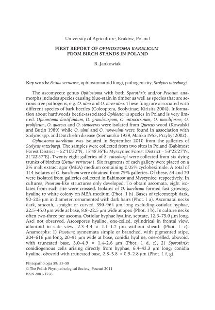
Phot. 1. Ophiostoma <strong>karelicum</strong> on 2% MEA: a – ascoma with neck, b – apex <strong>of</strong> the ascomatal neck,<br />
c – ascospores, d – Pesotum anamorph, synnemata, e – Pesotum anamorph, conidia, f – Sporothrix<br />
anamorph, conidiogenous cells, g – Sporothrix anamorph, conidia, h – 14-day-old colony.<br />
Phots. c, d–f have been done using contrast phase (photo by R. Jankowiak)
Short communications 57<br />
PCR amplicons <strong>of</strong> two isolates (009/O.k, 022/O.k) generated using primers<br />
ITS 1F and ITS 4 were sequenced (White et al. 1990) and found identical to those<br />
<strong>of</strong> O. <strong>karelicum</strong> <strong>from</strong> the GenBank database (Acc. No. EF486693, EF032478).<br />
A preliminary inoculation test was conducted with two randomly chosen isolates<br />
<strong>of</strong> O. <strong>karelicum</strong> obtained and identified in this study (009/O.k, 022/O.k). Inoculations<br />
followed the procedure described by Krokene and Solheim (1998). On the<br />
3 th <strong>of</strong> January 2011, the stems <strong>of</strong> two-year-old B. verrucosa seedlings were superficially<br />
wounded by removing a bark flap (4 × 7 mm). For inoculation, a disc (3 mm<br />
diameter) was cut <strong>from</strong> a margin <strong>of</strong> a 12-day-old culture <strong>of</strong> the test isolates, placed<br />
on each wound and then covered by the bark flap and a Parafilm ® strip. Fifty seedlings<br />
(25 per each isolate) were inoculated with the O. <strong>karelicum</strong>, whereas 25 plants<br />
were inoculated with sterile MEA as control treatment. After 11 weeks <strong>of</strong> incubation,<br />
inoculations with O. <strong>karelicum</strong> resulted in long necrotic lesions in the phloem<br />
and sapwood <strong>of</strong> the test seedlings (Phot. 2 a, b). The average lesion length for O.<br />
<strong>karelicum</strong> was 57.8 mm (18–120 mm) and for control 3.03 (0–7 mm). Both isolates<br />
<strong>of</strong> O. <strong>karelicum</strong> caused blue-stain <strong>of</strong> the sapwood in the two-year-old seedlings. The<br />
average depth <strong>of</strong> sapwood penetration was 1.06 mm (0.2–1.8 mm; Phot. 2 c, d).<br />
None <strong>of</strong> control plants or plants inoculated with O. <strong>karelicum</strong> died during the<br />
11-week-long observation period.<br />
Phot. 2. Pathogenicity test results. Lesions on the surface <strong>of</strong> stem caused by Ophiostoma <strong>karelicum</strong><br />
(a) and control (b). The lesions were observed 11 weeks after inoculation.<br />
Cross section through a <strong>birch</strong> stem in the inoculations points: c – O. <strong>karelicum</strong>, d – control.<br />
The arrow indicates blue-stain in the sapwood (photo by R. Jankowiak)
58 Short communications<br />
This is the <strong>first</strong> finding <strong>of</strong> O. <strong>karelicum</strong> in Poland. It has also been only <strong>report</strong>ed<br />
<strong>from</strong> the same host (Linnakoski et al. 2008), and <strong>from</strong> Pinus sylvestris and Picea abies<br />
in Finland and Russia (Linnakoski et al. 2010). In the present study, O. <strong>karelicum</strong><br />
was unable to cause mortality <strong>of</strong> any <strong>of</strong> the test seedlings. However, this fungus<br />
generated serious necrotic lesions in the phloem and sapwood <strong>of</strong> two-year-old B.<br />
verrucosa seedlings. This suggests that O. <strong>karelicum</strong> is a pathogen with medium level<br />
<strong>of</strong> virulence to B. verrucosa.<br />
Acknowledgements<br />
The author gratefully acknowledges Miroslav Kola ik (Institute <strong>of</strong> Microbiology,<br />
Prague, Czech Republic) for identification <strong>of</strong> O. <strong>karelicum</strong> using molecular<br />
techniques.<br />
Literature<br />
Kirisits T., 2004: Fungal associates <strong>of</strong> European bark beetles with special emphasis on the<br />
<strong>ophiostoma</strong>toid fungi. In: Bark and wood boring insects in living trees in Europe, a synthesis. Eds.<br />
F. Lieutier, K.R. Day, A. Battisti, J.C. Grégoire, H. Evans. Kluwer, Dordrecht: 185–223.<br />
Kowalski T., Butin H., 1989: Taxonomie bekannter und neuer Ceratocystis-Arten an Eiche (Quercus<br />
robur L.). J. Phytopathol. 124: 236–248.<br />
Krokene P., Solheim H., 1998: Assessing the virulence <strong>of</strong> four bark beetle-associated bluestain fungi using<br />
Norway spruce seedlings. Plant Pathol. 47: 537–540.<br />
Linnakoski R., de Beer Z.W., Ahtiainen J., Sidorov E., Niemelä P., Pappinen A., Wingfield M.J., 2010:<br />
Ophiostoma spp. associated with pine- and spruce-infesting bark beetles in Finland and Russia.<br />
Persoonia 25: 72–93.<br />
Linnakoski R., de Beer Z.W., Rousi M., Niemelä P., Pappinen A., Wingfield M.J., 2008: Fungi, including<br />
Ophiostoma <strong>karelicum</strong> sp. nov., associated with Scolytus ratzeburgii infesting <strong>birch</strong> in Finland and<br />
Russia. Mycol. Res. 112: 1475–1488.<br />
Mańka K., 1953: O przebiegu holenderskiej choroby wiązów (Ceratostomella ulmi (Schw.) Buisman) na<br />
terenie miasta Poznania. Acta Soc. Bot. Pol. 22, 2: 355–378.<br />
Przybył K., 2002: Variation <strong>of</strong> Dutch elm disease pathogen in west and north Poland. Phytopathol. Pol.<br />
24: 27–34.<br />
Siemaszko W., 1939: Zespoły grzybów towarzyszących kornikom polskim. Planta Pol. 7, 3.<br />
White T.J., Bruns T., Lee S., Taylor J., 1990: Amplification and direct sequencing <strong>of</strong> fungal ribosomal<br />
RNA genes for phylogenetics. In: PCR protocols: a guide to method and applications. Eds. M.A.<br />
Innis, D.H. Gelfand, J.J. Sninsky, T.J. White. Academic Press, New York: 315–321.<br />
Author’s address:<br />
Dr. Robert Jankowiak, Department <strong>of</strong> Forest Pathology, University <strong>of</strong><br />
Agriculture, Al. 29 Listopada 46, 31-425 Kraków, Poland, e-mail:<br />
rljankow@cyf-kr.edu.pl<br />
Accepted for publication: 12.03.2011