Infection of Protoplasts from Chenopodium quinoa with Cowpea ...
Infection of Protoplasts from Chenopodium quinoa with Cowpea ...
Infection of Protoplasts from Chenopodium quinoa with Cowpea ...

Create successful ePaper yourself
Turn your PDF publications into a flip-book with our unique Google optimized e-Paper software.
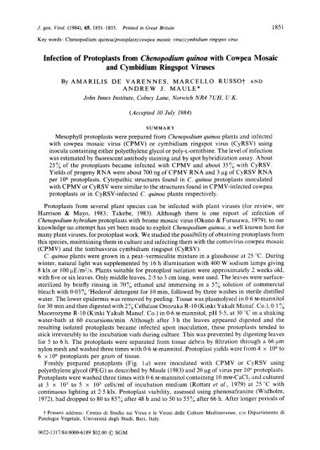
J. gen. Virol. (1984), 65, 1851-1855. Printed in Great Britain<br />
Key words: <strong>Chenopodium</strong> <strong>quinoa</strong>/protoplasts/cowpea mosaic virus~cymbidium ringspot virus<br />
<strong>Infection</strong> <strong>of</strong> <strong>Protoplasts</strong> <strong>from</strong> <strong>Chenopodium</strong> <strong>quinoa</strong> <strong>with</strong> <strong>Cowpea</strong> Mosaic<br />
and Cymbidium Ringspot Viruses<br />
By AMARILIS DE VARENNES, MARCELLO RUSSO'~<br />
ANDREW J. MAULE*<br />
John Innes Institute, Colney Lane, Norwich NR4 7UH, U.K.<br />
(Accepted I0 July 1984)<br />
SUMMARY<br />
AND<br />
Mesophyll protoplasts were prepared <strong>from</strong> <strong>Chenopodium</strong> <strong>quinoa</strong> plants and infected<br />
<strong>with</strong> cowpea mosaic virus (CPMV) or cymbidium ringspot virus (CyRSV) using<br />
inocula containing either polyethylene glycol or poly-L-ornithine. The level <strong>of</strong> infection<br />
was estimated by fluorescent antibody staining and by spot hybridization assay. About<br />
25~ <strong>of</strong> the protoplasts became infected <strong>with</strong> CPMV and about 3590 <strong>with</strong> CyRSV.<br />
Yields <strong>of</strong> progeny RNA were about 700 ng <strong>of</strong> CPMV RNA and 3 ~g <strong>of</strong> CyRSV RNA<br />
per 106 protoplasts. Cytopathic structures found in C. <strong>quinoa</strong> protoplasts inoculated<br />
<strong>with</strong> CPMV or CyRSV were similar to the structures found in CPMV-infected cowpea<br />
protoplasts or in CyRSV-infected C. <strong>quinoa</strong> plants respectively.<br />
<strong>Protoplasts</strong> <strong>from</strong> several plant species can be infected <strong>with</strong> plant viruses (for review, see<br />
Harrison & Mayo, 1983; Takebe, 1983). Although there is one report <strong>of</strong> infection <strong>of</strong><br />
<strong>Chenopodium</strong> hybridum protoplasts <strong>with</strong> brome mosaic virus (Okuno & Furusawa, 1979), to our<br />
knowledge no attempt has yet been made to exploit <strong>Chenopodium</strong> <strong>quinoa</strong>, a well known host for<br />
many plant viruses, for protoplast work. We studied the possibility <strong>of</strong> obtaining protoplasts <strong>from</strong><br />
this species, maintaining them in culture and infecting them <strong>with</strong> the comovirus cowpea mosaic<br />
(CPMV) and the tombusvirus cymbidium ringspot (CyRSV).<br />
C. <strong>quinoa</strong> plants were grown in a peat-vermiculite mixture in a glasshouse at 25 °C. During<br />
winter, natural light was supplemented by 16 h illumination <strong>with</strong> 400 W sodium lamps giving<br />
8 klx or 100 ~tE/mZ/s. Plants suitable for protoplast isolation were approximately 2 weeks old,<br />
<strong>with</strong> five or six leaves. Only middle leaves, 2-5 to 3 cm long, were used. The leaves were surface-<br />
sterilized by briefly rinsing in 70% ethanol and immersing in a 59o solution <strong>of</strong> commercial<br />
bleach <strong>with</strong> 0"03~o 'Hederol' detergent for l0 min, followed by three washes in sterile distilled<br />
water. The lower epidermis was removed by peeling. Tissue was plasmolysed in 0.6 M-mannitol<br />
for 30 min and then digested <strong>with</strong> 2~ Cellulase Onozuka R-10 (Kinki Yakult Manuf. Co.), 0.1 o~<br />
Macerozyme R-10 (Kinki Yakult Manuf. Co.) in 0-6 i-mannitol, pH 5.5, at 30 ~C in a shaking<br />
water-bath at 60 excursions/min. Although after 3 h the leaves appeared digested and the<br />
resulting isolated protoplasts became infected upon inoculation, these protoplasts tended to<br />
stick irreversibly to the incubation vials during culture. This was prevented by digesting leaves<br />
for 5 to 6 h. The protoplasts were separated <strong>from</strong> tissue debris by filtration through a 66 ~tm<br />
nylon mesh and washed three times <strong>with</strong> 0.6 M-mannitol. Protoplast yields were <strong>from</strong> 4 × 10⁶ to<br />
6 × 10⁶ protoplasts per gram <strong>of</strong> tissue.<br />
Freshly prepared protoplasts (Fig. t a) were inoculated <strong>with</strong> CPMV or CyRSV using<br />
polyethylene glycol (PEG) as described by Maule (1983) and 20 ~g <strong>of</strong> virus per 10 ~ protoplasts.<br />
<strong>Protoplasts</strong> were washed three times <strong>with</strong> 0-6 M-mannitol containing 10 mM-CaCI, and cultured<br />
at 3 × 10⁵ to 5 × l05 cells/ml <strong>of</strong> incubation medium (Rottier et al., 1979) at 25 °C <strong>with</strong><br />
continuous lighting at 2-5 klx. Protoplast viability, assessed using phenosafranine (Widholm,<br />
1972), had dropped to 80 to 85% after 48 h and to 50 to 55% after 66 h. After longer periods <strong>of</strong><br />
t Present address: Centro di Studio sui Virus e le Virosi delle Colture Mediterranee, c/o Dipartimento di<br />
Patologia Vegetale, Universitfi degli Studi, Bari, Italy.<br />
0022-1317/84/0000-6189 $02.00 © SGM<br />
1851
1852 Short communication<br />
Fig. 1, (a) C. <strong>quinoa</strong> protoplasts resuspended in 0.6 M-mannitol. (b, c) Fluorescence micrographs <strong>of</strong> C.<br />
<strong>quinoa</strong> protoplasts infected <strong>with</strong> CyRSV (b) or CPMV (c) and stained <strong>with</strong> fluorescent antibody. (d, e)<br />
Electron micrographs <strong>of</strong> C. <strong>quinoa</strong> protoplasts infected <strong>with</strong> (d) CPMV, showing part <strong>of</strong> an inclusion<br />
body (IB), and (e) CyRSV, showing two multivesicular bodies (MVB) and a vesiculated mitochondrion<br />
(inset). Bar markers represent 20 ~tm for (a) and (b), 15 ~tm for (c) and 200 nm for (d), (e) and inset.<br />
incubation the protoplasts were extensively clumped. Virus infection was assessed by indirect<br />
fluorescent antibody staining (Maule et al., 1980) and by spot hybridization (Thomas, 1980).<br />
C. <strong>quinoa</strong> protoplasts could also be infected <strong>with</strong> CPMV and CyRSV when poly-t-ornithine<br />
(PLO) was included in the inocula. Under the conditions tested (1 rtg/ml PLO, 10 ~tg/ml virus<br />
and 10 raM-potassium citrate, pH 5-2, in 0.6 M-mannitol), the proportion <strong>of</strong> protoplasts infected<br />
using inocula containing PLO was similar to that using inocuta containing PEG. Generally, the<br />
PEG method was used in preference because <strong>of</strong> its simplicity.<br />
The proportion <strong>of</strong> protoplasts stained <strong>with</strong> fluorescent antibody increased <strong>from</strong> 1 to 5°/o 12 h<br />
post-inoculation to about 25~ 36 to 60 h post-inoculation for CPMV, and <strong>from</strong> 15~ at 12 h to<br />
about 35~o 48 to 60 h post-inoculation for CyRSV, The appearance <strong>of</strong> the fluorescence in the
(a) ~ ~ ~"<br />
Short communication 1853<br />
0.8<br />
O<br />
0 ~ 0.6<br />
12 O<br />
2 4 0 0<br />
×<br />
~0-4<br />
36 Oe<br />
z<br />
(b)<br />
I I I I<br />
I -4<br />
--4 3<br />
48 O e >0-2 ~<br />
rj<br />
60<br />
I<br />
0 12 24 36 48 60<br />
Time post-inoculation (h)<br />
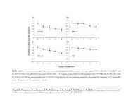
Fig. 2. Spot hybridization assay <strong>of</strong> CPMV and CyRSV RNA in extracts <strong>of</strong> infected protoplasts.<br />
Variation in the concentration <strong>of</strong> viral RNA is shown at various times (h) after inoculation <strong>with</strong> CPMV<br />
(T) or CyRSV (11), as an autoradiograph (a) or as a graphical representation (b) using the calculated<br />
concentration <strong>of</strong> viral RNA. Values are the mean <strong>of</strong> duplicate spots. Each spot is equivalent to 2.5 ×<br />
105 protoplasts.<br />
stained protoplasts differed strikingly between infections by the two viruses. Fluorescing<br />
material in protoplasts infected <strong>with</strong> CyRSV was diffusely spread in the cytoplasm forming a<br />
network between the chloroplasts (Fig. 1 b). In protoplasts infected <strong>with</strong> CPMV, fluorescent<br />
masses were localized in discrete areas <strong>of</strong> the cytoplasm (Fig. 1 c) and only occasionally, late in<br />
infection, was the fluorescence distributed throughout the cytoplasm as described for CPMV-<br />
infected cowpea protoplasts by Hibi et al. (1975).<br />
RNA was extracted <strong>from</strong> infected protoplasts disrupted in the presence <strong>of</strong> 5~ Sarkosyl<br />
(Sigma) in 40 mM-Tris-HC1, 80 mM-NaC1, 4 mM-EDTA, pH 8.5, by treatment <strong>with</strong> a buffer-<br />
saturated mixture <strong>of</strong> phenol and chlor<strong>of</strong>orm (8 : 1, v/v) and recovered by ethanol precipitation.<br />
When RNA was spotted onto nitrocellulose filters (Thomas, 1980) and probed <strong>with</strong> 32p-labelled<br />
cDNA (Maule et al., 1983) prepared to each viral RNA by random priming (Taylor et al., 1976),<br />
it was observed that the accumulation <strong>of</strong> viral RNA paralleled the accumulation <strong>of</strong> virus antigen<br />
(as assessed by fluorescent antibody staining). For quantification <strong>of</strong> viral RNA, the radioactive<br />
areas were cut <strong>from</strong> the nitrocellulose filters, assayed by scintillation counting, and compared<br />
<strong>with</strong> blots <strong>of</strong> known quantities <strong>of</strong> purified RNA. As shown in Fig. 2, CPMV-infected<br />
protoplasts contained about 700 ng <strong>of</strong> progeny viral RNA per 106 protoplasts and CyRSV-in-<br />
fected protoptasts, more than 3 ~tg <strong>of</strong> progeny RNA per 106 protoplasts. The molecular weight<br />
distribution <strong>of</strong> virus RNA detected in this assay was assessed using Northern blots (Thomas,<br />
1980). RNA extracted <strong>from</strong> protoplasts was denatured <strong>with</strong> 70~ (w/v) formamide in the pres-<br />
ence <strong>of</strong> t M-glyoxal (Covey et aL, 1983) and electrophoresed in 1X agarose gels. After<br />
electrophoresis the gels were blotted onto nitrocellulose filters and probed <strong>with</strong> 32p-labelled<br />
cDNA. Both species <strong>of</strong> CPMV RNA (B-RNA and" M-RNA) were detected in RNA <strong>from</strong><br />
CPMV-infected protoplasts (Fig. 3 a); a major species, corresponding to the genomic RNA, was<br />
detected in RNA <strong>from</strong> protoplasts infected <strong>with</strong> CyRSV (Fig. 3b), together <strong>with</strong> smaller species<br />
whose significance is at present under investigation (D. Gallitelli, personal communication).<br />
Samples <strong>of</strong> healthy and infected protoplasts were collected by centrifugation, resuspended in<br />
4~ glutaraldehyde in 0.05 M-phosphate buffer pH 7.0, containing 0.6M-mannitol, and<br />
processed for electron microscopy essentially as described by Martelli & Russo (1981). Thin<br />
sections <strong>of</strong> CPMV-infected protoplasts (Fig. I d) showed the presence <strong>of</strong> the cytopathic structure<br />
typical <strong>of</strong> infections by this virus both in cowpea mesophyll tissue (De Zoeten et al., 1974) and<br />
protoplasts (Hibi et al., 1975). In thin sections <strong>of</strong> CyRSV-infected protoplasts (Fig. l e)<br />
e~<br />
X<br />
<<br />
2;
1854 Short communication<br />
(a) 0 12 24 36 48 60 72 H (b) H 0 12 24 36 48<br />
M~<br />
il ,," '<br />
g<br />
L<br />
RNA<br />
Fig. 3. Northern blot <strong>of</strong> CPMV RNA (a) or CyRSV RNA (b) in infected protoplasts, taken at various<br />
times (h) after inoculation. H, Healthy protoplasts. Positions <strong>of</strong> the genomic RNAs <strong>of</strong> CPMV (B, M)<br />
and CyRSV (arrow) are marked.<br />
peroxisomes appeared to have been transformed into multivesicular bodies (MVB), a<br />
characteristic cytopathological feature <strong>of</strong> infections by this virus (Russo et aI., 1983). Also in<br />
these protoplasts, a small proportion <strong>of</strong> the mitochondria contained fibril-containing vesicles<br />
between the two mitochondrial membranes (Fig. 1 e, inset), a feature peculiar to CyRSV<br />
infection <strong>of</strong> C. <strong>quinoa</strong>, also found in leaf tissue, but not in several other plants infected <strong>with</strong> the<br />
same virus (A. Di Franco & M. Russo, unpublished results).<br />
In conclusion, we have shown that protoplasts <strong>from</strong> C. <strong>quinoa</strong> can be used for virus infection<br />
studies, including those <strong>of</strong> CyRSV for which C. <strong>quinoa</strong> is a local lesion host. C. <strong>quinoa</strong> protoplasts<br />
are easily produced and are stable. They look promising as an experimental system to be tried<br />
<strong>with</strong> other viruses belonging to different groups.<br />
We thank Dr J. Davies for helpful discussions. A. de Varennes holds a studentship <strong>from</strong> the National Institute<br />
<strong>of</strong> Scientific Research, Portugal.<br />
REFERENCES<br />
COVEY, S. N., TURNER, D. & MULDER, G. (1983). A small DNA molecule containing covalentty-linked<br />
ribonucleotides originates <strong>from</strong> the large intergenic region <strong>of</strong> the cauliflower mosaic virus genome. Nucleic<br />
Acids Research 11, 251-264.<br />
DE ZOETEN, G, A., ASSINK, A. M. & VAN KAMMEN, A. (1974). Association <strong>of</strong> cowpea mosaic virus induced double-<br />
stranded RNA <strong>with</strong> cytopathic structures in infected cells. Virology 59, 341-355.<br />
HARRISON, B. D. & MAYO, M. A. (1983). The use <strong>of</strong> protoplasts in plant virus research. In Use <strong>of</strong> Tissue Culture and<br />
<strong>Protoplasts</strong> in Plant Pathology, pp. 69-137. Edited by J. P. Helgeson & B. J. Deverall. London: Academic<br />
Press.<br />
HIBI, T., REZELMAN, G. & VAN KAMMEN, A. (1975). <strong>Infection</strong> <strong>of</strong> cowpea mesophyll protoplasts <strong>with</strong> cowpea mosaic<br />
virus. Virology 64, 308-318.<br />
MARTELLI, . P. & RUSSO, M. (1981). The fine structure <strong>of</strong> Cymbidium ringspot virus in host tissues. I. Electron<br />
microscopy <strong>of</strong> systemic infections. Journal <strong>of</strong> Ultrastructure Research 77, 93-104.<br />
MAULE, A. J. (1983). <strong>Infection</strong> <strong>of</strong> protoplasts <strong>from</strong> several Brassica species <strong>with</strong> cauliflower mosaic virus following<br />
inoculation using polyethylene glycol. Journal <strong>of</strong> General Virology 64, 2655-2660.<br />
MAULE, A, J., BOULTON, M. L & WOOD, K. R. (1980). An improved method for the infection <strong>of</strong> cucumber leaf<br />
protoplasts <strong>with</strong> cucumber mosaic virus. Phytopathologische Zeitschrifi 97, 118=126.<br />
MAULE, A. J., HULL, R. & DONSON, J. (1983). The application <strong>of</strong> spot hybridization to the detection <strong>of</strong> DNA and<br />
RNA viruses in plant tissues. Journal <strong>of</strong> Virological Methods 6, 215-224.<br />
OKUNO, T. & FURUSAWA, I. (1979). RNA polymerase activity and protein synthesis in brome mosaic virus-infected<br />
protoplasts. Virology 99, 2t8-225.<br />
ROTTIER, P. J. M., REZELMAN, G. & VAN KAMMEN, A. (1979). The inhibition <strong>of</strong> cowpea mosaic virus replication by<br />
actinomycin D. Virology 92, 299-309.
Short communication 1855<br />
RUSSO, M., DI FRANCO, A. & MARTELLI, G. P. (1983). The fine structure <strong>of</strong> Cymbidium ringspot virus infections in host<br />
tissues. III. Role <strong>of</strong> peroxisomes in the genesis <strong>of</strong> multivesicular bodies. Journal <strong>of</strong> Ultrastructure Research 82,<br />
52-63.<br />
TAKEBE, I. (1983). <strong>Protoplasts</strong> in plant virus research. International Reriew <strong>of</strong> Cytology suppl. 16, 89 111.<br />
TAYLOR, J. M., ILLMENSEE, R. & SUMMERS, J. (1976). Efficient transcription <strong>of</strong> RNA into DNA by avian sarcoma<br />
virus polymerase. Biochimica et biophysica aeta 442, 324-330.<br />
THOMAS, P. S. (1980). Hybridization <strong>of</strong> denatured RNA and small DNA fragments transferred to nitrocellulose.<br />
Proceedings <strong>of</strong> the National Academy <strong>of</strong> Sciences, USA 77, 5201-5205.<br />
WIDHOLM, J. M. (1972). The use <strong>of</strong> fluorescein diacetate and phenosafranine for determining the viability <strong>of</strong><br />
cultured plants cells. Stain Technology 47, 189 194.<br />
(Received 14 May 1984)