Page 1 of 22 The vaccinia virus A56 protein: a multifunctional ...
Page 1 of 22 The vaccinia virus A56 protein: a multifunctional ...
Page 1 of 22 The vaccinia virus A56 protein: a multifunctional ...

You also want an ePaper? Increase the reach of your titles
YUMPU automatically turns print PDFs into web optimized ePapers that Google loves.
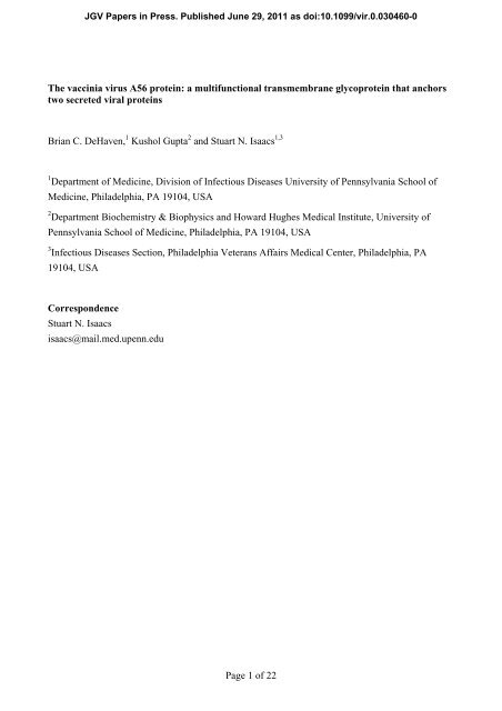
<strong>The</strong> <strong>vaccinia</strong> <strong>virus</strong> <strong>A56</strong> <strong>protein</strong>: a <strong>multifunctional</strong> transmembrane glyco<strong>protein</strong> that anchors<br />
two secreted viral <strong>protein</strong>s<br />
Brian C. DeHaven, 1 Kushol Gupta 2 and Stuart N. Isaacs 1,3<br />
1 Department <strong>of</strong> Medicine, Division <strong>of</strong> Infectious Diseases University <strong>of</strong> Pennsylvania School <strong>of</strong><br />
Medicine, Philadelphia, PA 19104, USA<br />
2 Department Biochemistry & Biophysics and Howard Hughes Medical Institute, University <strong>of</strong><br />
Pennsylvania School <strong>of</strong> Medicine, Philadelphia, PA 19104, USA<br />
3 Infectious Diseases Section, Philadelphia Veterans Affairs Medical Center, Philadelphia, PA<br />
19104, USA<br />
JGV Papers in Press. Published June 29, 2011 as doi:10.1099/vir.0.030460-0<br />
Correspondence<br />
Stuart N. Isaacs<br />
isaacs@mail.med.upenn.edu<br />
<strong>Page</strong> 1 <strong>of</strong> <strong>22</strong>
<strong>The</strong> <strong>vaccinia</strong> <strong>virus</strong> <strong>A56</strong> <strong>protein</strong> was one <strong>of</strong> the earliest-described pox<strong>virus</strong> <strong>protein</strong>s<br />
with an identifiable activity. While originally characterized as a haemagglutinin<br />
<strong>protein</strong>, <strong>A56</strong> has other functions as well. <strong>A56</strong> is capable <strong>of</strong> binding two viral<br />
<strong>protein</strong>s, a serine protease inhibitor (K2) and the <strong>vaccinia</strong> <strong>virus</strong> complement<br />
control <strong>protein</strong> (VCP), and anchoring them to the surface <strong>of</strong> infected cells. This is<br />
important; while both <strong>protein</strong>s have biologically relevant functions at the cell<br />
surface, neither one can locate there on its own. <strong>The</strong> <strong>A56</strong>–K2 complex reduces the<br />
amount <strong>of</strong> <strong>virus</strong> superinfecting an infected cell and also prevents the formation <strong>of</strong><br />
syncytia by infected cells; the <strong>A56</strong>–VCP complex can protect infected cells from<br />
complement attack. Deletion <strong>of</strong> the <strong>A56</strong>R gene results in varying effects on<br />
<strong>vaccinia</strong> <strong>virus</strong> virulence. In addition, since the gene encoding the <strong>A56</strong> <strong>protein</strong> is<br />
non-essential, it can be used as an insertion point for foreign genes and has been<br />
deleted in some <strong>virus</strong>es that are in clinical development as oncolytic agents.<br />
<strong>Page</strong> 2 <strong>of</strong> <strong>22</strong>
Introduction – the <strong>A56</strong> <strong>protein</strong><br />
Orthopox<strong>virus</strong>es are some <strong>of</strong> the most complex <strong>virus</strong>es infecting humans and include variola <strong>virus</strong>,<br />
the causative agent <strong>of</strong> smallpox, and <strong>vaccinia</strong> <strong>virus</strong>, which is used as a live vaccine. Although<br />
smallpox has been eradicated, <strong>vaccinia</strong> <strong>virus</strong> (VACV) is still studied as a model organism to<br />
understand basic aspects <strong>of</strong> pox virology, as a vaccine vector for immunizations against other<br />
infectious agents and for oncolytic cancer therapy. It is also studied because human infections with<br />
zoonotic pox<strong>virus</strong>es like monkeypox <strong>virus</strong> still occur. While VACV is a large <strong>virus</strong>, containing a<br />
genome <strong>of</strong> nearly 200 kbp that encodes more than 200 <strong>protein</strong>s, the genome is still small compared<br />
with that <strong>of</strong> the host cell. For this reason, VACV encodes a number <strong>of</strong> multi-functional <strong>protein</strong>s,<br />
one <strong>of</strong> which is <strong>A56</strong>. <strong>The</strong> <strong>A56</strong> <strong>protein</strong> is able to bind two other viral <strong>protein</strong>s, a serine protease<br />
inhibitor (K2) and the <strong>vaccinia</strong> <strong>virus</strong> complement-control <strong>protein</strong> (VCP), and express them at the<br />
surface <strong>of</strong> the infected cell. <strong>The</strong> <strong>A56</strong>–K2 complex binds to the entry–fusion machinery <strong>of</strong> VACV;<br />
this reduces superinfection and prevents cell–cell fusion <strong>of</strong> infected cells. <strong>The</strong> <strong>A56</strong>–VCP complex<br />
protects infected cells from complement attack and contributes to viral virulence. <strong>The</strong>se complexes<br />
were only discovered recently, but the history <strong>of</strong> <strong>A56</strong> <strong>protein</strong> studies extends much farther back in<br />
time.<br />
<strong>The</strong> <strong>A56</strong> <strong>protein</strong> was one <strong>of</strong> the first VACV genes to be identified and studied (Nagler, 1942). This<br />
was because what is now called the <strong>A56</strong> <strong>protein</strong> had haemagglutination activity; it was called<br />
VACV haemagglutinin (HA) from the 1940s until near the end <strong>of</strong> the 20th century. <strong>The</strong> presence <strong>of</strong><br />
a VACV <strong>protein</strong> with HA activity was believed to be important because several other <strong>virus</strong>es such<br />
as influenza, measles and mumps <strong>virus</strong>es contained <strong>protein</strong>s with HA activity. However, unlike<br />
these other viral envelope <strong>protein</strong>s, which were important for viral entry, a biologically relevant<br />
function could not ultimately be ascribed to the VACV HA activity. Nevertheless, the HA activity<br />
<strong>of</strong> pox<strong>virus</strong>es allowed the classification <strong>of</strong> pox<strong>virus</strong>es into those that had HA activity and those that<br />
did not. Members <strong>of</strong> the orthopox<strong>virus</strong> genus were the only ones that had HA activity (Fenner et al.,<br />
1988). Furthermore, haemabsorption and HA-inhibition assays were early methods used to<br />
differentiate between various orthopox<strong>virus</strong>es and variola <strong>virus</strong> isolates from various parts <strong>of</strong> the<br />
world (Fenner et al., 1988). Later, when the <strong>protein</strong> (now called ‘<strong>A56</strong>’) and the gene (designated<br />
‘<strong>A56</strong>R’ but also designated VACWR181 in VACV strain WR) responsible for this activity was first<br />
identified (Ichihashi, 1977; Shida, 1986a), the <strong>A56</strong>R ORF was used to build phylogenetic trees that<br />
showed the genetic relationships between the members <strong>of</strong> the genus Orthopox<strong>virus</strong> (Hutin et al.,<br />
2001).<br />
<strong>The</strong> ability to track the <strong>protein</strong> by its haemagglutinating activity also provided a means to show that<br />
the <strong>protein</strong> is found on the cell membrane <strong>of</strong> the infected cell (Blackman & Bubel, 1972), but not on<br />
mature <strong>virus</strong> (MV), the most abundant form <strong>of</strong> <strong>virus</strong> made during an infection (Fig. 1). While the<br />
<strong>protein</strong> is not found on MV, it is present on another infectious form <strong>of</strong> <strong>virus</strong> called the extracellular<br />
<strong>Page</strong> 3 <strong>of</strong> <strong>22</strong>
<strong>virus</strong> (EV) (Payne & Norrby, 1976). <strong>The</strong> EV is critical for the efficient spread <strong>of</strong> the <strong>virus</strong> in vitro<br />
and in vivo. However, unlike the other glyco<strong>protein</strong>s found on EV, the <strong>A56</strong> <strong>protein</strong> has considerably<br />
more sequence variability between different orthopox<strong>virus</strong>es. This may be because, unlike the other<br />
EV glyco<strong>protein</strong>s, <strong>A56</strong> is not essential for formation <strong>of</strong> the EV. As mentioned earlier, this sequence<br />
variability, initially recognized in HA-inhibition assays, made <strong>A56</strong>R a useful ORF for building<br />
phylogenetic trees.<br />
Based on the <strong>A56</strong> <strong>protein</strong> sequence, the calculated molecular mass <strong>of</strong> the <strong>protein</strong> is ~35 kDa, but<br />
the <strong>protein</strong> contains putative sites for both N- and O-glycosylation and runs at an apparent<br />
molecular mass <strong>of</strong> 85 kDa (Brown et al., 1991; Payne, 1992; Shida & Dales, 1981). Thus, <strong>A56</strong> is<br />
heavily glycosylated and this glycosylation is linked to its HA activity. <strong>The</strong> positions <strong>of</strong> the five<br />
predicted N-linked glycosylation sites are shown in Fig. 2. Blocking N-linked glycosylation with<br />
tunicamycin results in an apparent molecular mass <strong>of</strong> 62 kDa and this moiety retains its HA<br />
activity. In the presence <strong>of</strong> tunicamycin, <strong>A56</strong> is still able to incorporate labelled glucosamine,<br />
confirming that the <strong>protein</strong> also undergoes O-linked glycosylation. Because HA activity is lost<br />
when the <strong>protein</strong> is completely deglycosylated, this implies that O-linked glycans are involved in<br />
the HA activity (Shida & Dales, 1981). A prior review on <strong>A56</strong> by Hisatoshi Shida (Shida, 1989)<br />
pointed out that <strong>A56</strong> was the first viral <strong>protein</strong> shown to be O-glycosylated, and that this <strong>protein</strong><br />
provided additional evidence that O-linked glycosylation occurs as a <strong>protein</strong> transits through the<br />
Golgi apparatus (Shida & Dales, 1981; Spiro, 1966). <strong>The</strong>re are predicted to be as many as 23 Olinked<br />
glycosylation sites, which all fall between residues 149 and 255 (with 20 falling between<br />
residues 175 and 230). <strong>The</strong> heavy glycosylation <strong>of</strong> the <strong>A56</strong> <strong>protein</strong> may be the reason why anti-<strong>A56</strong><br />
antibodies cannot neutralize EV or protect from infection (Galmiche et al., 1999; Pulford et al.,<br />
2004). Interestingly, completely unglycosylated <strong>A56</strong> <strong>protein</strong> migrates with an apparent molecular<br />
mass <strong>of</strong> 58 kDa; this is different from the 35 kDa calculated from the sequence <strong>of</strong> the ORF (Shida,<br />
1986b). Shida pointed out that this discrepancy is not because <strong>of</strong> other post-translational<br />
modifications because in vitro translation <strong>of</strong> the <strong>A56</strong> ORF resulted in a <strong>protein</strong> that migrates at ~60<br />
kDa (Shida, 1989). Thus, this is a clear example <strong>of</strong> a difference between predicted and observed<br />
molecular masses, <strong>of</strong> the type that are known to occur. Additionally, the <strong>A56</strong>R gene has two<br />
separate promoters for early and late gene expression. Interestingly, late expression also produces a<br />
smaller, 68 kDa form <strong>of</strong> the <strong>protein</strong> (Brown et al., 1991), although the biological significance <strong>of</strong> this<br />
smaller <strong>protein</strong> is not known. Both the 85 and 68 kDa forms <strong>of</strong> the <strong>protein</strong> can be seen by using<br />
Western blotting at late time points.<br />
A schematic diagram <strong>of</strong> the <strong>A56</strong> <strong>protein</strong> is shown in Fig. 2. <strong>The</strong> ~100 aa N-terminal region <strong>of</strong> <strong>A56</strong><br />
(after the signal sequence) has sequence similarities with the immunoglobulin (Ig) superfamily.<br />
This region <strong>of</strong> the <strong>protein</strong> is more highly conserved among orthopox<strong>virus</strong>es than the rest <strong>of</strong> the <strong>A56</strong><br />
ectodomain (Aguado et al., 1992; Cavallaro & Esposito, 1992). In this Ig domain, it was<br />
<strong>Page</strong> 4 <strong>of</strong> <strong>22</strong>
hypothesized that the two cysteines in the ectodomain form an intramolecular disulfide bridge (Jin<br />
et al., 1989). Following on from this, modern computer modelling <strong>of</strong> this portion <strong>of</strong> the <strong>A56</strong> <strong>protein</strong><br />
supports formation <strong>of</strong> an Ig domain structure, shows the first two cysteines <strong>of</strong> <strong>A56</strong> to be in close<br />
proximity and predicts that these cysteines form an intramolecular bond (Fig. 3). While not<br />
experimentally proven, but based on phenotypes <strong>of</strong> mutated <strong>virus</strong>es, the Ig domain may be involved<br />
in the observed cell–cell fusion regulatory activity. An analysis <strong>of</strong> VACV mutants produced by<br />
chemical mutagenesis (Shida & Matsumoto, 1983) revealed that the cell–cell fusion regulatory<br />
properties and haemabsorption properties <strong>of</strong> the <strong>A56</strong> <strong>protein</strong> are separate (Seki et al., 1990). While<br />
<strong>A56</strong>R gene-knockout <strong>virus</strong>es cannot perform either function, one point mutant (Glu121 to Lys) was<br />
found to be HA negative but could still prevent syncytia formation. Since the exact mechanism <strong>of</strong><br />
haemagglutination is not known, it is difficult to speculate as to why this mutation causes a loss <strong>of</strong><br />
activity, especially when the O-linked glycosylation was linked to HA activity. Additionally, a <strong>virus</strong><br />
containing a Cys103 to Tyr mutation did not have HA activity or the ability to prevent syncytia. As<br />
discussed earlier, this cysteine is predicted to form an intramolecular disulfide bond (Figs 2 and 3),<br />
so this mutation is likely to destabilize the entire domain, resulting in an abnormally folded <strong>protein</strong><br />
that has been reported to make it to the cell surface, but which does not have HA activity or prevent<br />
syncytia formation (Seki et al., 1990). As discussed later, a third cysteine at position 162 (just<br />
before the first tandem repeat) has recently been shown to form a disulfide bridge with VCP<br />
(DeHaven et al., 2010). Between the Ig domain and the transmembrane domain there are two<br />
tandem-repeat motifs, which start near residue 170 and continue until residue 238 (Jin et al., 1989).<br />
<strong>The</strong>se repeats, which are predicted to be heavily modified by O-glycosylation, do not share<br />
sequence similarity with other <strong>protein</strong>s. While the function <strong>of</strong> the tandem repeats in the <strong>A56</strong> <strong>protein</strong><br />
are unknown, tandem repeats are <strong>of</strong>ten important in <strong>protein</strong> structure and function. <strong>The</strong><br />
transmembrane domain, which anchors the <strong>protein</strong> in membranes, has been implicated as an<br />
important domain that results in <strong>A56</strong> interacting with the F13 <strong>protein</strong> (also called VP37) (Oie et al.,<br />
1990). Since the F13 <strong>protein</strong> is a key <strong>protein</strong> in the formation <strong>of</strong> EV (Blasco & Moss, 1991), F13<br />
may help direct the incorporation <strong>of</strong> <strong>A56</strong> into the EV envelope. A short intracellular domain <strong>of</strong> ~12<br />
aa that is present at the C terminus <strong>of</strong> the <strong>protein</strong> may be responsible for trafficking <strong>A56</strong> <strong>protein</strong> out<br />
<strong>of</strong> the endoplasmic reticulum (ER) and into the Golgi apparatus (Shida & Matsumoto, 1983).<br />
Analysis <strong>of</strong> a panel <strong>of</strong> <strong>virus</strong>es with various HA and syncytia-inducing phenotypes revealed that an<br />
<strong>A56</strong> <strong>protein</strong> lacking the cytoplasmic tail was transported from the ER to the Golgi apparatus more<br />
slowly but could still make it to the cytoplasmic membrane, while a <strong>protein</strong> with an aberrant<br />
cytoplasmic tail [owing to an upstream nucleotide insertion(s)] interfered with transport <strong>of</strong> the<br />
<strong>protein</strong> out <strong>of</strong> the ER.<br />
<strong>Page</strong> 5 <strong>of</strong> <strong>22</strong>
<strong>A56</strong> <strong>protein</strong> inhibits spontaneous cell–cell fusion <strong>of</strong> VACV infected cells<br />
by forming a complex with K2<br />
<strong>The</strong> ability to haemagglutinate was not the only early function ascribed to <strong>A56</strong>. Cells infected with<br />
certain strains <strong>of</strong> VACV can result in cell–cell fusion, and in 1971 this was directly linked to a lack<br />
<strong>of</strong> <strong>A56</strong> <strong>protein</strong> (Ichihashi & Dales, 1971). Interestingly, the loss <strong>of</strong> another viral <strong>protein</strong>, K2, also<br />
causes infected cells to fuse. This was reported by three different groups in 1992 (Law & Smith,<br />
1992; Turner & Moyer, 1992; Zhou et al., 1992). Recently, it has been shown that the <strong>A56</strong> and K2<br />
<strong>protein</strong>s form a complex on the surface <strong>of</strong> infected cells, and that this complex is responsible for<br />
preventing syncytia formation (Turner & Moyer, 2006). <strong>The</strong> K2 <strong>protein</strong> is also found on EV<br />
particles, as observed by electron microscopy and proteomics studies (Brum et al., 2003; Manes et<br />
al., 2008). K2, also known as serine protease inhibitor (SPI)-3, is one <strong>of</strong> a family <strong>of</strong> pox<strong>virus</strong><br />
<strong>protein</strong>s that share sequence similarities with serpin <strong>protein</strong>s (Boursnell et al., 1988). K2 <strong>protein</strong><br />
(SPI-3) has in vitro protease-inhibition activity (Turner et al., 2000; Wang et al., 2000), but this<br />
activity is not related to its ability to prevent syncytia formation (Turner & Moyer, 1995). While<br />
regions <strong>of</strong> K2 that are important for its interaction with <strong>A56</strong> have been identified (Turner & Moyer,<br />
1995), the domain <strong>of</strong> <strong>A56</strong> that interacts with K2 has not been identified. In a transient transfection<br />
system, <strong>A56</strong> mutated at either Cys34 or Cys103 was able to be expressed on the cell surface (and<br />
bind VCP) (DeHaven et al., 2010) but could not bind K2 (B. C. DeHaven & S. N. Isaacs,<br />
unpublished), again implying that the Ig domain is involved in interacting with K2.<br />
<strong>The</strong> mechanism by which the <strong>A56</strong>/K2 complex inhibits spontaneous fusion <strong>of</strong> infected cells was not<br />
clear until the discovery that the <strong>virus</strong> encoded multi-subunit entry fusion complex (EFC), which<br />
was found on the MV membrane (Senkevich et al., 2005), interacted with the <strong>A56</strong>–K2 complex<br />
(Wagenaar & Moss, 2007). It was further shown that the <strong>A56</strong>–K2 complex interacts directly with<br />
the A16 and G9 <strong>protein</strong>s, two <strong>of</strong> the <strong>protein</strong>s that are part <strong>of</strong> the EFC (Wagenaar et al., 2008). This<br />
finding led to the hypothesis that the <strong>A56</strong>–K2 complex on infected cells could prevent reinfection<br />
<strong>of</strong> already infected cells (Moss, 2006). Support for this hypothesis was obtained experimentally<br />
when recombinant VACVs with deletions <strong>of</strong> one <strong>of</strong> the genes encoding the <strong>A56</strong>–K2 complex were<br />
used to infect cells, followed by superinfection with VACV carrying a luciferase reporter gene.<br />
Superinfection <strong>of</strong> cells that were infected by wild-type <strong>virus</strong> expressing the <strong>A56</strong>–K2 complex had<br />
significantly lower luciferase levels than cells infected with a <strong>virus</strong> expressing a mutated <strong>A56</strong>–K2<br />
complex (Turner & Moyer, 2008). It has also been shown that transfecting the <strong>A56</strong>R and K2L genes<br />
into cells is sufficient to diminish both infection and virion induced cell–cell fusion (Wagenaar &<br />
Moss, 2009). Antibodies against either the K2 (Turner & Moyer, 2006, 2008) or <strong>A56</strong> <strong>protein</strong>s<br />
(Wagenaar & Moss, 2009) increase the amount <strong>of</strong> superinfection and cell fusion seen. This may be<br />
because <strong>of</strong> the antibodies either blocking the <strong>A56</strong>–K2 complex from interacting with the MV EFC<br />
or perhaps by causing the displacement <strong>of</strong> K2 from <strong>A56</strong>, rendering the complex non-functional.<br />
<strong>Page</strong> 6 <strong>of</strong> <strong>22</strong>
Since syncytia formation <strong>of</strong> infected cells is mediated by EV, it appears that the <strong>A56</strong>—K2 complex<br />
is inhibiting syncytia formation by ‘inhibiting’ EV re-entry into an infected cell. This would occur<br />
first by the non-fusogenic dissolution <strong>of</strong> the EV outer envelope (Law et al., 2006) that exposes the<br />
EFC machinery on the MV, and then entry into an already infected cell is inhibited by engagement<br />
with <strong>A56</strong>–K2. Many other <strong>virus</strong>es have evolved mechanisms to prevent superinfection prior to<br />
entry (Berngruber et al., 2010). For example, the influenza neuraminidase <strong>protein</strong> cleaves the entry<br />
receptor as new <strong>virus</strong> is released, preventing superinfection (Huang et al., 2008), while retro<strong>virus</strong>es<br />
can downregulate entry receptors (Lindwasser et al., 2007; Wildum et al., 2006). Other <strong>virus</strong>es,<br />
such as vesicular stomatitis <strong>virus</strong>, prevent superinfection by slowing endocytosis <strong>of</strong> new <strong>virus</strong><br />
(Simon et al., 1990). Thus, while other <strong>virus</strong>es prevent superinfection prior to entry, none use a<br />
mechanism that actually interferes with their own entry <strong>protein</strong>s like orthopox<strong>virus</strong>es.<br />
<strong>The</strong> <strong>A56</strong>–K2 complex on the surface <strong>of</strong> infected cells prevents superinfection by incoming MV<br />
particles. This should not be confused with the ability <strong>of</strong> another set <strong>of</strong> pox<strong>virus</strong> <strong>protein</strong>s (A33 and<br />
A36) on infected cells that repel EV away from newly infected cells (Doceul et al., 2010). By<br />
repelling EV away from a newly infected cell, <strong>virus</strong> spread to distant uninfected cells is enhanced.<br />
Thus, VACV has evolved a way to prevent or at least reduce superinfection by MV and EV prior to<br />
<strong>virus</strong> entry. Given the vital role that EV plays in the spread <strong>of</strong> <strong>virus</strong> in vitro and in vivo, we<br />
postulate that the repulsion <strong>of</strong> EV away from newly infected cells has a more important function<br />
than the <strong>A56</strong>–K2 complex preventing superinfection by MV. However, the <strong>A56</strong>–K2 complex may<br />
play a valuable role early in the initial spread <strong>of</strong> <strong>virus</strong> away from the very first cells infected.<br />
<strong>The</strong> interaction <strong>of</strong> <strong>A56</strong> and K2 has been shown to occur in VACV and cowpox <strong>virus</strong> (CPXV), and<br />
homologues <strong>of</strong> both <strong>protein</strong>s are present in all sequenced members <strong>of</strong> the genus Orthopox<strong>virus</strong>.<br />
<strong>The</strong>re was a curious report <strong>of</strong> an ectromelia <strong>virus</strong> (ECTV) that caused syncytia formation despite<br />
the presence <strong>of</strong> the ECTV homologues <strong>of</strong> <strong>A56</strong> and K2 (Erez et al., 2009). However, cell–cell fusion<br />
is a complicated process, and other factors may be at work. <strong>The</strong> phenotype described for this ECTV<br />
might be explained by the presence <strong>of</strong> mutations in other viral genes. Also <strong>of</strong> note is that Chang et<br />
al. have recently shown that MV surface <strong>protein</strong>s A25 and A26 are fusion suppressors (Chang et<br />
al., 2010). <strong>The</strong>y found that mutated VACVs that contain deletions <strong>of</strong> these <strong>protein</strong>s cause<br />
spontaneous fusion at neutral pH. This may explain the syncytia-forming phenotype reported by<br />
Erez et al. (2009).<br />
Cell surface expression <strong>of</strong> VCP through interactions with <strong>A56</strong><br />
K2 is not the only viral <strong>protein</strong> that directly interacts with the ectodomain <strong>of</strong> the <strong>A56</strong> <strong>protein</strong>.<br />
Recently, a new interaction was discovered between <strong>A56</strong> and VCP. VCP is a 35 kDa soluble<br />
<strong>protein</strong> that was previously characterized as being the major secreted <strong>protein</strong> from VACV-infected<br />
cells (Isaacs et al., 1992; Kotwal & Moss, 1988; Kotwal et al., 1990). VCP has the ability to inhibit<br />
<strong>Page</strong> 7 <strong>of</strong> <strong>22</strong>
multiple steps <strong>of</strong> the complement cascade (Bernet et al., 2004; Kotwal et al., 1990; Liszewski et al.,<br />
2006, 2009; McKenzie et al., 1992; Miller et al., 1997; Mullick et al., 2005; Rosengard et al., 1999,<br />
2002; Sahu et al., 1998; Sfyroera et al., 2005). Research has shown that VCP-deletion <strong>virus</strong>es are<br />
mildly attenuated in vivo (DeHaven et al., 2010; Isaacs et al., 1992), possibly owing to an improved<br />
adaptive immune response in the absence <strong>of</strong> complement inhibition (Girgis et al., 2011). Besides<br />
being secreted, VCP was found to be expressed on the surface <strong>of</strong> infected cells (Girgis et al., 2008).<br />
Surface expression required the presence <strong>of</strong> the free N-terminal cysteine <strong>of</strong> VCP and the <strong>A56</strong><br />
<strong>protein</strong> (Girgis et al., 2008). Furthermore, it was shown recently that the N-terminal free cysteine <strong>of</strong><br />
VCP forms a covalent bond with the unpaired cysteine <strong>of</strong> the ectodomain <strong>of</strong> the <strong>A56</strong> <strong>protein</strong><br />
(Cys162) (DeHaven et al., 2010). This results in expression <strong>of</strong> VCP on the surface <strong>of</strong> infected cells.<br />
It is not yet known whether the K2 <strong>protein</strong> and VCP can interact with the same <strong>A56</strong> molecule.<br />
Based on the domains <strong>of</strong> <strong>A56</strong> that K2 and VCP interact with (Figs 2 and 3), it is possible that a<br />
heterotrimer could form. However, we favour a model where the <strong>A56</strong> <strong>protein</strong> is expressed at early<br />
and late time points as a monomer and some <strong>A56</strong> molecules interact with K2, while other molecules<br />
interact with VCP. We base this model on the temporal organization <strong>of</strong> VACV <strong>protein</strong> synthesis.<br />
<strong>The</strong> K2 <strong>protein</strong> is expressed from an early promoter (Turner & Moyer, 1995) while VCP is<br />
expressed from a late promoter (B. C. DeHaven & S. N. Isaacs, unpublished; Moulton et al., 2010).<br />
Thus, this would keep the K2 <strong>protein</strong> and VCP from competing for <strong>A56</strong> molecules.<br />
In contrast to the apparently unique function <strong>of</strong> the <strong>A56</strong>–K2 complex for preventing superinfection<br />
by interacting with the viral EFC, surface expression <strong>of</strong> a complement-control <strong>protein</strong> is seen in<br />
other <strong>virus</strong>es. Several herpes<strong>virus</strong>es and flavi<strong>virus</strong>es express complement regulators that are found<br />
on the surface <strong>of</strong> infected cells (Bernet et al., 2003; Chung et al., 2006; Friedman et al., 1984;<br />
Harris et al., 1990). This <strong>A56</strong>–VCP complex can protect cells from complement-mediated lysis <strong>of</strong><br />
infected cells, and surface-bound VCP may be important for full virulence in vivo (DeHaven et al.,<br />
2010; Girgis et al., 2008). Proteomics data on MV and EV particles from VACV indicate that VCP<br />
is found on EV (Manes et al., 2008) (Fig. 1). While this has not been confirmed experimentally, it is<br />
intriguing to speculate that VCP trafficked to the EV envelope provides additional protection from<br />
the host complement attack.<br />
<strong>The</strong> interaction between VCP and <strong>A56</strong> does not seem to be limited to VACV. <strong>The</strong> smallpox<br />
homologue <strong>of</strong> VCP, called SPICE (for smallpox inhibitor <strong>of</strong> complement enzymes), can also bind<br />
to the VACV <strong>A56</strong> <strong>protein</strong> when plasmids expressing both <strong>protein</strong>s are transfected into cells. Also,<br />
immun<strong>of</strong>luorescence staining <strong>of</strong> ECTV-infected cells shows surface expression <strong>of</strong> its complementcontrol<br />
<strong>protein</strong> (called EMICE). Interestingly, in transfection studies, EMICE does not interact with<br />
the VACV <strong>A56</strong> <strong>protein</strong>, but is able to bind to the ECTV <strong>A56</strong> <strong>protein</strong> (DeHaven et al., 2010). This<br />
suggests co-evolution <strong>of</strong> these <strong>protein</strong>s might have occurred. It is unclear which <strong>protein</strong> might be<br />
driving this co-evolution, but, given the greater differences between orthopox<strong>virus</strong> <strong>A56</strong> <strong>protein</strong>s in<br />
<strong>Page</strong> 8 <strong>of</strong> <strong>22</strong>
the domain where the complement control <strong>protein</strong> binds, we hypothesize that <strong>A56</strong> may be driving<br />
this co-evolution. It is unclear whether there is evidence <strong>of</strong> similar co-evolution between <strong>A56</strong> and<br />
K2. Despite only 83% identity between the CPXV and VACV <strong>A56</strong> <strong>protein</strong>s, the CPXV K2 <strong>protein</strong><br />
can interact with the VACV <strong>A56</strong> <strong>protein</strong> and inhibit syncytia formation (Turner & Moyer, 1995). If<br />
the K2 <strong>protein</strong> interacts with the Ig domain <strong>of</strong> <strong>A56</strong> this region has less sequence divergence<br />
(Aguado et al., 1992; Cavallaro & Esposito, 1992) and thus little co-evolution has been required.<br />
<strong>A56</strong> and VACV virulence<br />
<strong>The</strong> <strong>A56</strong> <strong>protein</strong> is not needed for viral replication in cell culture. However, the contributions <strong>of</strong><br />
<strong>A56</strong> to VACV virulence have not been fully elucidated and the data are inconsistent (see Table 1).<br />
While work with a plaque-purified <strong>virus</strong> (NYCBH strain) showed significant attenuation with an<br />
<strong>A56</strong>R gene-knockout <strong>virus</strong> in intracranial and intranasal challenge models (Lee et al., 1992), it was<br />
not compared to a gene-rescue <strong>virus</strong> and the knockout contained the �-galactosidase marker gene.<br />
Similarly, work with Western Reserve (WR)-strain-based VACV <strong>virus</strong>, in which the <strong>A56</strong>R gene<br />
was deleted, showed a small amount <strong>of</strong> attenuation when given by the intracranial route in young<br />
mice (Flexner et al., 1987). An additional study using WR showed an increase in LD50 levels in<br />
both intracranial and intraperitoneal models when <strong>A56</strong> is knocked out (Shida et al., 1988).<br />
However, in this same study, no attenuation was seen when inserting a foreign gene [the human T-<br />
lymphotropic <strong>virus</strong> (HTLV) glyco<strong>protein</strong>] in place <strong>of</strong> <strong>A56</strong> in the LC16mO or LO strains; deleting<br />
<strong>A56</strong> resulted in little-to-no attenuation in both intracranial and intraperitoneal models (Shida et al.,<br />
1988). It should be noted that the LO and LC16mO strains are more attenuated than the WR strain.<br />
Based on these imperfect studies, it appears that deletion <strong>of</strong> the <strong>A56</strong>R gene does attenuate the <strong>virus</strong><br />
in some cases, and this attenuation is more readily seen when starting with a virulent <strong>virus</strong>.<br />
Deletion <strong>of</strong> the VACV <strong>protein</strong>s that bind the <strong>A56</strong> <strong>protein</strong> provides a clearer, if unfinished picture <strong>of</strong><br />
the contribution <strong>of</strong> <strong>A56</strong> to VACV virulence, as these mutants appear to result in different levels <strong>of</strong><br />
virulence in vivo (Table 1). <strong>The</strong> deletion <strong>of</strong> the K2L ORF does not appear to markedly attenuate<br />
<strong>vaccinia</strong>. No differences were seen when comparing a wild-type VACV with a K2L-knockout<br />
VACV, which were given to mice by the intranasal route <strong>of</strong> infection (Law & Smith, 1992).<br />
Similarly, intranasal infection <strong>of</strong> mice with a K2L-knockout or wild-type CPXV showed nearly<br />
identical LD50 (Thompson et al., 1993). Since deletion <strong>of</strong> the K2L gene results in the same syncytia-<br />
producing phenotype as deletion <strong>of</strong> the <strong>A56</strong>R gene, these findings appear to indicate that the <strong>virus</strong>es<br />
that do not form a functional <strong>A56</strong>–K2 complex remain virulent in mice. <strong>The</strong>se <strong>virus</strong>es may even<br />
compensate for the loss <strong>of</strong> superinfection control by increased pathogenesis because <strong>of</strong> syncytia<br />
formation.<br />
As previously mentioned, VCP is needed for full VACV (strain WR) virulence, as knockout <strong>virus</strong>es<br />
are attenuated in an intradermal rabbit model (Isaacs et al., 1992), and both intradermal and<br />
<strong>Page</strong> 9 <strong>of</strong> <strong>22</strong>
intranasal mouse models (DeHaven et al., 2010; Girgis et al., 2011). Importantly, a VCP mutant<br />
(VCPmut) that cannot form the <strong>A56</strong>–VCP complex (owing to mutation <strong>of</strong> the unpaired N-terminal<br />
cysteine <strong>of</strong> VCP) is also attenuated after intranasal and intradermal challenge (DeHaven et al.,<br />
2010). Taken as a whole, these experiments seem to suggest that the <strong>A56</strong>–VCP complex is more<br />
important to virulence in mouse models than the <strong>A56</strong>–K2 complex. However, direct comparisons <strong>of</strong><br />
mutations that affect these two complexes and appropriate rescue <strong>virus</strong>es have not been carried out.<br />
Gene screening using <strong>A56</strong>R-knockout <strong>virus</strong>es and oncolytic vectors<br />
Because the <strong>A56</strong>R gene is non-essential for <strong>virus</strong> replication, it has been used as a region <strong>of</strong> the<br />
VACV genome suitable for insertion and expression <strong>of</strong> foreign genes (Shida, 1989). <strong>The</strong> HA<br />
properties <strong>of</strong> the <strong>A56</strong> <strong>protein</strong> allow for easy identification <strong>of</strong> recombinant plaques with foreign<br />
genes inserted into the <strong>A56</strong>R locus, even in the absence <strong>of</strong> a selection marker. That is, the addition<br />
<strong>of</strong> chicken erythrocytes to an infected cell monolayer will cause wild-type plaques to appear red,<br />
while recombinant <strong>virus</strong>es with <strong>A56</strong> deleted will remain white (Oda, 1965; Shida & Matsumoto,<br />
1983). Another application <strong>of</strong> <strong>A56</strong> mutants has been in the development <strong>of</strong> VACVs as oncolytic<br />
cancer vectors. Recently, a new vector was developed, GLV-1h68, which includes the deletion <strong>of</strong><br />
<strong>A56</strong> along with a series <strong>of</strong> other gene deletions [thymidine kinase (J2R) and F14.5L, a non-essential<br />
MV <strong>protein</strong> (Izmailyan & Chang, 2008)]. When compared with the parental <strong>virus</strong> where only J2R<br />
and F14.5 were deleted, the additional <strong>A56</strong> knockout resulted in a <strong>virus</strong> that was further attenuated<br />
and able to reduce the size <strong>of</strong> breast cancer tumours in a nude mouse model. Why including an<br />
<strong>A56</strong>R gene deletion in this oncolytic <strong>virus</strong> improved the survival <strong>of</strong> tumour-bearing mice is<br />
unknown, but it is intriguing to speculate upon the potential contribution <strong>of</strong> infected tumour cells<br />
that did not express the K2 <strong>protein</strong> or VCP on their surfaces. GLV-1h68 has since been successfully<br />
used to shrink tumours in several xenograft mouse models (Gentschev et al., 2010; Lin et al., 2008;<br />
Worschech et al., 2009; Yu et al., 2009a, b).<br />
Conclusions<br />
<strong>The</strong> history <strong>of</strong> <strong>A56</strong> is can be divided into two acts. In the first act, the <strong>protein</strong> was discovered and<br />
many basic characteristics about the <strong>protein</strong> were described. After a number <strong>of</strong> years <strong>of</strong> diminished<br />
work on <strong>A56</strong>, the last decade has seen a burst <strong>of</strong> studies that focus on the <strong>A56</strong> <strong>protein</strong>. During this<br />
second act important discoveries about interactions with other viral <strong>protein</strong>s and their relevant<br />
biological functions have been made. <strong>The</strong> <strong>A56</strong> <strong>protein</strong> is found in multiple locations during an<br />
infection (Fig. 1), and its interactions with other viral <strong>protein</strong>s produce multiple effects. While <strong>A56</strong><br />
is a ‘non-essential’ <strong>protein</strong>, it is an important one: through its interaction with the K2 <strong>protein</strong> it is<br />
involved in preventing reinfection <strong>of</strong> already-infected cells, which may promote viral spread as well<br />
as inhibiting syncytia formation. Through its interactions with VCP, it is involved with defending<br />
infected cells from the immune response <strong>of</strong> the host. Research on <strong>A56</strong> has also been incorporated<br />
<strong>Page</strong> 10 <strong>of</strong> <strong>22</strong>
into new oncolytic vectors, and new panels <strong>of</strong> mutant <strong>virus</strong>es are in progress that may assist in<br />
further elucidating the role <strong>A56</strong> plays in pathogenesis.<br />
Acknowledgements<br />
<strong>The</strong> authors would like to thank Joe Esposito for helpful discussions and the reviewers <strong>of</strong> the<br />
submitted manuscript whose comments helped us improve this mini-review. S.N.I. is supported in<br />
part by Public Health Service grants U01-AI077913, U01-AI066333 and U54-AI057168 (Middle<br />
Atlantic Regional Center <strong>of</strong> Excellence in Biodefense and Emerging Infectious Diseases) from the<br />
National Institute <strong>of</strong> Allergy and Infectious Disease. P.C.D is supported by institutional training<br />
grants T32 AI07324 and U01-AI066333.<br />
<strong>Page</strong> 11 <strong>of</strong> <strong>22</strong>
References<br />
Aguado, B., Selmes, I. P. & Smith, G. L. (1992). Nucleotide sequence <strong>of</strong> 21.8 kbp <strong>of</strong> variola<br />
major <strong>virus</strong> strain Harvey and comparison with <strong>vaccinia</strong> <strong>virus</strong>. J Gen Virol 73, 2887–2902.<br />
doi:10.1099/00<strong>22</strong>-1317-73-11-2887 Medline<br />
Altschul, S. F., Madden, T. L., Schäffer, A. A., Zhang, J., Zhang, Z., Miller, W. & Lipman, D.<br />
J. (1997). Gapped BLAST and PSI-BLAST: a new generation <strong>of</strong> <strong>protein</strong> database search<br />
programs. Nucleic Acids Res 25, 3389–3402. doi:10.1093/nar/25.17.3389 Medline<br />
Bernet, J., Mullick, J., Singh, A. K. & Sahu, A. (2003). Viral mimicry <strong>of</strong> the complement system.<br />
J Biosci 28, 249–264. doi:10.1007/BF02970145 Medline<br />
Bernet, J., Mullick, J., Panse, Y., Parab, P. B. & Sahu, A. (2004). Kinetic analysis <strong>of</strong> the<br />
interactions between <strong>vaccinia</strong> <strong>virus</strong> complement control <strong>protein</strong> and human complement<br />
<strong>protein</strong>s C3b and C4b. J Virol 78, 9446–9457. doi:10.1128/JVI.78.17.9446-9457.2004<br />
Medline<br />
Berngruber, T. W., Weissing, F. J. & Gandon, S. (2010). Inhibition <strong>of</strong> superinfection and the<br />
evolution <strong>of</strong> viral latency. J Virol 84, 10200–10208. doi:10.1128/JVI.00865-10 Medline<br />
Blackman, K. E. & Bubel, H. C. (1972). Origin <strong>of</strong> the <strong>vaccinia</strong> <strong>virus</strong> hemagglutinin. J Virol 9,<br />
290–296. Medline<br />
Blasco, R. & Moss, B. (1991). Extracellular <strong>vaccinia</strong> <strong>virus</strong> formation and cell-to-cell <strong>virus</strong><br />
transmission are prevented by deletion <strong>of</strong> the gene encoding the 37,000-Dalton outer<br />
envelope <strong>protein</strong>. J Virol 65, 5910–5920. Medline<br />
Boursnell, M. E., Foulds, I. J., Campbell, J. I. & Binns, M. M. (1988). Non-essential genes in<br />
the <strong>vaccinia</strong> <strong>virus</strong> HindIII K fragment: a gene related to serine protease inhibitors and a gene<br />
related to the 37K <strong>vaccinia</strong> <strong>virus</strong> major envelope antigen. J Gen Virol 69, 2995–3003.<br />
doi:10.1099/00<strong>22</strong>-1317-69-12-2995 Medline<br />
Brown, C. K., Turner, P. C. & Moyer, R. W. (1991). Molecular characterization <strong>of</strong> the <strong>vaccinia</strong><br />
<strong>virus</strong> hemagglutinin gene. J Virol 65, 3598–3606. Medline<br />
Brum, L. M., Turner, P. C., Devick, H., Baquero, M. T. & Moyer, R. W. (2003). Plasma<br />
membrane localization and fusion inhibitory activity <strong>of</strong> the cowpox <strong>virus</strong> serpin SPI-3<br />
require a functional signal sequence and the <strong>virus</strong> encoded hemagglutinin. Virology 306,<br />
289–302. doi:10.1016/S0042-68<strong>22</strong>(02)00017-X Medline<br />
Brünger, A. T., Adams, P. D., Clore, G. M., DeLano, W. L., Gros, P., Grosse-Kunstleve, R.<br />
W., Jiang, J. S., Kuszewski, J., Nilges, M. & other authors (1998). Crystallography &<br />
<strong>Page</strong> 12 <strong>of</strong> <strong>22</strong>
NMR system: A new s<strong>of</strong>tware suite for macromolecular structure determination. Acta<br />
Crystallogr D Biol Crystallogr 54, 905–921. doi:10.1107/S0907444998003254 Medline<br />
Cavallaro, K. F. & Esposito, J. J. (1992). Sequences <strong>of</strong> the raccoon pox<strong>virus</strong> hemagglutinin<br />
<strong>protein</strong>. Virology 190, 434–439. doi:10.1016/0042-68<strong>22</strong>(92)91<strong>22</strong>9-N Medline<br />
Chang, S. J., Chang, Y. X., Izmailyan, R., Tang, Y. L. & Chang, W. (2010). Vaccinia <strong>virus</strong> A25<br />
and A26 <strong>protein</strong>s are fusion suppressors for mature virions and determine strain-specific<br />
<strong>virus</strong> entry pathways into HeLa, CHO-K1, and L cells. J Virol 84, 84<strong>22</strong>–8432.<br />
doi:10.1128/JVI.00599-10 Medline<br />
Chen, V. B., Arendall, W. B., III, Headd, J. J., Keedy, D. A., Immormino, R. M., Kapral, G.<br />
J., Murray, L. W., Richardson, J. S. & Richardson, D. C. (2010). MolProbity: all-atom<br />
structure validation for macromolecular crystallography. Acta Crystallogr D Biol<br />
Crystallogr 66, 12–21. doi:10.1107/S0907444909042073 Medline<br />
Chung, K. M., Liszewski, M. K., Nybakken, G., Davis, A. E., Townsend, R. R., Fremont, D.<br />
H., Atkinson, J. P. & Diamond, M. S. (2006). West Nile <strong>virus</strong> nonstructural <strong>protein</strong> NS1<br />
inhibits complement activation by binding the regulatory <strong>protein</strong> factor H. Proc Natl Acad<br />
Sci U S A 103, 19111–19116. doi:10.1073/pnas.0605668103 Medline<br />
DeHaven, B. C., Girgis, N. M., Xiao, Y., Hudson, P. N., Olson, V. A., Damon, I. K. & Isaacs, S.<br />
N. (2010). Pox<strong>virus</strong> complement control <strong>protein</strong>s are expressed on the cell surface through<br />
an intermolecular disulfide bridge with the viral <strong>A56</strong> <strong>protein</strong>. J Virol 84, 11245–11254.<br />
doi:10.1128/JVI.00372-10 Medline<br />
Doceul, V., Hollinshead, M., van der Linden, L. & Smith, G. L. (2010). Repulsion <strong>of</strong><br />
superinfecting virions: a mechanism for rapid <strong>virus</strong> spread. Science 327, 873–876.<br />
doi:10.1126/science.1183173 Medline<br />
Erez, N., Paran, N., Maik-Rachline, G., Politi, B., Israely, T., Schnider, P., Fuchs, P.,<br />
Melamed, S. & Lustig, S. (2009). Induction <strong>of</strong> cell-cell fusion by ectromelia <strong>virus</strong> is not<br />
inhibited by its fusion inhibitory complex. Virol J 6, 151. doi:10.1186/1743-4<strong>22</strong>X-6-151<br />
Medline<br />
Eswar, N., Webb, B., Marti-Renom, M. A., Madhusudhan, M. S., Eramian, D., Shen, M. Y.,<br />
Pieper, U. & Sali, A. (2006). Comparative <strong>protein</strong> structure modeling using Modeller. Curr<br />
Protoc Bioinformatics 5, Unit 5.6. Medline<br />
Fenner, F., Henderson, D. A., Arita, I., Jezek, Z. & Ladnyi, D. (1988). Smallpox and Its<br />
Eradication. Geneva: WHO.<br />
<strong>Page</strong> 13 <strong>of</strong> <strong>22</strong>
Flexner, C., Hügin, A. & Moss, B. (1987). Prevention <strong>of</strong> <strong>vaccinia</strong> <strong>virus</strong> infection in<br />
immunodeficient mice by vector-directed IL-2 expression. Nature 330, 259–262.<br />
doi:10.1038/330259a0 Medline<br />
Friedman, H. M., Cohen, G. H., Eisenberg, R. J., Seidel, C. A. & Cines, D. B. (1984).<br />
Glyco<strong>protein</strong> C <strong>of</strong> herpes simplex <strong>virus</strong> 1 acts as a receptor for the C3b complement<br />
component on infected cells. Nature 309, 633–635. doi:10.1038/309633a0 Medline<br />
Galmiche, M. C., Goenaga, J., Wittek, R. & Rindisbacher, L. (1999). Neutralizing and<br />
protective antibodies directed against <strong>vaccinia</strong> <strong>virus</strong> envelope antigens. Virology 254, 71–<br />
80. doi:10.1006/viro.1998.9516 Medline<br />
Gentschev, I., Ehrig, K., Donat, U., Hess, M., Rudolph, S., Chen, N., Yu, Y. A., Zhang, Q.,<br />
Bullerdiek, J. & other authors (2010). Significant growth inhibition <strong>of</strong> canine mammary<br />
carcinoma xenografts following treatment with oncolytic <strong>vaccinia</strong> <strong>virus</strong> GLV-1h68. J Oncol<br />
2010, 736907. Medline<br />
Girgis, N. M., Dehaven, B. C., Fan, X., Viner, K. M., Shamim, M. & Isaacs, S. N. (2008). Cell<br />
surface expression <strong>of</strong> the <strong>vaccinia</strong> <strong>virus</strong> complement control <strong>protein</strong> is mediated by<br />
interaction with the viral <strong>A56</strong> <strong>protein</strong> and protects infected cells from complement attack. J<br />
Virol 82, 4205–4214. doi:10.1128/JVI.02426-07 Medline<br />
Girgis, N. M., Dehaven, B. C., Xiao, Y., Alexander, E., Viner, K. M. & Isaacs, S. N. (2011). <strong>The</strong><br />
<strong>vaccinia</strong> <strong>virus</strong> complement control <strong>protein</strong> modulates adaptive immune responses during<br />
infection. J Virol 85, 2547–2556. doi:10.1128/JVI.01474-10 Medline<br />
Gras, S., Burrows, S. R., Kjer-Nielsen, L., Clements, C. S., Liu, Y. C., Sullivan, L. C., Bell, M.<br />
J., Brooks, A. G., Purcell, A. W. & McCluskey, J. (2009). <strong>The</strong> shaping <strong>of</strong> T cell receptor<br />
recognition by self-tolerance. Immunity 30, 193–203. doi:10.1016/j.immuni.2008.11.011<br />
Medline<br />
Harris, S. L., Frank, I., Yee, A., Cohen, G. H., Eisenberg, R. J. & Friedman, H. M. (1990).<br />
Glyco<strong>protein</strong> C <strong>of</strong> herpes simplex <strong>virus</strong> type 1 prevents complement-mediated cell lysis and<br />
<strong>virus</strong> neutralization. J Infect Dis 162, 331–337. doi:10.1093/infdis/162.2.331 Medline<br />
Huang, I. C., Li, W., Sui, J., Marasco, W., Choe, H. & Farzan, M. (2008). Influenza A <strong>virus</strong><br />
neuraminidase limits viral superinfection. J Virol 82, 4834–4843. doi:10.1128/JVI.00079-08<br />
Medline<br />
Hutin, Y. J., Williams, R. J., Malfait, P., Pebody, R., Loparev, V. N., Ropp, S. L., Rodriguez,<br />
M., Knight, J. C., Tshioko, F. K. & other authors (2001). Outbreak <strong>of</strong> human<br />
monkeypox, Democratic Republic <strong>of</strong> Congo, 1996 to 1997. Emerg Infect Dis 7, 434–438.<br />
Medline<br />
<strong>Page</strong> 14 <strong>of</strong> <strong>22</strong>
Ichihashi, Y. (1977). Vaccinia-specific hemagglutinin. Virology 76, 527–538. doi:10.1016/0042-<br />
68<strong>22</strong>(77)90235-5 Medline<br />
Ichihashi, Y. & Dales, S. (1971). Biogenesis <strong>of</strong> pox<strong>virus</strong>es: interrelationship between<br />
hemagglutinin production and polykaryocytosis. Virology 46, 533–543. doi:10.1016/0042-<br />
68<strong>22</strong>(71)90057-2 Medline<br />
Isaacs, S. N., Kotwal, G. J. & Moss, B. (1992). Vaccinia <strong>virus</strong> complement-control <strong>protein</strong><br />
prevents antibody-dependent complement-enhanced neutralization <strong>of</strong> infectivity and<br />
contributes to virulence. Proc Natl Acad Sci U S A 89, 628–632. doi:10.1073/pnas.89.2.628<br />
Medline<br />
Izmailyan, R. & Chang, W. (2008). Vaccinia <strong>virus</strong> WR53.5/F14.5 <strong>protein</strong> is a new component <strong>of</strong><br />
intracellular mature <strong>virus</strong> and is important for calcium-independent cell adhesion and<br />
<strong>vaccinia</strong> <strong>virus</strong> virulence in mice. J Virol 82, 10079–10087. doi:10.1128/JVI.00816-08<br />
Medline<br />
Jin, D. Y., Li, Z. L., Jin, Q., Hao, Y. W. & Hou, Y. D. (1989). Vaccinia <strong>virus</strong> hemagglutinin. A<br />
novel member <strong>of</strong> the immunoglobulin superfamily. J Exp Med 170, 571–576.<br />
doi:10.1084/jem.170.2.571 Medline<br />
Kotwal, G. J. & Moss, B. (1988). Vaccinia <strong>virus</strong> encodes a secretory polypeptide structurally<br />
related to complement control <strong>protein</strong>s. Nature 335, 176–178. doi:10.1038/335176a0<br />
Medline<br />
Kotwal, G. J., Isaacs, S. N., McKenzie, R., Frank, M. M. & Moss, B. (1990). Inhibition <strong>of</strong> the<br />
complement cascade by the major secretory <strong>protein</strong> <strong>of</strong> <strong>vaccinia</strong> <strong>virus</strong>. Science 250, 827–830.<br />
doi:10.1126/science.<strong>22</strong>37434 Medline<br />
Law, K. M. & Smith, G. L. (1992). A <strong>vaccinia</strong> serine protease inhibitor which prevents <strong>virus</strong>-<br />
induced cell fusion. J Gen Virol 73, 549–557. doi:10.1099/00<strong>22</strong>-1317-73-3-549 Medline<br />
Law, M., Carter, G. C., Roberts, K. L., Hollinshead, M. & Smith, G. L. (2006). Ligand-induced<br />
and nonfusogenic dissolution <strong>of</strong> a viral membrane. Proc Natl Acad Sci U S A 103, 5989–<br />
5994. doi:10.1073/pnas.0601025103 Medline<br />
Lee, M. S., Roos, J. M., McGuigan, L. C., Smith, K. A., Cormier, N., Cohen, L. K., Roberts, B.<br />
E. & Payne, L. G. (1992). Molecular attenuation <strong>of</strong> <strong>vaccinia</strong> <strong>virus</strong>: mutant generation and<br />
animal characterization. J Virol 66, 2617–2630. Medline<br />
Lin, S. F., Price, D. L., Chen, C. H., Brader, P., Li, S., Gonzalez, L., Zhang, Q., Yu, Y. A.,<br />
Chen, N. & other authors (2008). Oncolytic <strong>vaccinia</strong> virotherapy <strong>of</strong> anaplastic thyroid<br />
cancer in vivo. J Clin Endocrinol Metab 93, 4403–4407. doi:10.1210/jc.2008-0316 Medline<br />
<strong>Page</strong> 15 <strong>of</strong> <strong>22</strong>
Lindwasser, O. W., Chaudhuri, R. & Bonifacino, J. S. (2007). Mechanisms <strong>of</strong> CD4<br />
downregulation by the Nef and Vpu <strong>protein</strong>s <strong>of</strong> primate immunodeficiency <strong>virus</strong>es. Curr<br />
Mol Med 7, 171–184. doi:10.2174/156652407780059177 Medline<br />
Liszewski, M. K., Leung, M. K., Hauhart, R., Buller, R. M., Bertram, P., Wang, X.,<br />
Rosengard, A. M., Kotwal, G. J. & Atkinson, J. P. (2006). Structure and regulatory<br />
pr<strong>of</strong>ile <strong>of</strong> the monkeypox inhibitor <strong>of</strong> complement: comparison to homologs in <strong>vaccinia</strong> and<br />
variola and evidence for dimer formation. J Immunol 176, 3725–3734. Medline<br />
Liszewski, M. K., Leung, M. K., Hauhart, R., Fang, C. J., Bertram, P. & Atkinson, J. P.<br />
(2009). Smallpox inhibitor <strong>of</strong> complement enzymes (SPICE): dissecting functional sites and<br />
abrogating activity. J Immunol 183, 3150–3159. doi:10.4049/jimmunol.0901366 Medline<br />
Manes, N. P., Estep, R. D., Mottaz, H. M., Moore, R. J., Clauss, T. R., Monroe, M. E., Du, X.,<br />
Adkins, J. N., Wong, S. W. & Smith, R. D. (2008). Comparative proteomics <strong>of</strong> human<br />
monkeypox and <strong>vaccinia</strong> intracellular mature and extracellular enveloped virions. J<br />
Proteome Res 7, 960–968. doi:10.1021/pr070432+ Medline<br />
McKenzie, R., Kotwal, G. J., Moss, B., Hammer, C. H. & Frank, M. M. (1992). Regulation <strong>of</strong><br />
complement activity by <strong>vaccinia</strong> <strong>virus</strong> complement-control <strong>protein</strong>. J Infect Dis 166, 1245–<br />
1250. doi:10.1093/infdis/166.6.1245 Medline<br />
Miller, C. G., Shchelkunov, S. N. & Kotwal, G. J. (1997). <strong>The</strong> cowpox <strong>virus</strong>-encoded homolog <strong>of</strong><br />
the <strong>vaccinia</strong> <strong>virus</strong> complement control <strong>protein</strong> is an inflammation modulatory <strong>protein</strong>.<br />
Virology <strong>22</strong>9, 126–133. doi:10.1006/viro.1996.8396 Medline<br />
Moss, B. (2006). Pox<strong>virus</strong> entry and membrane fusion. Virology 344, 48–54.<br />
doi:10.1016/j.virol.2005.09.037 Medline<br />
Moulton, E. A., Bertram, P., Chen, N., Buller, R. M. & Atkinson, J. P. (2010). Ectromelia <strong>virus</strong><br />
inhibitor <strong>of</strong> complement enzymes protects intracellular mature <strong>virus</strong> and infected cells from<br />
mouse complement. J Virol 84, 9128–9139. doi:10.1128/JVI.02677-09 Medline<br />
Mullick, J., Bernet, J., Panse, Y., Hallihosur, S., Singh, A. K. & Sahu, A. (2005). Identification<br />
<strong>of</strong> complement regulatory domains in <strong>vaccinia</strong> <strong>virus</strong> complement control <strong>protein</strong>. J Virol 79,<br />
12382–12393. doi:10.1128/JVI.79.19.12382-12393.2005 Medline<br />
Nagler, F. P. O. (1942). Application <strong>of</strong> Hirst's phenomenon to the titration <strong>of</strong> <strong>vaccinia</strong> <strong>virus</strong> and<br />
<strong>vaccinia</strong> immune serum. Med J Aust 1, 281–283.<br />
Oda, M. (1965). Rescue <strong>of</strong> dermo<strong>vaccinia</strong> abortive infection by neuro<strong>vaccinia</strong> <strong>virus</strong> in L cells.<br />
Virology 25, 664–666. doi:10.1016/0042-68<strong>22</strong>(65)90096-6 Medline<br />
<strong>Page</strong> 16 <strong>of</strong> <strong>22</strong>
Oie, M., Shida, H. & Ichihashi, Y. (1990). <strong>The</strong> function <strong>of</strong> the <strong>vaccinia</strong> hemagglutinin in the<br />
proteolytic activation <strong>of</strong> infectivity. Virology 176, 494–504. doi:10.1016/0042-<br />
68<strong>22</strong>(90)90019-N Medline<br />
Payne, L. G. (1992). Characterization <strong>of</strong> <strong>vaccinia</strong> <strong>virus</strong> glyco<strong>protein</strong>s by monoclonal antibody<br />
precipitation. Virology 187, 251–260. doi:10.1016/0042-68<strong>22</strong>(92)90313-E Medline<br />
Payne, L. G. & Norrby, E. (1976). Presence <strong>of</strong> haemagglutinin in the envelope <strong>of</strong> extracellular<br />
<strong>vaccinia</strong> <strong>virus</strong> particles. J Gen Virol 32, 63–72. doi:10.1099/00<strong>22</strong>-1317-32-1-63 Medline<br />
Pruitt, K. D., Tatusova, T. & Maglott, D. R. (2007). NCBI reference sequences (RefSeq): a<br />
curated non-redundant sequence database <strong>of</strong> genomes, transcripts and <strong>protein</strong>s. Nucleic<br />
Acids Res 35 (Database issue), D61–D65. doi:10.1093/nar/gkl842 Medline<br />
Pulford, D. J., Gates, A., Bridge, S. H., Robinson, J. H. & Ulaeto, D. (2004). Differential<br />
efficacy <strong>of</strong> <strong>vaccinia</strong> <strong>virus</strong> envelope <strong>protein</strong>s administered by DNA immunisation in<br />
protection <strong>of</strong> BALB/c mice from a lethal intranasal pox<strong>virus</strong> challenge. Vaccine <strong>22</strong>, 3358–<br />
3366. doi:10.1016/j.vaccine.2004.02.034 Medline<br />
Rosengard, A. M., Alonso, L. C., Korb, L. C., Baldwin, W. M., III, Sanfilippo, F., Turka, L. A.<br />
& Ahearn, J. M. (1999). Functional characterization <strong>of</strong> soluble and membrane-bound forms<br />
<strong>of</strong> <strong>vaccinia</strong> <strong>virus</strong> complement control <strong>protein</strong> (VCP). Mol Immunol 36, 685–697.<br />
doi:10.1016/S0161-5890(99)00081-4 Medline<br />
Rosengard, A. M., Liu, Y., Nie, Z. & Jimenez, R. (2002). Variola <strong>virus</strong> immune evasion design:<br />
expression <strong>of</strong> a highly efficient inhibitor <strong>of</strong> human complement. Proc Natl Acad Sci U S A<br />
99, 8808–8813. Medline<br />
Sahu, A., Isaacs, S. N., Soulika, A. M. & Lambris, J. D. (1998). Interaction <strong>of</strong> <strong>vaccinia</strong> <strong>virus</strong><br />
complement control <strong>protein</strong> with human complement <strong>protein</strong>s: factor I-mediated<br />
degradation <strong>of</strong> C3b to iC3b1 inactivates the alternative complement pathway. J Immunol<br />
160, 5596–5604. Medline<br />
Seki, M., Oie, M., Ichihashi, Y. & Shida, H. (1990). Hemadsorption and fusion inhibition<br />
activities <strong>of</strong> hemagglutinin analyzed by <strong>vaccinia</strong> <strong>virus</strong> mutants. Virology 175, 372–384.<br />
doi:10.1016/0042-68<strong>22</strong>(90)904<strong>22</strong>-N Medline<br />
Senkevich, T. G., Ojeda, S., Townsley, A., Nelson, G. E. & Moss, B. (2005). Pox<strong>virus</strong><br />
multi<strong>protein</strong> entry-fusion complex. Proc Natl Acad Sci U S A 102, 18572–18577.<br />
doi:10.1073/pnas.0509239102 Medline<br />
<strong>Page</strong> 17 <strong>of</strong> <strong>22</strong>
Sfyroera, G., Katragadda, M., Morikis, D., Isaacs, S. N. & Lambris, J. D. (2005). Electrostatic<br />
modeling predicts the activities <strong>of</strong> orthopox<strong>virus</strong> complement control <strong>protein</strong>s. J Immunol<br />
174, 2143–2151. Medline<br />
Shida, H. (1986a). Nucleotide sequence <strong>of</strong> the <strong>vaccinia</strong> <strong>virus</strong> hemagglutinin gene. Virology 150,<br />
451–462. doi:10.1016/0042-68<strong>22</strong>(86)90309-0 Medline<br />
Shida, H. (1986b). Variants <strong>of</strong> <strong>vaccinia</strong> <strong>virus</strong> hemagglutinin altered in intracellular transport. Mol<br />
Cell Biol 6, 3734–3745. Medline<br />
Shida, H. (1989). Vaccinia <strong>virus</strong> hemagglutinin. Subcell Biochem 15, 405–440. Medline<br />
Shida, H. & Dales, S. (1981). Biogenesis <strong>of</strong> <strong>vaccinia</strong>: carbohydrate <strong>of</strong> the hemagglutinin<br />
molecules. Virology 111, 56–72. doi:10.1016/0042-68<strong>22</strong>(81)90653-X Medline<br />
Shida, H. & Matsumoto, S. (1983). Analysis <strong>of</strong> the hemagglutinin glyco<strong>protein</strong> from mutants <strong>of</strong><br />
<strong>vaccinia</strong> <strong>virus</strong> that accumulates on the nuclear envelope. Cell 33, 423–434.<br />
doi:10.1016/0092-8674(83)90424-5 Medline<br />
Shida, H., Hinuma, Y., Hatanaka, M., Morita, M., Kidokoro, M., Suzuki, K., Maruyama, T.,<br />
Takahashi-Nishimaki, F., Sugimoto, M. & other authors (1988). Effects and virulences<br />
<strong>of</strong> recombinant <strong>vaccinia</strong> <strong>virus</strong>es derived from attenuated strains that express the human T-<br />
cell leukemia <strong>virus</strong> type I envelope gene. J Virol 62, 4474–4480. Medline<br />
Simon, K. O., Cardamone, J. J., Jr, Whitaker-Dowling, P. A., Youngner, J. S. & Widnell, C.<br />
C. (1990). Cellular mechanisms in the superinfection exclusion <strong>of</strong> vesicular stomatitis <strong>virus</strong>.<br />
Virology 177, 375–379. doi:10.1016/0042-68<strong>22</strong>(90)90494-C Medline<br />
Spiro, R. G. (1966). Characterization <strong>of</strong> carbohydrate units <strong>of</strong> glyco<strong>protein</strong>s. Methods Enzymol 8,<br />
26–52. doi:10.1016/0076-6879(66)08006-6<br />
Thompson, J. P., Turner, P. C., Ali, A. N., Crenshaw, B. C. & Moyer, R. W. (1993). <strong>The</strong> effects<br />
<strong>of</strong> serpin gene mutations on the distinctive pathobiology <strong>of</strong> cowpox and rabbitpox <strong>virus</strong><br />
following intranasal inoculation <strong>of</strong> Balb/c mice. Virology 197, 328–338.<br />
doi:10.1006/viro.1993.1594 Medline<br />
Turner, P. C. & Moyer, R. W. (1992). An orthopox<strong>virus</strong> serpinlike gene controls the ability <strong>of</strong><br />
infected cells to fuse. J Virol 66, 2076–2085. Medline<br />
Turner, P. C. & Moyer, R. W. (1995). Orthopox<strong>virus</strong> fusion inhibitor glyco<strong>protein</strong> SPI-3 (open<br />
reading frame K2L) contains motifs characteristic <strong>of</strong> serine <strong>protein</strong>ase inhibitors that are not<br />
required for control <strong>of</strong> cell fusion. J Virol 69, 5978–5987. Medline<br />
<strong>Page</strong> 18 <strong>of</strong> <strong>22</strong>
Turner, P. C. & Moyer, R. W. (2006). <strong>The</strong> cowpox <strong>virus</strong> fusion regulator <strong>protein</strong>s SPI-3 and<br />
hemagglutinin interact in infected and uninfected cells. Virology 347, 88–99.<br />
doi:10.1016/j.virol.2005.11.012 Medline<br />
Turner, P. C. & Moyer, R. W. (2008). <strong>The</strong> <strong>vaccinia</strong> <strong>virus</strong> fusion inhibitor <strong>protein</strong>s SPI-3 (K2) and<br />
HA (<strong>A56</strong>) expressed by infected cells reduce the entry <strong>of</strong> superinfecting <strong>virus</strong>. Virology 380,<br />
<strong>22</strong>6–233. doi:10.1016/j.virol.2008.07.020 Medline<br />
Turner, P. C., Baquero, M. T., Yuan, S., Thoennes, S. R. & Moyer, R. W. (2000). <strong>The</strong> cowpox<br />
<strong>virus</strong> serpin SPI-3 complexes with and inhibits urokinase-type and tissue-type plasminogen<br />
activators and plasmin. Virology 272, 267–280. doi:10.1006/viro.2000.0377 Medline<br />
Wagenaar, T. R. & Moss, B. (2007). Association <strong>of</strong> <strong>vaccinia</strong> <strong>virus</strong> fusion regulatory <strong>protein</strong>s with<br />
the multicomponent entry/fusion complex. J Virol 81, 6286–6293. doi:10.1128/JVI.00274-<br />
07 Medline<br />
Wagenaar, T. R. & Moss, B. (2009). Expression <strong>of</strong> the <strong>A56</strong> and K2 <strong>protein</strong>s is sufficient to inhibit<br />
<strong>vaccinia</strong> <strong>virus</strong> entry and cell fusion. J Virol 83, 1546–1554. doi:10.1128/JVI.01684-08<br />
Medline<br />
Wagenaar, T. R., Ojeda, S. & Moss, B. (2008). Vaccinia <strong>virus</strong> <strong>A56</strong>/K2 fusion regulatory <strong>protein</strong><br />
interacts with the A16 and G9 subunits <strong>of</strong> the entry fusion complex. J Virol 82, 5153–5160.<br />
doi:10.1128/JVI.00162-08 Medline<br />
Wang, Y. X., Turner, P. C., Ness, T. L., Moon, K. B., Schoeb, T. R. & Moyer, R. W. (2000).<br />
<strong>The</strong> cowpox <strong>virus</strong> SPI-3 and myxoma <strong>virus</strong> SERP1 serpins are not functionally<br />
interchangeable despite their similar <strong>protein</strong>ase inhibition pr<strong>of</strong>iles in vitro. Virology 272,<br />
281–292. doi:10.1006/viro.2000.0378 Medline<br />
Wildum, S., Schindler, M., Münch, J. & Kirchh<strong>of</strong>f, F. (2006). Contribution <strong>of</strong> Vpu, Env, and<br />
Nef to CD4 down-modulation and resistance <strong>of</strong> human immunodeficiency <strong>virus</strong> type 1-<br />
infected T cells to superinfection. J Virol 80, 8047–8059. doi:10.1128/JVI.00252-06<br />
Medline<br />
Worschech, A., Chen, N., Yu, Y. A., Zhang, Q., Pos, Z., Weibel, S., Raab, V., Sabatino, M.,<br />
Monaco, A. & other authors (2009). Systemic treatment <strong>of</strong> xenografts with <strong>vaccinia</strong> <strong>virus</strong><br />
GLV-1h68 reveals the immunologic facet <strong>of</strong> oncolytic therapy. BMC Genomics 10, 301.<br />
doi:10.1186/1471-2164-10-301 Medline<br />
Yu, Y. A., Galanis, C., Woo, Y., Chen, N., Zhang, Q., Fong, Y. & Szalay, A. A. (2009a).<br />
Regression <strong>of</strong> human pancreatic tumor xenografts in mice after a single systemic injection<br />
<strong>of</strong> recombinant <strong>vaccinia</strong> <strong>virus</strong> GLV-1h68. Mol Cancer <strong>The</strong>r 8, 141–151. doi:10.1158/1535-<br />
7163.MCT-08-0533 Medline<br />
<strong>Page</strong> 19 <strong>of</strong> <strong>22</strong>
Yu, Z., Li, S., Brader, P., Chen, N., Yu, Y. A., Zhang, Q., Szalay, A. A., Fong, Y. & Wong, R.<br />
J. (2009b). Oncolytic <strong>vaccinia</strong> therapy <strong>of</strong> squamous cell carcinoma. Mol Cancer 8, 45.<br />
doi:10.1186/1476-4598-8-45 Medline<br />
Zhou, J., Sun, X. Y., Fernando, G. J. & Frazer, I. H. (1992). <strong>The</strong> <strong>vaccinia</strong> <strong>virus</strong> K2L gene<br />
encodes a serine protease inhibitor which inhibits cell-cell fusion. Virology 189, 678–686.<br />
doi:10.1016/0042-68<strong>22</strong>(92)90591-C Medline<br />
<strong>Page</strong> 20 <strong>of</strong> <strong>22</strong>
Fig. 1. Diagram <strong>of</strong> the location <strong>of</strong> the <strong>A56</strong> <strong>protein</strong> in a VACV-infected cell. <strong>A56</strong>, K2 and VCP all<br />
have signal sequences that result in their being trafficked through the endoplasmic reticulum (ER).<br />
Presumably, initial <strong>protein</strong>–<strong>protein</strong> interactions take place here, followed by movement through the<br />
Golgi apparatus and to the cytoplasmic membrane. As a transmembrane <strong>protein</strong>, <strong>A56</strong> is found on<br />
the surface <strong>of</strong> the infected cell, where it is present as a monomer, as well as in complex with the K2<br />
<strong>protein</strong> and with VCP. Since <strong>A56</strong> is also one <strong>of</strong> the glyco<strong>protein</strong>s found on the extracellular <strong>virus</strong><br />
(EV), the <strong>A56</strong>–K2 complex (and probably the <strong>A56</strong>–VCP complex) is also expressed on the EV<br />
outer membrane. EV is formed by a small portion <strong>of</strong> MV interacting with the Golgi apparatus,<br />
where the MV picks up an additional membrane that contains an additional set <strong>of</strong> membrane<br />
<strong>protein</strong>s, including <strong>A56</strong>.<br />
Fig. 2. Schematic map <strong>of</strong> the domains <strong>of</strong> the <strong>A56</strong> <strong>protein</strong>. After a cleaved signal sequence (aa 1–<br />
16), there is an Ig domain (aa 17–105), which includes a predicted intramolecular disulfide bridge<br />
(between cysteines 34 and 103), a stalk region (aa 121–275) containing tandem repeats (aa 170–<br />
240, striped boxes) that are highly modified by O-linked glycosylation, a transmembrane domain<br />
(aa 276–303, white box), and a short cytoplasmic tail (aa 304–315, grey box). <strong>The</strong> Ig domain is<br />
believed to be involved in binding the K2 <strong>protein</strong>. Cysteine 162 forms a disulfide bridge with the<br />
free N-terminal cysteine <strong>of</strong> VCP. Also shown are the approximate locations <strong>of</strong> the predicted Nlinked<br />
glycosylation sites (grey lollipops) at aa 37, 69, 112, 161 and 254.<br />
Fig. 3. Predicted structure <strong>of</strong> the N terminus <strong>of</strong> the <strong>A56</strong> <strong>protein</strong> and homology modelling <strong>of</strong> the Ig<br />
domain <strong>of</strong> <strong>A56</strong>. Predicted beta sheets are coloured yellow and predicted alpha helices are coloured<br />
red; random coils are coloured green. <strong>The</strong> predicted disulfide bridge between cysteine residues 34<br />
and 102 is indicated by orange spheres. A buried tryptophan residue at aa 29 that is conserved<br />
among IgG-like domains is coloured blue. Outside <strong>of</strong> the model <strong>of</strong> the Ig domain, the unpaired<br />
cysteine at aa 162 is shown. <strong>The</strong> IgG-like domain <strong>of</strong> <strong>A56</strong> was used as a query sequence for five<br />
iterations <strong>of</strong> PSI–BLAST (Altschul et al., 1997) searching <strong>of</strong> the National Center for Biotechnology<br />
Information (NCBI) RefSeq database (Pruitt et al., 2007). A large number <strong>of</strong> <strong>protein</strong>s with sequence<br />
homology were identified; the best scoring <strong>protein</strong> from this query for which an atomic structure<br />
has been determined is the T-cell-receptor alpha-chain IgG domain from CF34 [PDB 3FFC, chain<br />
D (Gras et al., 2009)], with an E score <strong>of</strong> 9.02×10 �27 and <strong>22</strong>% sequence identity. <strong>The</strong> <strong>A56</strong> sequence<br />
was threaded onto this three-dimensional structure using the homology modelling program<br />
MODELLER 9v8 (Eswar et al., 2006). <strong>The</strong> resulting model was then energy minimized by using the<br />
program CNS version 1.3 (Brünger et al., 1998). <strong>The</strong> overall quality <strong>of</strong> this minimized model was<br />
evaluated using the MOLPROBITY server (Chen et al., 2010). It was found that 98.9% <strong>of</strong> the residues<br />
resided in the allowable regions <strong>of</strong> a Ramachandran plot and had overall root-mean-square<br />
deviations for bonds and angles <strong>of</strong> 0.004 and 0.671, respectively. <strong>The</strong> model was rendered using the<br />
program PYMOL.<br />
<strong>Page</strong> 21 <strong>of</strong> <strong>22</strong>
Table 1. Summary <strong>of</strong> in vivo work with <strong>A56</strong> and/or VCP and K2 mutant <strong>virus</strong>es<br />
C, Intracranial; IN, intranasal; ID, intradermal; IP, intraperitoneal; WT, wild type; K2ko, K2L ORF deletion; VCPko, VCP ORF deletion;<br />
VCPmut, mutated N-terminal cysteine that results in VCP that does not form a homodimer and does interact with <strong>A56</strong>. <strong>The</strong> relative degree<br />
<strong>of</strong> attenuation observed is indicated by the number <strong>of</strong> plus (+) signs.<br />
Virus strain or gene mutation<br />
<strong>A56</strong>-knockout <strong>virus</strong>es<br />
Route Attenuation Notes References<br />
NYCBH IC ++ LD50=1.6×10 5 p.f.u. versus 1.9×10 2 NYCBH IN +++<br />
p.f.u. for WT<br />
LD50 >1×10<br />
Lee et al. (1992)<br />
8 p.f.u. versus 2.5×10 4 WR IC +<br />
p.f.u. for WT<br />
100% death at 17 days; 100% death for WT was 8 days<br />
Lee et al. (1992)<br />
Flexner et al. (1987)<br />
WR IP ++ LD50=7.8×10 7 p.f.u. versus 9.3×10 5 p.f.u. for WT (<strong>A56</strong>R ORF was replaced with<br />
HTLV glyco<strong>protein</strong>)<br />
Shida et al. (1988)<br />
WR IC ++ LD50=1.5×10 2 p.f.u. versus
<strong>A56</strong>/K2<br />
<strong>A56</strong><br />
<strong>A56</strong>/VCP<br />
ER/secretory<br />
pathway Viral factory<br />
EV<br />
MV<br />
Nucleus
NH2-<br />
34<br />
162 -<br />
170 -<br />
240 -<br />
275 -<br />
304 -<br />
COOH -<br />
—C—C—<br />
103<br />
120 -<br />
-C<br />
Ig Domain<br />
C103Y mutant – HA negative; syncytia forming<br />
E121K mutant – HA negative only<br />
VCP-binding cysteine<br />
Tandem repeats<br />
Transmembrane region<br />
Intracellular domain
C103<br />
N<br />
C162<br />
C<br />
C34