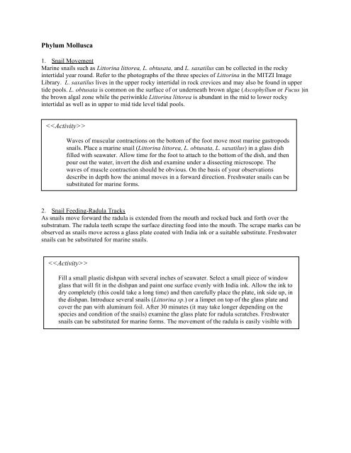
Phylum Mollusca
Phylum Mollusca
Phylum Mollusca

You also want an ePaper? Increase the reach of your titles
YUMPU automatically turns print PDFs into web optimized ePapers that Google loves.
<strong>Phylum</strong> <strong>Mollusca</strong><br />
1. Snail Movement<br />
Marine snails such as Littorina littorea, L. obtusata, and L. saxatilus can be collected in the rocky<br />
intertidal year round. Refer to the photographs of the three species of Littorina in the MITZI Image<br />
Library. L. saxatilus lives in the upper rocky intertidal in rock crevices and may also be found in upper<br />
tide pools. L. obtusata is common on the surface of or underneath brown algae (Ascophyllum or Fucus )in<br />
the brown algal zone while the periwinkle Littorina littorea is abundant in the mid to lower rocky<br />
intertidal as well as in upper to mid tide level tidal pools.<br />
<br />
Waves of muscular contractions on the bottom of the foot move most marine gastropods<br />
snails. Place a marine snail (Littorina littorea, L. obtusata, L. saxatilus) in a glass dish<br />
filled with seawater. Allow time for the foot to attach to the bottom of the dish, and then<br />
pour out the water, invert the dish and examine under a dissecting microscope. The<br />
waves of muscle contraction should be obvious. On the basis of your observations<br />
describe in depth how the animal moves in a forward direction. Freshwater snails can be<br />
substituted for marine forms.<br />
2. Snail Feeding-Radula Tracks<br />
As snails move forward the radula is extended from the mouth and rocked back and forth over the<br />
substratum. The radula teeth scrape the surface directing food into the mouth. The scrape marks can be<br />
observed as snails move across a glass plate coated with India ink or a suitable substitute. Freshwater<br />
snails can be substituted for marine snails.<br />
<br />
Fill a small plastic dishpan with several inches of seawater. Select a small piece of window<br />
glass that will fit in the dishpan and paint one surface evenly with India ink. Allow the ink to<br />
dry completely (this could take a long time) and then carefully place the plate, ink side up, in<br />
the dishpan. Introduce several snails (Littorina sp.) or a limpet on top of the glass plate and<br />
cover the pan with aluminum foil. After 30 minutes (it may take longer depending on the<br />
species and condition of the snails) examine the glass plate for radula scratches. Freshwater<br />
snails can be substituted for marine forms. The movement of the radula is easily visible with<br />
a magnifying glass as the snails move over the walls of the aquarium.
3. Snail Anatomy<br />
Refer to the labeled pictures of the periwinkle Littorina littorea in the MITZI Image Library.<br />
<br />
Examine the structure of the shell. Note the concentric growth rings. During the colder part of<br />
the year the rings will be crowded together because the animal can’t feed as often.<br />
• Can you determine how old your snail is?<br />
Crack open a periwinkle shell and carefully remove the animal whole and place in a small<br />
glass dish filled with seawater. Locate the head, tentacles, eyes, operculum, columnar muscle<br />
(attaches the animal to the shell) and liver. Refer to the labeled photograph of Littorina littorea.<br />
Locate the sac just above the head.<br />
This is called the mantle cavity<br />
where the gills and anus are located.<br />
Examine your snail for parasites as<br />
directed below.<br />
a) Snail Parasites -Can you see any<br />
actively moving larval stages<br />
emerging from the liver tissue of<br />
Littorina littorea? If the animal is<br />
parasitized there is a good chance<br />
that you will see the parasitic larvae.<br />
After a few minutes in the dish, the transparent larvae usually fall from the liver to the sides of<br />
the animal.The liver of the snail Littorina littorea is often infested with larval stages of the bird<br />
fluke (flatworm) Cryptocotyle lingua. The adult lives parasitically in the bird’s intestine<br />
(commonly found in the Herring Gull Larus harengus) and the fertilized eggs leave the<br />
intestine and develop into a larval stage (miracidium) that infect snails. In the snail, the<br />
miracidium forms a larval stage called a sporocyst that internally forms several larval stages<br />
termed redia. Each redia in turn forms several cercaria. The cercaria leaves the snail and attach<br />
to fish where they form a cyst (metacercaria). Birds contract the parasite by consuming<br />
infected fish. The metcercaria leaves its cyst and develops into the adult parasite in the bird<br />
intestine. Be sure to have students wash hands thoroughly after the activity.<br />
4. Movement of snails on a vertical surface<br />
Refer to photographs of all three species (Littorina littorea, L. obtusata, and L. saxatilus) in the MITZI<br />
Image Library. All three species are available, year round, on most rocky shores. L. saxatilus lives in the<br />
upper rocky intertidal in rock crevices and may also be found in upper tide pools. L. obtusata is common<br />
on the surface of or underneath brown algae (Ascophyllum or Fucus ) in the brown algal zone while the<br />
periwinkle Littorina littorea is abundant in the mid to lower rocky intertidal as well as in upper to mid<br />
tide level tidal pools.<br />
<br />
All three species can be kept in a covered container with a small amount of seawater in the<br />
refrigerator for one week. Place several snails in a tall glass container (a 1000ml graduated cylinder<br />
is ideal) with a small amount of seawater on the bottom. As snails climb upward trace their path with<br />
a wax pencil or marking pen and the time it takes them to get there. Next, cover the glass containers<br />
completely with aluminum foil and repeat. Discuss the following questions:<br />
• Which species climbs the highest in the least amount of time?<br />
• Is this related to where the snails live?<br />
• Does absence of light affect snail movement?
5. Shell Adaptation in the Dogwhelk<br />
Nucella lapillus, an exercise using statistical analysis. Littorina littorea, the periwinkle can be substituted<br />
for the dogwhelk. The dogwhelk Nucella lapillus is a carnivorous snail (<strong>Phylum</strong> <strong>Mollusca</strong>, Class<br />
Gastropoda) living in northern temperate waters both in the United States and Europe. It feeds almost<br />
exclusively on barnacles and blue mussels by chemically and mechanically drilling a hole through the<br />
shell and then consuming the flesh beneath.<br />
• Extension question: Are snails larger in sites protected from wave action than at locations<br />
exposed to wave action?<br />
<br />
Nucella lapillus were collected, at low tide (12:15 PM), from three sites at Rye Harbor State<br />
Park, Rye, New Hampshire on 11/20/2000 by Robert Zottoli.<br />
Site 1 (S1) is a boulder field on the northern side of the entrance to Rye Harbor. It is<br />
directly exposed to incoming waves. The boulders were generally between 0.25 and 1 meter<br />
in length and rested either on other boulders or on a layer of peat formed from a sunken<br />
forest. Snails were found either on or underneath exposed rock surfaces.<br />
Site 2 (S2) is on the ocean side of the jetty protecting Rye Harbor. It is also exposed<br />
to incoming waves. The large granite slabs that make up the jetty, are placed haphazardly,<br />
and presumably offer more protection from incoming waves.<br />
Site 3 (S3) is a boulder field, on the northern side, within the harbor, adjacent to the<br />
inner side of the jetty. The boulders are about the same size as those in Site 1. Approximately<br />
30 of the largest specimens encountered within each site were collected at about the time of<br />
low tide.<br />
• Do you think this is a valid way of collecting specimens? Explain.<br />
The shells of the snails from S1 were marked with red fingernail polish while those from S2 and S3<br />
were marked respectively with pink and black fingernail polish. Snails were then boiled for 3 minutes<br />
and removed from their shells with a toothpick or nail.<br />
Shell height, shell width, and shell aperture were all measured for each site to the nearest 0.1mm with<br />
sliding Vernier calipers. See the data below. Calculate the height/aperture ratio for each specimen<br />
and enter the data in the appropriate column on the blackboard. Calculate mean values for each<br />
measurement. Enter the data into a statistical program and compare mean values between each of the<br />
three populations using the t-test.<br />
Length<br />
Width<br />
Aperature<br />
Discussion:<br />
• Is there a significant difference between the<br />
mean values of shell height; shell width; or<br />
aperture width between S1 and S2?<br />
• Between S1 and S3? Between S2 and S3?<br />
• Does the data support your original<br />
hypothesis?<br />
• What can you conclude from these results?<br />
• Can you think of a way to improve this<br />
exercise?
Measurements (mm) of Length, Width, and Aperture width of Nucella lapillus from three sites at<br />
Rye Harbor, New Hampshire. Descriptions of sites 1-3 are discussed above.<br />
Key to data chart L = length, W= Width, AP= Aperture.<br />
Site 1 is exposed; Site 2 is intermediate; Site 3 is protected.<br />
Site 1 Site 2 Site 3 Site 1 Site 2 Site 3 Site 1 Site 2 Site 3<br />
L L L W W W AP AP AP<br />
27 26.4 30.5 26 16.6 15.8 24.3 16.3 19.4<br />
30 25 32.5 10.6 15 17.4 20 15.3 22.5<br />
27 26 31.9 16.5 11.2 18.9 13.5 11.1 22.3<br />
23.5 25.3 32.4 14 15.6 17.3 16 15.1 19.1<br />
27 25.6 28.2 15 15 15.5 12.3 15.1 19<br />
31 21 28.3 17.4 10.6 15.3 13 14 18.4<br />
25.5 24.6 28.7 14 14 15.9 15.8 15.4 18.9<br />
24 22.4 30 14 12.5 16.8 14.8 14.6 20.3<br />
27.5 24.5 29.5 15.6 13.3 16.9 19.7 15 20.2<br />
28 26.5 33.5 15 14.5 18.4 20 20 20.9<br />
28.5 27 30.7 15.2 15.2 18 18 15.6 16.7<br />
26.3 25.6 29.7 16 14.4 18.2 15.5 15.7 17.4<br />
31.3 25.5 27 18 14.5 17 20.4 16.7 17.6<br />
28.5 21.1 31.2 14.6 11.2 18.3 20 14.7 19.3<br />
27 26.6 29.8 15 14.4 15.8 18 16.4 15.3<br />
24 27.8 28.2 14.6 14.7 16.3 16 16.1 16.6<br />
23.5 27.5 28.4 15.7 14.1 16.8 14.6 17.1 17.6<br />
25.6 26.7 31.5 15.5 14.7 17.8 17 18.3 19<br />
23.8 24.4 31.3 14 12.4 19.3 15.8 17.1 19.7<br />
23.4 24.7 34 15 13.8 19.4 16 16.3 20.8<br />
30.3 26.5 33.5 20.4 15.4 18.3 20.9 14.3 22.7<br />
30.1 25.5 33.1 14 13.3 18.8 15.2 14.6 20.4<br />
20.4 12.1 36.5 14 12.5 21.9 15.2 14.7 23.1<br />
29 12.3 32.5 17.3 14.1 19.2 19.4 14.4 20.1<br />
30 12.1 32.2 15.5 14.4 19.2 15.8 12.8 22.1<br />
25 22.6 29.1 18.7 11.4 17.2 18.8 12.2 21<br />
28.8 22.9 27.2 17.3 12.7 16.1 18.4 15.1 17.3<br />
24.4 21.3 31.4 14.1 12.1 18.3 15.2 12.4 19.9<br />
23.2 25.1 32.3 14.3 14.4 19.5 16.7 24.5 21.1<br />
26.5 22.2 30.2 15.5 13.1 18.2 16.4 14.2 20.4
6. Blue Mussel (Mytilus edulis) Anatomy<br />
Refer to the labeled photograph of the blue mussel in the MITZI Image Library. Blue mussels can be<br />
collected on intertidal rock surfaces or on the sides of marina or commercial fishing floats. They are also<br />
available commercially in many Maine supermarkets or seafood stores.<br />
<br />
Examine the external surface of the shell. Note the concentric external growth rings. In<br />
places where the rings are crowded together, slower growth took place.<br />
• How old do you think your specimen is?<br />
• What is the advantage of a heavy exoskeleton?<br />
• Does it hinder the animal in any way?<br />
The oldest part of the shell is the broad dorsal umbo. The elastic hinge ligament attaches the<br />
two shells dorsally.<br />
• What function does the hinge ligament perform?<br />
Right vs. Left Valve - Place the valve in your hand with the dorsal surface facing toward you<br />
and the umbo pointing up. If the umbo points toward the right, it is the left valve, and if the<br />
umbo is toward the left, it is the right valve. Remove the right valve by first cracking it with<br />
a hammer and then carefully remove the shell pieces. You must cut the adductor muscles that<br />
hold the shells together. Place the animal in a dish of seawater. The mantle tissue,<br />
responsible for secreting the shell along its free edge, covers the surface immediately under<br />
the shell. Remove the mantle tissue. It may already have been torn loose. Examine the foot.<br />
The byssal threads that attach the animal to the substratum should be visible at the base of<br />
the foot.<br />
Find the pair of gills exposed when the mantle tissue is removed. Water is drawn by ciliary<br />
action through the inhalant ventral siphon, passes upward through the gill and out through<br />
the exhalent siphon. Place some Congo red-stained yeast (red food coloring will also work)<br />
or a small amount of mud from the animals’ habitat on the surface of the gill and describe its<br />
movement. Food is eventually passed from the gill to the labial palps. There is a food groove<br />
between the palps that leads into the mouth.
With a scalpel or dissecting knife, remove a filament (dorsal to ventral) from the gill and<br />
examine under the microscope. Describe what you see and make a sketch of the gill tissue<br />
paying special attention to the placement of cilia. Note the heart just below the hinge<br />
ligament the holds the shells together. The ventricle surrounds the white intestine and has<br />
two auricles leading from it.<br />
• Is it still beating? Record the number of beats per minute.<br />
The main purpose of the heart is to move blood through the gills.<br />
• Why is this essential?<br />
Mussels have an open circulatory system.<br />
• What does that mean?<br />
The dark reddish kidney can be seen below the heart.<br />
The visceral mass lies under the gill. It contains the dark green digestive gland (located<br />
near the labial palps), the gonad and the intestine. Using a diagram as a guide, carefully<br />
slice away the epidermis until you can see the digestive gland, gonad, and portions of the<br />
intestine. It’s unlikely that you will find the entire digestive tract.