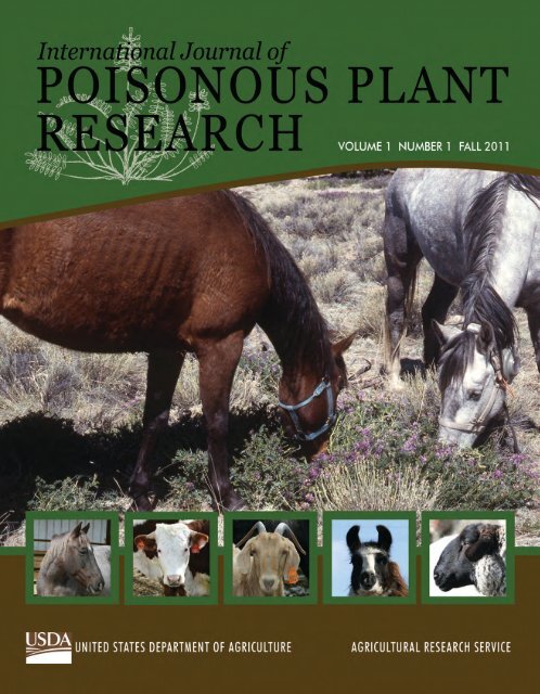
International Journal of Poisonous Plant Research - Agricultural ...
International Journal of Poisonous Plant Research - Agricultural ...
International Journal of Poisonous Plant Research - Agricultural ...

Create successful ePaper yourself
Turn your PDF publications into a flip-book with our unique Google optimized e-Paper software.
<strong>International</strong> <strong>Journal</strong> <strong>of</strong> <strong>Poisonous</strong> <strong>Plant</strong> <strong>Research</strong><br />
A <strong>Journal</strong> for <strong>Research</strong> and Investigation <strong>of</strong> <strong>Poisonous</strong> <strong>Plant</strong>s<br />
Tom Vilsack, Secretary<br />
U.S. Department <strong>of</strong> Agriculture<br />
Catherine E. Woteki, Under Secretary<br />
<strong>Research</strong>, Education and Economics<br />
Edward B. Knipling, Administrator<br />
<strong>Agricultural</strong> <strong>Research</strong> Service<br />
Sandy Miller Hays, Director<br />
Information Staff<br />
Christo Botha<br />
South Africa<br />
Peter Cheeke<br />
USA<br />
Steven Colegate<br />
USA<br />
John Edgar<br />
Australia<br />
Editors-in-Chief<br />
Kip E. Panter James A. Pfister<br />
USDA-ARS <strong>Poisonous</strong> <strong>Plant</strong> <strong>Research</strong> Lab<br />
Logan, UT<br />
Dale Gardner<br />
USA<br />
Editorial Board<br />
Silvana Lima Gorniak<br />
Brazil<br />
Jeff Hall<br />
USA<br />
Gonzalo Diaz<br />
Colombia<br />
Anthony Knight<br />
USA<br />
Franklin Riet-Correa<br />
Brazil<br />
Bryan Stegelmeier<br />
USA<br />
Kevin Welch<br />
USA<br />
ISSN 2154-3216
Chemistry<br />
Stephen Lee<br />
USA<br />
Immunology<br />
Isis Hueza<br />
Brazil<br />
Molecular Biology/Biochemistry<br />
Zane Davis<br />
USA<br />
Editorial Advisory Board<br />
Pathology<br />
Claudio SL Barros<br />
Brazil<br />
Pharmacology<br />
Benedict Green<br />
USA<br />
<strong>Plant</strong> Physiology<br />
Daniel Cook<br />
USA<br />
Assistant Editor<br />
Terrie Wierenga<br />
USDA-ARS <strong>Poisonous</strong> <strong>Plant</strong> <strong>Research</strong> Lab<br />
Logan, UT<br />
Range Science/Botany<br />
Michael Ralphs<br />
USA<br />
Toxicology<br />
Eduardo J Gimeno<br />
Argentina<br />
Veterinary Science<br />
Jeremy Allen<br />
Australia<br />
Aim and Scope<br />
The <strong>International</strong> <strong>Journal</strong> <strong>of</strong> <strong>Poisonous</strong> <strong>Plant</strong> <strong>Research</strong> publishes original papers on all aspects <strong>of</strong> poisonous plants<br />
including identification, analyses <strong>of</strong> toxins, case reports, control methods, and ecology.<br />
Access and Subscription<br />
The <strong>International</strong> <strong>Journal</strong> <strong>of</strong> <strong>Poisonous</strong> <strong>Plant</strong> <strong>Research</strong> is published twice a year (spring and fall) by the U.S.<br />
Department <strong>of</strong> Agriculture. All journal contents are available online without access fee or password control.<br />
Visit the <strong>Journal</strong> at http://www.ars.usda.gov/is/np/<strong>Poisonous</strong><strong>Plant</strong>s/<strong>Poisonous</strong><strong>Plant</strong><strong>Research</strong><strong>Journal</strong>Intro.htm.<br />
Full-text articles in PDF format also are freely available online.<br />
Submission Information<br />
To obtain submission instructions and contributor information, contact Editors-in-Chief Kip E. Panter<br />
(kip.panter@ars.usda.gov) or James A. Pfister (jim.pfister@ars.usda.gov).<br />
The <strong>International</strong> <strong>Journal</strong> <strong>of</strong> <strong>Poisonous</strong> <strong>Plant</strong> <strong>Research</strong> is published by the U.S. Department <strong>of</strong> Agriculture and thus<br />
is not subject to copyright protection. All contents <strong>of</strong> the <strong>Journal</strong> except where otherwise noted are in the public<br />
domain and may be freely reprinted or redistributed. However, proper citations are recommended.<br />
The <strong>Journal</strong> may report research involving pesticides. It does not imply that uses discussed herein have been<br />
registered. All uses <strong>of</strong> pesticides must be registered by appropriate State, territorial, and/or Federal agencies before<br />
they can be recommended.
Mention <strong>of</strong> trade names or commercial products in the <strong>Journal</strong> is solely for the purpose <strong>of</strong> providing specific<br />
information and does not imply recommendation or endorsement by the U.S. Department <strong>of</strong> Agriculture.<br />
The U.S. Department <strong>of</strong> Agriculture (USDA) prohibits discrimination in all its programs and activities on the basis<br />
<strong>of</strong> race, color, national origin, age, disability, and where applicable, sex, marital status, familial status, parental<br />
status, religion, sexual orientation, genetic information, political beliefs, reprisal, or because all or part <strong>of</strong> an<br />
individual's income is derived from any public assistance program. (Not all prohibited bases apply to all programs.)<br />
Persons with disabilities who require alternative means for communication <strong>of</strong> program information (Braille, large<br />
print, audiotape, etc.) should contact USDA's TARGET Center at (202) 720-2600 (voice and TDD). To file a<br />
complaint <strong>of</strong> discrimination, write to USDA, Director, Office <strong>of</strong> Civil Rights, 1400 Independence Avenue, S.W.,<br />
Washington, D.C. 20250-9410, or call (800) 795-3272 (voice) or (202) 720-6382 (TDD). USDA is an equal<br />
opportunity provider and employer.<br />
October 2011
Preface<br />
We are very pleased to publish this inaugural issue <strong>of</strong> a new all-electronic journal,<br />
<strong>International</strong> <strong>Journal</strong> <strong>of</strong> <strong>Poisonous</strong> <strong>Plant</strong> <strong>Research</strong> (IJPPR), through the<br />
assistance <strong>of</strong> the USDA-ARS Information Staff, Beltsville, MD. IJPPR is an<br />
online-only, peer-reviewed journal published semi-annually (two issues per<br />
calendar year). The primary objective <strong>of</strong> IJPPR is to provide timely research<br />
results and new technology to all those working in various disciplines involving<br />
poisonous plants. The <strong>Journal</strong> will encompass all aspects <strong>of</strong> poisonous plant<br />
research including original research, case reports, field observations in domestic<br />
and wild animals, and scientific reviews. There currently is not any journal,<br />
electronic or printed, that specifically targets toxic plant research, and IJPPR aims<br />
to fill this critical gap. We’ve employed an electronic publication system to make<br />
the papers as widely accessible as possible on the USDA-ARS website. It is our<br />
sincere hope that IJPPR will contribute in a meaningful manner toward<br />
facilitating and enhancing poisonous plant research and communication around<br />
the globe.<br />
This inaugural issue <strong>of</strong> IJPPR consists <strong>of</strong> papers submitted by various research<br />
scientists around the world, reflecting our objective <strong>of</strong> making IJPPR a truly<br />
international journal. We thank those who have assisted in the production <strong>of</strong> this<br />
inaugural issue, particularly Terrie Wierenga, Editorial Assistant, USDA-ARS<br />
<strong>Poisonous</strong> <strong>Plant</strong> <strong>Research</strong> Laboratory, Logan, UT; Sandy Miller Hays, Director,<br />
USDA-ARS Information Staff, Beltsville, MD; and Mina Chung, Supervisory<br />
Editor, USDA-ARS Information Staff, Beltsville, MD. We also thank all the<br />
individuals who have agreed to serve on the IJPPR Editorial Board and Editorial<br />
Advisory Board.<br />
James A. Pfister<br />
Kip E. Panter<br />
Editors-in-Chief<br />
USDA-ARS <strong>Poisonous</strong> <strong>Plant</strong> <strong>Research</strong> Laboratory<br />
Logan, UT
Contents<br />
1 Toxic <strong>Plant</strong>s <strong>of</strong> Veterinary and <strong>Agricultural</strong> Interest in Colombia<br />
Gonzalo J. Diaz<br />
20 Analysis <strong>of</strong> the Toxic Amino Acid Indospicine by Liquid Chromatography-Tandem Mass<br />
Spectrometry<br />
Dale R. Gardner and Franklin Riet-Correa<br />
28 Clinical and Pathological Aspects and Cerebellar Lectin Binding in Cattle Poisoned With<br />
Solanum fastigiatum var. fastigiatum and Solanum bonariense<br />
Fabiano J.F. Sant’Ana, Claudio G. Barbeito, Fabian Nishida, Eduardo J. Gimeno,<br />
José M. Verdes, Daniel Battes, Antonio Moraña, and Claudio S.L. Barros<br />
35 Fetotoxicity <strong>of</strong> Astragalus lentiginosus (Locoweed) in Spanish Goats<br />
Stella Furlan, Kip E. Panter, James A. Pfister, and Bryan L. Stegelmeier<br />
41 Locoweed Poisoning in the Native Grasslands <strong>of</strong> China<br />
Zhao Meng-Li, Gao Xinlei, and Han Bing<br />
47 Locoweed Toxicity, Ecology, Control, and Management<br />
Michael H. Ralphs and Bryan L. Stegelmeier<br />
65 Pathological Effects <strong>of</strong> Short-Term Crotalaria retusa Ingestion by Guinea Fowl (Numida<br />
meleagris)<br />
Windleyanne Gonçalves Amorim Bezerra, Milzete Alves de Souza, Ruben Horn<br />
Vasconcelos, Luis Augusto Vieira Cordeiro, Jael Soares Batista,<br />
and Benito Soto-Blanco<br />
70 Short Communication—Lithium Carbonate as a Potential Aversive Agent<br />
Leendert D. Snyman and R. Anitra Schultz<br />
72 Western Juniper-Induced Abortions in Beef Cattle<br />
Kevin D. Welch, Dale R. Gardner, Kip E. Panter, Bryan L. Stegelmeier,<br />
Cory Parsons, James A. Pfister, and Daniel Cook
Toxic <strong>Plant</strong>s <strong>of</strong> Veterinary and <strong>Agricultural</strong> Interest in<br />
Colombia<br />
Gonzalo J. Diaz<br />
Toxicology Laboratory, Faculty <strong>of</strong> Veterinary Medicine and Animal Science, National University<br />
<strong>of</strong> Colombia, Bogotá, Colombia<br />
Corresponding author: Gonzalo Diaz, gjdiazg@bt.unal.edu.co<br />
Abstract<br />
Colombia has the second largest plant biodiversity <strong>of</strong> any country in the world, with<br />
about 25,000 species <strong>of</strong> vascular plants. This is due in part to its equatorial location, and<br />
large variation in elevation and associated gradients in temperatures and rainfall.<br />
Livestock in Colombia graze vast tracts <strong>of</strong> land with a wide variety <strong>of</strong> herbaceous and<br />
woody plants. Although the annual cattle mortality from plant poisoning in Colombia is<br />
estimated at 130,000, the economic impact on the entire livestock industry has not been<br />
fully evaluated. Information on toxic plants is scarce in Colombia, and livestock<br />
poisoning by plants is seldom documented. This review presents the current knowledge<br />
on the identity <strong>of</strong> plants known to have poisoned livestock in Colombia and on research<br />
conducted into these toxic plants. To the extent known, the toxic component(s), major<br />
clinical signs and circumstances <strong>of</strong> poisoning, location, and environmental factors are<br />
discussed. Many <strong>of</strong> the plants identified in Colombia are considered toxic on the basis <strong>of</strong><br />
world literature, but toxicosis in Colombia has not always been documented. The<br />
information on toxic plant chemistry in Colombia is mostly limited to the plant’s nitrate<br />
or cyanide content. <strong>Research</strong> is needed to determine not only which plants represent a<br />
potential risk for animal health and production but also their phytochemistry and<br />
toxicology. It is strongly recommended that veterinarians document plant poisoning cases<br />
through government reporting services and that university and government veterinarians,<br />
scientists, and extension agents investigate episodes <strong>of</strong> plant toxicosis and publish their<br />
findings. This would help identify toxic species for further phytochemical and<br />
toxicological studies and possibly pharmacological activity.<br />
Keywords: toxic plants, Colombia, plant poisoning, livestock, pets<br />
Introduction<br />
Toxic plants affecting both large and small animals<br />
are a major concern for the practicing veterinarian<br />
and livestock producer in every country. In countries<br />
with higher plant biodiversity, the number <strong>of</strong><br />
problematic toxic plants may be greater. <strong>Plant</strong><br />
biodiversity in Colombia is very high, as there are<br />
about 25,000 species <strong>of</strong> vascular plants in Colombia,<br />
both native and naturalized (Bernal et al. 2006). This<br />
biodiversity corresponds to about 8 percent <strong>of</strong> the<br />
total vascular plants on earth, which makes the<br />
country the second largest in plant biodiversity in<br />
the world, the largest being Brazil. However,<br />
information on toxic plants in Colombia is scarce<br />
and is usually published only in local, Spanish-<br />
1
Diaz: Toxic <strong>Plant</strong>s <strong>of</strong> Colombia<br />
language journals. Further, it is not customary<br />
among local veterinarians to write case reports, thus<br />
most <strong>of</strong> the plant poisonings that occur in Colombia<br />
are not documented in the literature.<br />
The impact <strong>of</strong> toxic plants on Colombian<br />
livestock production has not been fully evaluated. It<br />
is estimated that more than 40 million hectares <strong>of</strong> the<br />
country are used for livestock production, with a<br />
bovine population <strong>of</strong> about 26 million animals<br />
(Ministerio de Agricultura y Desarrollo Rural,<br />
2005). Colombia ranked ninth in the world for cattle<br />
population. However, livestock production is mostly<br />
extensive with a very low population <strong>of</strong> animals kept<br />
under intensive production systems. Land used for<br />
cattle grazing contains complex mixes <strong>of</strong> native and<br />
invasive plants (Peña et al. 1980), which may<br />
increase the risk <strong>of</strong> exposure to toxic plants, many <strong>of</strong><br />
which have not been characterized or have only been<br />
partially characterized. Conservative estimates<br />
indicate that in Colombia, toxic plants cause an<br />
annual mortality rate <strong>of</strong> about 0.5 percent (Peña et<br />
al. 1980), which is currently equivalent to about<br />
130,000 cattle. This percentage is in agreement to<br />
that reported by Tokarnia et al. (2002) in Brazil, who<br />
estimated that between 800,000 to 1,120,000 cattle<br />
<strong>of</strong> the 160-million population die annually from<br />
plant poisoning, a mortality rate corresponding to<br />
0.5 to 0.7 percent.<br />
The aim <strong>of</strong> the present review is to briefly<br />
describe the most important native and introduced<br />
toxic plants present in Colombia that affect animals<br />
and to summarize the published research on these<br />
toxic plants, highlighting research conducted in<br />
Colombia.<br />
Major Toxic <strong>Plant</strong>s Affecting Animal Health<br />
and Production in Colombia<br />
<strong>Plant</strong>s were grouped based on the major organ<br />
system affected by consumption <strong>of</strong> the plant.<br />
Common names given to these plants in Colombia<br />
are provided in parentheses after the Latin botanical<br />
name or indicated in one <strong>of</strong> the tables. It is important<br />
to note that Colombia is a tropical country located<br />
on the Equator, which means that there are no<br />
seasons such as winter, spring, summer, or fall, and<br />
that annual plants behave as perennials under these<br />
conditions. The average environmental temperature<br />
is mostly determined by elevation with distinct<br />
temperature gradients from low- to high-elevation<br />
rangelands. There are, however, “dry” and “rainy”<br />
seasons when low or high precipitation is expected<br />
2<br />
every year. The times <strong>of</strong> the year with higher<br />
precipitation rates are April through May and<br />
October through November. The start <strong>of</strong> the rainy<br />
period is accompanied by intense growth <strong>of</strong> some<br />
plant species used for animal feed (especially<br />
grasses), a situation that is usually associated with<br />
increased accumulation <strong>of</strong> potentially toxic<br />
compounds such as nitrates. However, some plants<br />
accumulate more toxins during the dry periods, for<br />
example, the native plant Mascagnia concinna,<br />
which accumulates more cyanogenic glycosides<br />
during these periods. Colombia is politically divided<br />
into “departments” (states), and sometimes this word<br />
is used to indicate a specific geographical region.<br />
<strong>Plant</strong>s That Affect the Digestive System<br />
<strong>Plant</strong>s That Cause Irritation <strong>of</strong> the Oral Cavity<br />
Many plants belonging to the Araceae family<br />
contain needle-shaped calcium oxalate crystals in<br />
their leaves. These crystals are known as raphides<br />
and are housed inside specialized cells known as<br />
idioblasts (Genua and Hillson 1985). When the plant<br />
leaves are chewed, the idioblasts are broken and the<br />
oxalate crystals are expelled, causing an immediate<br />
burning sensation in the oral cavity tissues. <strong>Plant</strong>s<br />
that accumulate calcium oxalates are fairly common<br />
in Colombia and some <strong>of</strong> them are even native to the<br />
country, such as Dieffenbachia picta (cucaracho),<br />
recognized as the most toxic <strong>of</strong> all Araceae plants<br />
(Cao 2003). The genus Dieffenbachia comprises<br />
about 135 species, most <strong>of</strong> them present in South<br />
America. Colombia has the highest biodiversity with<br />
37 species, followed by Ecuador with 34, Peru with<br />
30, Brazil with 27, Panama with 20, and Costa Rica<br />
with 13 (Croat 2004). Although toxicosis by D. picta<br />
in livestock is rare, the ingestion <strong>of</strong> its leaves has<br />
caused intoxication in humans and pets. In dogs, the<br />
oxalates <strong>of</strong> D. picta can cause severe inflammation<br />
and necrosis <strong>of</strong> the epithelium <strong>of</strong> the tongue and oral<br />
cavity and may even cause death (Loretti et al.<br />
2003). Besides calcium oxalates, D. picta contains<br />
proteolytic enzymes that induce histamine release<br />
causing a severe inflammatory response that may<br />
lead to asphyxia and death (Loretti et al. 2003).<br />
Other plants <strong>of</strong> the Araceae family common in<br />
Colombia are Alocasia macrorrhiza (rascadera,<br />
bore, taro gigante), Caladium spp. (caladio,<br />
rascadera), Monstera deliciosa (abalazo, balazo),<br />
and Philodendron spp. (balazo). With the exception<br />
<strong>of</strong> Alocasia macrorrhiza, these plants are all native
to tropical America. These plants contain reduced<br />
concentrations <strong>of</strong> calcium oxalate compared with D.<br />
picta, and they are rarely associated with<br />
toxicological problems.<br />
<strong>Plant</strong>s That Affect the Gastrointestinal Tract<br />
Ricinus communis (castor, higuerilla, palmacristi,<br />
ricino) is a naturalized plant common in Colombia<br />
from sea level to 2600 m elevation. R. communis<br />
seeds contain ricin, one <strong>of</strong> the most potent lectins<br />
known. In general, lectins cause necrosis <strong>of</strong> the cells<br />
lining the gastrointestinal tract. Ricin is comprised<br />
<strong>of</strong> two subunits: Unit B (for binding) is the actual<br />
lectin that binds to galactosyl residues in cellular<br />
membranes, whereas unit A (for activity) is an<br />
enzyme capable <strong>of</strong> inactivating ribosomes in<br />
eukaryotic cells (Barbieri et al. 1993). All animal<br />
species are sensitive to the effects <strong>of</strong> ricin. The<br />
toxicosis, however, is uncommon and it is usually<br />
associated with feeding garden clippings or with<br />
contamination <strong>of</strong> forage grasses with R. communis<br />
trimmings (Aslani et al. 2007). Clinical signs include<br />
weakness, salivation, pr<strong>of</strong>use aqueous diarrhea,<br />
dehydration, mydriasis, teeth grinding, hypothermia,<br />
and recumbence; the major postmortem finding is<br />
severe gastroenteritis (Aslani et al. 2007). Other<br />
plants present in Colombia that contain potentially<br />
toxic lectins in their seeds are Jatropha curcas<br />
(piñón de fraile, purga de fraile), Abrus precatorius<br />
(chochos de pinta negra, jetiriquí) and Canavalia<br />
ensiformis (canavalia, fríjol blanco). The lectins<br />
present in these plants correspond to curcin, abrin,<br />
and concanavalin A, respectively. However,<br />
toxicosis with these plants has not been documented<br />
in Colombia.<br />
Another plant compound highly irritating to the<br />
gastrointestinal mucosa is ricinoleic acid, a fatty acid<br />
present in Ricinus communis seeds, considered to be<br />
responsible for the cathartic properties <strong>of</strong> ricin oil.<br />
Ricinoleic acid is an irritant that alters the intestinal<br />
epithelium causing loss <strong>of</strong> water and electrolytes,<br />
increased loss <strong>of</strong> luminal DNA, and decreased<br />
enzymatic activity <strong>of</strong> enterocytes (Bretagne et al.<br />
1981).<br />
<strong>Plant</strong>s That Affect the Blood<br />
<strong>Plant</strong>s Causing Hemolytic Anemia<br />
Feeding culled onions has been associated with<br />
hemolytic anemia in cattle and other animal species.<br />
IJPPR, vol. 1, Fall 2011<br />
Allium cepa, which includes all types <strong>of</strong> onions, is<br />
capable <strong>of</strong> causing toxicosis in both large and small<br />
animals due to its content <strong>of</strong> organic sulfoxides,<br />
especially alkyl or alkenyl cysteinyl sulfoxides (Rae<br />
1999, Parton 2000). After ingestion, the organosulfoxides<br />
are transformed into a complex mixture <strong>of</strong><br />
organic sulfur compounds, some <strong>of</strong> which are<br />
capable <strong>of</strong> causing intravascular hemolysis in cattle,<br />
sheep, and horses. Onion toxicosis, which occurs<br />
sporadically in cattle in Colombia, has been<br />
extensively documented in the literature with the<br />
first case reported in 1909 (Goldsmith 1909). The<br />
toxicosis occurs because cattle readily eat onions<br />
and usually prefer them to high-quality forages or<br />
grains (Rae 1999). The excessive intake <strong>of</strong> onions<br />
leads to hemolytic anemia and methemoglobinemia,<br />
which develops within a week <strong>of</strong> onion ingestion.<br />
Clinical signs in cattle include diarrhea, hemoglobinuria,<br />
ataxia, and coma. Cattle are more sensitive<br />
than horses, and goats are the most resistant. The<br />
hemolytic anemia caused by onion ingestion can<br />
also occur in dogs and cats (Parton 2000).<br />
Another plant that causes intravascular<br />
hemolysis is Brassica oleracea (col silvestre),<br />
several varieties <strong>of</strong> which are used as forage for<br />
ruminants. B. oleracea contains the non-protein<br />
amino acid S-methyl cysteinyl sulfoxide (SMCO),<br />
which is reduced in the rumen to dimethyl disulfide,<br />
a hemolysin (Duncan and Milne 1993). The anemia<br />
induced by the intravascular hemolysis may be lethal<br />
in cattle, which are very sensitive to the hemolytic<br />
effects <strong>of</strong> SMCO (Prache 1994).<br />
<strong>Plant</strong>s Causing Methemoglobinemia<br />
The nitrite ion, which is formed by bacteria in the<br />
rumen from plant nitrate, is the major cause <strong>of</strong><br />
methemoglobinemia in ruminants. Methemoglobin<br />
is an abnormal form <strong>of</strong> hemoglobin in which its<br />
normal ferrous moiety (Fe 2+ ) oxidized to the<br />
abnormal ferric form (Fe 3+ ). The oxidized form is<br />
not capable <strong>of</strong> transporting oxygen and there is a<br />
decrease in the oxygenation capacity <strong>of</strong> the blood.<br />
The severity <strong>of</strong> the clinical signs and effects depends<br />
on the amount <strong>of</strong> methemoglobin formed. Signs <strong>of</strong><br />
hypoxia develop when 20 to 30 percent <strong>of</strong> the<br />
hemoglobin is converted to methemogloblin, and<br />
death can occur at 70 to 80 percent methemoglobin<br />
levels (Vermunt and Visser 1987). Many plants have<br />
been identified as accumulators <strong>of</strong> toxic nitrate<br />
levels in Colombia (table 1), this being one <strong>of</strong> the<br />
most common plant toxicosis recognized in cattle.<br />
3
Diaz: Toxic <strong>Plant</strong>s <strong>of</strong> Colombia<br />
As shown in table 1, the most important group <strong>of</strong><br />
plants responsible for nitrate poisoning in cattle are<br />
forage grasses with at least nine species known to be<br />
associated with nitrate poisoning. An example <strong>of</strong> the<br />
high nitrate levels present in Colombian grasses is<br />
the study <strong>of</strong> Trheebilcock et al. (1978), who<br />
analyzed samples <strong>of</strong> Panicum maximum from the<br />
northern part <strong>of</strong> the country (Departments <strong>of</strong><br />
Córdoba and Sucre). The average nitrate levels<br />
found were 1209 and 5260 ppm for fresh plants<br />
collected during the dry season and after the onset <strong>of</strong><br />
the rainy season, respectively. Also in Colombia,<br />
Guzmán et al. (1978) reported a case in the Valle del<br />
Cauca Department that caused acute mortality in 19<br />
<strong>of</strong> 64 steers that were fed cut Pennisetum<br />
purpureum. The grass was found to contain 445 ppm<br />
nitrate and 971 ppm nitrite; the high nitrite content<br />
was attributed to microbial processes.<br />
The Amaranthacea family also contains plants<br />
associated with nitrate poisoning in cattle.<br />
Amaranthus dubius and A. spinosus are two species<br />
<strong>of</strong> Amaranthus common in Colombia that have been<br />
associated with nitrate intoxication, especially<br />
during the transition between the dry and the wet<br />
seasons (Torres 1984a). Another Amaranthacea is<br />
Chenopodium album, a plant recently reported in<br />
Colombia (Fernández-Alonso and Hernández-<br />
Schmidt 2007), which can cause lethal intoxication<br />
in ruminants because <strong>of</strong> its high nitrate levels<br />
(although it can also accumulate soluble oxalates).<br />
Levels <strong>of</strong> 2,500 ppm nitrate-nitrogen were reported<br />
in Chenopodium album hay associated with<br />
4<br />
mortality in cattle (Ozmen et al. 2003). Other<br />
Chenopodium spp. such as C. ambrosoides and C.<br />
quinoa are considered native to Colombia but have<br />
not been reported as toxic.<br />
Another plant associated with high nitrate<br />
content is Mascagnia concinna, a vine <strong>of</strong> the<br />
Malpighiaceae family native to the Magdalena<br />
Valley <strong>of</strong> Colombia. Nitrate concentrations ranging<br />
from 5,300 to 29,200 ppm dry matter were reported<br />
by Torres (1984a) and from 1,555 to 10,763 in fresh<br />
material by Trheebilcok et al. (1978).<br />
The Phytolaccaceae Petiveria alliacea can also<br />
contain toxic levels <strong>of</strong> nitrate. Studies conducted in<br />
Colombia with fresh plants showed that during the<br />
dry season, the plant accumulates an average <strong>of</strong><br />
1,155 ppm nitrate but during the rainy season, the<br />
average levels are 7,867 ppm (Trheebilcock et al.<br />
1978). Heliotropium indicum is a Boraginaceae also<br />
known to accumulate toxic concentrations <strong>of</strong><br />
nitrates. In samples collected in the northern part <strong>of</strong><br />
the country, Trheebilcock et al. (1978) found<br />
average nitrate levels <strong>of</strong> 178 and 7,195 ppm in fresh<br />
material collected before the rainy season and<br />
immediately after the start <strong>of</strong> the rainy season,<br />
respectively.<br />
<strong>Plant</strong>s That Affect the Coagulation <strong>of</strong> Blood<br />
Cumarinic glycosides can be found in<br />
Anthoxanthum odoratum (Poaceae) and in Melilotus<br />
spp. (Fabaceae). Anthoxanthum odoratum (oloroso)<br />
was introduced in Colombia during colonial times,<br />
Table 1. Major nitrate-accumulating plants affecting livestock in Colombia<br />
Family Latin name Common name<br />
Amaranthaceae Amaranthus dubius Adormidera, bledo liso<br />
Amaranthus hybridus Amaranto, bledo chico<br />
Chenopodium album Quenopodio<br />
Boraginaceae Heliotropium indicum Verbena, rabo de alacrán<br />
Malpighiaceae Mascagnia concinna Mindaca, mataganado<br />
Poaceae Andropogon bicornis Barba de indio, cola de zorro<br />
Brachiaria mutica Pasto pará<br />
Lolium perenne Balico, raigrás inglés, raigrás perenne<br />
Panicum maximum Pasto guinea, siempreverde<br />
Paspalum paniculatum Paja brava, paja del camino<br />
Paspalum virgatum Gramalote, yerba peluda<br />
Penisetum purpureum Pasto elefante<br />
Sorghum bicolor Sorgo, sorgo forrajero<br />
Sorghum halepense Pasto Johnson, capim argentino<br />
Phytolaccaceae Petiveria alliacea Anamú<br />
Solanaceae Solanum nigrum Campano, yerbamora
and it is common in cold regions <strong>of</strong> the country at<br />
altitudes from 2,600 to 3,500 m above sea level<br />
(Fernández-Alonso and Hernández-Schmidt 2007).<br />
Melilotus albus and M. <strong>of</strong>ficinalis (trébol dulce)<br />
were also introduced, and they are currently<br />
considered part <strong>of</strong> the naturalized flora <strong>of</strong> Colombia<br />
(Bernal et al. 2006, Fernández-Alonso and<br />
Hernández-Schmidt 2007). When hay from these<br />
plants becomes moldy, the cumarinic glycosides can<br />
produce dicumarol, an anticoagulant that causes<br />
depletion <strong>of</strong> active vitamin K in the liver resulting in<br />
reduced clotting factors being released into the blood<br />
(Hallak and Wedlund 1991). The toxicosis by A.<br />
odoratum and Melilotus spp. in cattle has been well<br />
documented in the world literature (Pritchard et al.<br />
1983, Puschner et al. 1998, Runciman et al. 2002).<br />
Affected animals are weak and reluctant to move,<br />
show petechial hemorrhages in mucosal surfaces,<br />
may bleed from natural orifices, and show increased<br />
prothrombin and partial thromboplastin time. At<br />
postmortem examination, multiple petechial and<br />
ecchymotic hemorrhages are seen. Uncoagulated<br />
blood may also be seen in any <strong>of</strong> the body tissues<br />
and cavities including the chest and abdomen,<br />
depending on the activity <strong>of</strong> the animal.<br />
Cardiotoxic <strong>Plant</strong>s<br />
Cardiac glycosides are a specific type <strong>of</strong> toxic<br />
glycosides that affect the cardiac muscle, sometimes<br />
causing fatal toxicosis. Cardiac glycosides increase<br />
the contraction force <strong>of</strong> the heart by inhibiting the<br />
myocardial Na-K ATP-ase, which can lead to<br />
cardiac arrest (Poindexter et al. 2007). Two types <strong>of</strong><br />
cardiac glycosides are recognized depending on their<br />
chemical characteristics, namely, cardenolide and<br />
bufadienolide glycosides. At least four plants<br />
containing cardenolide cardiac glycosides are<br />
present in Colombia: Digitalis purpurea (dedalera,<br />
digital, guargüeron), Nerium oleander (oleander,<br />
delfa, adelfa, azuceno de La Habana), Thevetia<br />
peruviana (catapis, oleander amarillo), and<br />
Asclepias curassavica (bencenuco, mataganado). All<br />
have sporadically caused toxicosis in herbivores.<br />
Digitalis purpurea was introduced into<br />
Colombia as an ornamental by British engineers<br />
around 1856; and it currently grows wild in<br />
highlands, including the high plateau where Bogotá<br />
is located, at 2,640 m above sea level. D. purpurea<br />
contains cardiac glycosides in all parts <strong>of</strong> the plant<br />
but the concentration is higher in the leaves.<br />
IJPPR, vol. 1, Fall 2011<br />
Nerium oleander is a perennial bush native to<br />
the Mediterranean region and Asia but now is<br />
common in all tropical and subtropical regions <strong>of</strong> the<br />
world. In Colombia it is cultivated as an ornamental<br />
for its colourful flowers, which can be white, pink,<br />
yellow, or red. All parts <strong>of</strong> the plant contain cardiac<br />
glycosides with oleandrin being the most abundant.<br />
Toxicosis has occurred in horses and cattle usually<br />
due to contamination <strong>of</strong> pastures with plant clippings<br />
from N. oleander bushes. Thevetia peruviana (= T.<br />
nereifolia, Cascabela peruviana, and C. thevetia) is<br />
a bush native to South America, similar to Nerium<br />
oleander but smaller, with narrower leaves and only<br />
yellow flowers. This plant contains cardenolide<br />
cardiac glycosides, primarily thevetin A and thevetin<br />
B, especially in the seeds (Roberts et al. 2006).<br />
There are reports <strong>of</strong> human toxicosis caused by this<br />
plant, generally associated with the intake <strong>of</strong> the<br />
seeds. Intake <strong>of</strong> one or two seeds causes<br />
gastrointestinal symptoms, and intake <strong>of</strong> three or<br />
four seeds affects the heart and may cause death<br />
(Roberts et al. 2006). In the city <strong>of</strong> Medellín<br />
(Department <strong>of</strong> Antioquia), seeds <strong>of</strong> T. peruviana<br />
were sold as a weight loss aid and several women<br />
died after eating the seeds (López 2002).<br />
Asclepias curassavica is a plant native to the<br />
Caribbean but now is commonly found in Colombia<br />
at elevations up to 1,600 m. This plant contains<br />
asclepine, a cardiac glycoside with higher potency<br />
than strophantin, digoxine, digitoxine, and<br />
digitoxigenine (Patnaik and Köhler 1978), which are<br />
some <strong>of</strong> the most potent cardiac glycosides known.<br />
Even though the plant is not palatable for herbivores,<br />
it has been associated with sporadic cases <strong>of</strong><br />
toxicosis in cattle.<br />
Hepatotoxic <strong>Plant</strong>s<br />
The main hepatotoxic plants present in Colombia<br />
affect the liver by causing either hepatocellular<br />
necrosis or intrahepatic cholestasis. Pyrrolizidine<br />
alkaloids (PAs) are a large group <strong>of</strong> hepatotoxins<br />
characterized by the presence <strong>of</strong> a pyrrolizidine<br />
nucleus in their structure and are capable <strong>of</strong> causing<br />
hepatocellular necrosis. Compounds in plants known<br />
to cause intrahepatic cholestasis are the lantadenes<br />
from Lantana spp.; sporidesmin, a mycotoxin<br />
formed by a fungus on grasses; and the steroidal<br />
saponins present in several grasses. All hepatotoxins<br />
may cause secondary photosensitization in<br />
ruminants due to an alteration in the metabolism <strong>of</strong><br />
5
Diaz: Toxic <strong>Plant</strong>s <strong>of</strong> Colombia<br />
chlorophyll leading to skin damage when ruminants<br />
are exposed to the sun.<br />
<strong>Plant</strong>s Containing Substances That Cause<br />
Hepatocellular Necrosis<br />
The PAs are a large group <strong>of</strong> hepatotoxins present in<br />
plants found in Colombia, and PA toxicosis has been<br />
reported in livestock, poultry, pigs, and humans in<br />
Colombia. Extensive literature reviews on the<br />
chemistry, mechanism <strong>of</strong> action, and effects <strong>of</strong> PAs<br />
in animals and humans have been published<br />
(Mattocks 1986, Diaz 2001, Fu et al. 2004, Rietjens<br />
et al. 2005). In general, PAs induce hepatocyte<br />
necrosis that progresses to the destruction <strong>of</strong> the<br />
parenchymal cells <strong>of</strong> the organ and eventually to<br />
liver failure. PAs are also potent carcinogens at<br />
levels below those causing hepatic necrosis. Even<br />
though PAs are mainly hepatotoxic, some <strong>of</strong> them<br />
can also affect the lungs, especially monocrotaline.<br />
More than 6,000 plants are believed to contain<br />
PAs, many <strong>of</strong> which are present in Colombia in all<br />
kinds <strong>of</strong> ecosystems. The most important PAproducing<br />
plants from the toxicological standpoint<br />
belong to one <strong>of</strong> the families Asteracea, Fabaceae, or<br />
Boraginaceae. Table 2 summarizes the major PAcontaining<br />
plants present in Colombia.<br />
Among the Asteraceae family (formerly known<br />
as Compositae) the most important hepatotoxic<br />
genera are Senecio and Eupatorium. Two toxic<br />
species <strong>of</strong> Senecio common in Colombia are Senecio<br />
formosus and S. madagascariensis. The former is a<br />
plant native to the highlands between 3,000 and<br />
4,000 m above sea level and commonly found in the<br />
Colombian Andean regions <strong>of</strong> Cundinamarca,<br />
Cauca, and Nariño. There are no reports <strong>of</strong> toxicosis<br />
in animals caused by this plant; however, Senecio<br />
formosus has caused irreversible hepatic damage in<br />
human patients who ingested infusions made with its<br />
dry leaves. The clinical history, symptoms, signs,<br />
6<br />
lesions, and postmortem findings <strong>of</strong> almost 20 fatal<br />
cases reported in Bogotá were documented by Toro<br />
et al. (1997).<br />
Senecio madagascariensis is an annual or<br />
perennial herb native to South Africa reported for<br />
the first time in Colombia in the 1980s. It is an<br />
aggressive weed that propagates rapidly, and it has<br />
already colonized all the high plateau <strong>of</strong> the<br />
departments <strong>of</strong> Cundinamarca and Boyacá<br />
(Fernández-Alonso and Hernández-Schmidt 2007).<br />
Horses are highly sensitive to the PA <strong>of</strong> S.<br />
madagascariensis and can even be intoxicated in<br />
utero. In Australia, Small et al. (1993) reported a<br />
case where a foal exhibited growth retardation and<br />
jaundice at birth and died at 2 months <strong>of</strong> age with<br />
liver damage. During gestation the mare was kept in<br />
a field heavily infested with S. madagascariensis,<br />
which resulted in fetal exposure in utero. In<br />
Colombia, S. madagascariensis has been associated<br />
with sudden death in cows immediately after<br />
parturition. The cause <strong>of</strong> this sudden death syndrome<br />
is unknown, but it is possible that the metabolic<br />
changes associated with parturition and the onset <strong>of</strong><br />
lactation pose an extra load to a liver that has been<br />
severely affected by the chronic ingestion <strong>of</strong> the<br />
plant. Burgueño-Tapia et al. (2001) analyzed S.<br />
madagascariensis plants collected in Colombia and<br />
found that the plants contain chemical substances<br />
known as calolides. However, the toxicological with<br />
plants from Australia or Hawaii although the<br />
concentration was lower. The total concentration <strong>of</strong><br />
PAs in samples from Australia, Hawaii, and<br />
Colombia was 3,089, 2,133, and 805 µg/g,<br />
respectively. The major PAs found in the Colombian<br />
samples were senecivernine, senecionine,<br />
integerreimine, mucronatinine, and usaramine (D.R.<br />
Gardner and G.J. Diaz, 2009, unpublished data). The<br />
other genus <strong>of</strong> the Asteracea family reported to<br />
accumulate PA is Eupatorium. Several species <strong>of</strong><br />
this genus have been reported in Colombia (Powell<br />
Table 2. Major pyrrolizidine alkaloid-producing plants reported in Colombia<br />
Family Latin name Common name<br />
Asteraceae Eupatorium spp. Amarguero, chilico, hierba de chivo<br />
Senecio formosus Arnica, árnica de páramo, árnica de Bogotá<br />
Senecio madagascariensis Manzanilla del llano<br />
Boraginaceae Borago <strong>of</strong>ficinalis Borraja<br />
Cynoglossum spp. Cinoglosa, lengua de perro<br />
Heliotropium europeum, H. indicum Verbena, rabo de alacrán<br />
Symphytum <strong>of</strong>ficinale Consuelda, consuelda mayor<br />
Fabaceae Crotalaria spp. Crotalaria, cascabel, cascabelito
and King 1969), including E. inulaefolium, which<br />
has been reported as hepatotoxic for cattle in other<br />
countries (Sharma et al. 1998). Another toxic<br />
Eupatorium species in Colombia is E.<br />
stochaedifolium, whose leaves and flowers were<br />
reported by Pérez-Arbeláez (1931) as toxic.<br />
However, no information on the toxic components<br />
<strong>of</strong> the plant or its effects in animals or humans was<br />
provided.<br />
Within the Fabaceae family, the genus<br />
Crotalaria is notorious for the high PA content <strong>of</strong><br />
some <strong>of</strong> its plants. In Colombia, Crotalaria spp.<br />
grow from sea level to about 3,000 m above sea<br />
level, especially in areas with clearly defined dry<br />
periods such as the inter-Andean valleys, the<br />
northern part <strong>of</strong> the country, and the eastern<br />
savannas known as the llanos. These plants grow as<br />
weeds in well-fertilized soils used to grow corn,<br />
sorghum, or soybeans, and their seeds may<br />
contaminate these agricultural crops. At least 19<br />
species <strong>of</strong> Crotalaria are present in Colombia<br />
(Bernal 1986) and some are recognized as toxic,<br />
including C. spectabilis, C. retusa, C. sagittalis, and<br />
C. pallida. Crotalaria poisoning in Colombia has<br />
been reported in pigs, goats, laying hens, and broiler<br />
chickens. In 2001 large losses were caused to the<br />
poultry and pig industry when sorghum grain<br />
contaminated with C. retusa seeds was used to<br />
prepare mixed rations for monogastric animals. The<br />
level <strong>of</strong> contamination in sorghum lots with C.<br />
retusa seeds ranged from 2 to 5 percent (G.J. Diaz,<br />
2009, unpublished data). These levels are<br />
extraordinarily high since a level <strong>of</strong> just 0.05 percent<br />
(equivalent to one C. retusa seed per 65,000<br />
sorghum seeds) was associated with lethality in pigs<br />
(Hooper 1978). Crotalaria pallida is another toxic<br />
Crotalaria sp. present in Colombia. A natural<br />
outbreak <strong>of</strong> C. pallida poisoning was reported in<br />
goats in the Department <strong>of</strong> Santander (Canchila<br />
2001), and experimentally, C. pallida seeds were<br />
found to be highly toxic to broiler chickens (Diaz et<br />
al. 2003).<br />
The third family <strong>of</strong> plants known to accumulate<br />
high levels <strong>of</strong> PAs is the Boraginaceae . A total <strong>of</strong><br />
13 genera <strong>of</strong> this family have been reported in<br />
Colombia, including the toxic genera Heliotropium,<br />
Symphytum, and Cynoglossum (Barajas-Meneses et<br />
al. 2005). The Heliotropium genus is represented by<br />
at least 9 species, which are widely distributed from<br />
0 to 3,200 m above sea level (Barajas-Meneses et al.<br />
2005). The main toxic Heliotropium species reported<br />
IJPPR, vol. 1, Fall 2011<br />
in Colombia are the introduced species H.<br />
europaeum and H. indicum, the latter containing not<br />
only PAs but also toxic concentrations <strong>of</strong> nitrates.<br />
The other Heliotropium species present in Colombia<br />
(e.g. H. angiospermum, H. peruvianum, H.<br />
procumbens, H. salicioides, and H. ternatum.) have<br />
not been studied to determine their potential adverse<br />
effects in animals or humans. Another toxic<br />
Boraginaceae present in Colombia is Symphytum<br />
<strong>of</strong>ficinale, a perennial herb native to Europe and<br />
recently reported in Colombia (Fernández-Alonso et<br />
al. 2007). Similar to Senecio formosus, Symphyum<br />
<strong>of</strong>ficinale represents mainly a risk for humans and no<br />
cases <strong>of</strong> toxicosis in animals have been reported.<br />
The ingestion <strong>of</strong> tea made with S. <strong>of</strong>ficinale leaves<br />
has caused liver damage in human patients, and the<br />
sale <strong>of</strong> the dry leaves <strong>of</strong> the plant has been banned in<br />
countries such as Germany and Canada (Stickel and<br />
Seitz 2005). Cynoglossum <strong>of</strong>ficinale is another<br />
Boraginaceae known to contain hepatotoxic PA.<br />
Calves dosed with 60 mg/kg <strong>of</strong> PA from C.<br />
<strong>of</strong>ficinale died within 24 h with massive<br />
hepatocellular necrosis and liver hemorrhages<br />
(Baker et al. 1991). C. <strong>of</strong>ficinale has not been<br />
reported in Colombia but two other species <strong>of</strong><br />
Cynoglossum (C. amabile and C. trianaeum) were<br />
reported by Barajas-Meneses et al. (2005). The<br />
presence <strong>of</strong> C. <strong>of</strong>ficinale in Colombia cannot be<br />
ruled out because it is considered a cosmopolitan<br />
plant. The toxicology <strong>of</strong> C. amabile and C.<br />
trianaeum has not been investigated.<br />
<strong>Plant</strong>s That Cause Intrahepatic Cholestasis<br />
Lantana camara (venturosa, sanguinaria, lantana) is<br />
a tree or bush <strong>of</strong> the Verbenaceae family native to<br />
tropical America. In Colombia, it is a common plant<br />
in all ecosystems from sea level to 2,500 m<br />
elevation. The phytochemistry <strong>of</strong> L. camara is<br />
complex as it contains a wide variety <strong>of</strong> chemical<br />
substances, including triterpenes, mono and<br />
sesquiterpenes, iridoid and phenyl ethanoid<br />
glycosides, nafthoquinones, and flavonoids, among<br />
other compounds (Ghisalberti 2000, Sharma and<br />
Sharma 2007). The hepatotoxic action <strong>of</strong> L. camara<br />
has been attributed to two pentacyclic triterpenes<br />
known as lantadene A and B. The lantadene content<br />
in L. camara plants is variable, and potentially toxic<br />
plants contain at least 80 and 200 mg/kg <strong>of</strong><br />
lantadenes A and B, respectively (Ghisalberti 2000).<br />
In practice, this is equivalent to a dosage <strong>of</strong> 40 g <strong>of</strong><br />
7
Diaz: Toxic <strong>Plant</strong>s <strong>of</strong> Colombia<br />
fresh material per kilogram <strong>of</strong> weight. Lantadenes<br />
are biotransformed by hepatic cytochrome P-450<br />
enzymes into toxic compounds that damage the bile<br />
canaliculi, producing intrahepatic cholestasis and<br />
impairment <strong>of</strong> the normal flow <strong>of</strong> bile (Sharma and<br />
Sharma 2007). The primary toxic action <strong>of</strong> the<br />
lantadenes may result in secondary photosensitization<br />
due to the reduced excretion <strong>of</strong> phylloerythrin, a<br />
natural metabolite product <strong>of</strong> the anaerobic<br />
fermentation <strong>of</strong> chlorophyll and normally excreted in<br />
bile (Johnson 1982). Disruption in the biliary<br />
elimination <strong>of</strong> phylloerythrin increases its blood<br />
level and deposition in subcutaneous tissues. In nonpigmented<br />
areas <strong>of</strong> the skin or in areas without dark<br />
hair, phylloerythrin reacts with solar light, forming<br />
reactive molecules that damage the local tissue<br />
causing erythema, edema, inflammation, and<br />
necrosis <strong>of</strong> the epidermis. Lantana camara toxicosis<br />
can affect cattle, sheep, goats, horses, and buffaloes.<br />
Apart from L. camara, there are at least 14 species<br />
<strong>of</strong> Lantana present in Colombia (Bernal et al. 2006),<br />
whose toxicology and potential adverse effects in<br />
animals have not been investigated.<br />
<strong>Plant</strong>s that contain steroidal saponins may also<br />
cause intrahepatic cholestasis in cattle but through a<br />
different mechanism <strong>of</strong> action than lantadenes. The<br />
toxic effect <strong>of</strong> the steroidal saponins is related to<br />
their normal metabolism in the rumen (Graydon et<br />
al. 1991). The first step in the metabolism <strong>of</strong><br />
steroidal saponins is a rapid hydrolysis in the rumen<br />
that releases the corresponding sugars and aglycones<br />
(sapogenins). The sapogenins are then absorbed and<br />
transported to the liver where they are conjugated<br />
with glucuronic acid and excreted in the bile. Once<br />
in the bile, they form insoluble calcium salts <strong>of</strong><br />
sapogenin glucuronate that precipitate inside and<br />
around the biliary ducts (Graydon et al. 1991). These<br />
glucuronate crystals block the normal secretion <strong>of</strong><br />
bile, which in turn disrupts the normal secretion <strong>of</strong><br />
phylloerythrin, the compound responsible for the<br />
secondary photosensitization. The major sapogenin<br />
responsible for hepatogenous photosensitization is<br />
epismilagenine (Miles et al. 1992). Most <strong>of</strong> the<br />
plants that contain toxic levels <strong>of</strong> steroidal saponins<br />
in Colombia belong to the Poaceae family (grasses)<br />
and include Brachiaria brizantha (pasto alambre),<br />
Brachiaria decumbens (braquiaria), Panicum<br />
coloratum (pasto Klein), Panicum maximum (pasto<br />
guinea), and Pennisetum clandestinum (kikuyo).<br />
Toxic effects have been reported but not confirmed.<br />
Alternatively, B. brizantha and B. decumbens can<br />
also induce secondary photosensitization in cattle,<br />
8<br />
sheep, and goats due to hepatic damage from the<br />
hepatotoxic compound sporidesmin, a mycotoxin<br />
produced by the fungus Pithomyces chartarum. This<br />
toxicosis has been observed sporadically in<br />
Colombia. The mechanism <strong>of</strong> action <strong>of</strong> sporidesmin<br />
involves the formation <strong>of</strong> reactive oxygen species<br />
that damage the biliary canaliculi (Morris et al.<br />
2004). Sapindus saponaria (chambimbe, jaboncillo,<br />
pepo) is a tree native to the tropical humid forests <strong>of</strong><br />
Colombia (600-2,000 m above sea level) that grows<br />
up to 12 m in height. In Colombia, ingestion <strong>of</strong> S.<br />
saponaria by cattle has been associated with<br />
hepatotoxicity and photosensitization (Torres<br />
1984b), which could be explained by its content <strong>of</strong><br />
saponins (Tsuzuki et al. 2007). However, the toxic<br />
component <strong>of</strong> S. saponaria to cattle is still not<br />
confirmed. Phytochemical studies conducted by<br />
Wahab and Selim (1985) showed that this plant<br />
contains flavonoids (in leaves and twigs), tannins,<br />
essential oils, anthraquinones (in twigs), β-sitosterol,<br />
α and β-amirin (in seeds), rutin, luteolin, and 4’methoxyflavon<br />
(in seeds and leaves). The saponins<br />
<strong>of</strong> S. saponaria are toxic to fish and have<br />
traditionally been used by indigenous people for<br />
fishing (Quigley 1956).<br />
Trema micrantha (Ulmaceae), a plant reported<br />
as hepatotoxic for horses and ruminants in Brazil<br />
(Gava et al. 2010), occurs in Colombia where it is<br />
known as zurrumbo, majagua, verraquillo, and other<br />
names depending on the geographical region (Bernal<br />
et al. 2006). Xanthium spp. (Asteraceae) containing<br />
the hepatotoxic compound carboxyatractyloside<br />
(Witte et al. 1990) are also found in Colombia: X.<br />
cavanillesii (cadillo), X. spinosum (casamarucha),<br />
and X. strumarium (cadillo, cardo) (Bernal et al.<br />
2006). However, no cases <strong>of</strong> toxicosis associated<br />
with these plants have been documented.<br />
<strong>Plant</strong>s That Affect the Urinary System<br />
Urinary bladder tumors in cattle have been<br />
associated with the intake <strong>of</strong> Pteridium aquilinum<br />
(helecho macho, helecho liso). This weedy plant<br />
found worldwide grows in well-drained, acid soils<br />
and open lands and is common in the eastern part <strong>of</strong><br />
Colombia. Cattle readily eat the plant when it is still<br />
young; old plants are normally not eaten unless there<br />
are no other plants in the pasture. This plant contains<br />
at least two important toxic components: a<br />
thiaminase capable <strong>of</strong> destroying vitamin B1 and a<br />
mutagenic carcinogenic glycoside known as<br />
ptaquiloside (Smith 1997). In Colombia the toxicosis
y P. aquilinum has been mainly associated with a<br />
disease in cattle known as bovine enzootic<br />
hematuria, which causes economic losses in some<br />
Departments where dairy cattle are raised (Pedraza<br />
et al. 1983). The toxicosis results from the chronic<br />
intake <strong>of</strong> ptaquiloside and its major sign is hematuria<br />
caused by the development <strong>of</strong> multiple bleeding<br />
tumors in the bladder mucosa (Pedraza et al. 1983,<br />
Smith 1997). The glycoside can be excreted in the<br />
milk (Alonso-Amelot et al. 1997) and in Costa Rica<br />
and Venezuela, the intake <strong>of</strong> milk from cows feeding<br />
on P. aquilinum has been associated with an<br />
increased incidence <strong>of</strong> gastric cancer (Alonso-<br />
Amelot 1997). The incidence <strong>of</strong> gastric cancer in<br />
humans who consume milk from cows exposed to P.<br />
aquilinum has not been investigated in Colombia.<br />
High levels <strong>of</strong> soluble oxalates that chemically<br />
correspond to sodium or potassium salts <strong>of</strong> oxalic<br />
acid (Diaz 2001) are a common cause <strong>of</strong> plantinduced<br />
nephrotoxicity. Soluble oxalates are readily<br />
absorbed in the systemic circulation where they can<br />
react with blood calcium, causing hypocalcemia and<br />
tetania. Oxalates eventually form insoluble calcium<br />
oxalate crystals that block the renal tubules (James<br />
and Butcher 1972). Precipitation <strong>of</strong> calcium oxalate<br />
crystals in the kidney leads to anuria, uremia, and<br />
acute renal failure. Soluble oxalate toxicosis is more<br />
common in ruminants because the plants that contain<br />
them are usually more palatable and readily eaten<br />
compared with plants containing insoluble oxalates.<br />
At postmortem examination there are edema and<br />
hemorrhages <strong>of</strong> the ruminal mucosa and kidney<br />
inflammation (James and Butcher 1972). Most <strong>of</strong> the<br />
soluble oxalate-accumulating plants <strong>of</strong> toxicological<br />
interest in Colombia belong to the Poaceae (grasses),<br />
Amaranthaceae, and Polygonaceae families.<br />
Native or naturalized grasses known to<br />
accumulate potentially toxic levels <strong>of</strong> soluble<br />
oxalates include Brachiaria humidicola (braquiaria<br />
alambre), Cenchrus ciliaris (pasto buffel), Digitaria<br />
decumbens (pasto pangola), Panicum maximum<br />
(pasto guinea, india, siempreverde), Pennisetum<br />
clandestinum (kikuyo), Pennisetum purpureum<br />
(pasto elefante), and Setaria sphacelata (setaria,<br />
pasto miel). In horses, prolonged intake <strong>of</strong> tropical<br />
grasses containing soluble oxalates can lead to<br />
secondary hyperparathyroidism or osteodystrophia<br />
fibrosa (Cheeke 1995). This problem is caused by<br />
reduced calcium absorption from the gut due to the<br />
reaction <strong>of</strong> the soluble oxalate with the dietary<br />
calcium, forming calcium oxalate. Levels <strong>of</strong> 0.5<br />
percent or more soluble oxalate in forage grasses<br />
IJPPR, vol. 1, Fall 2011<br />
may cause nutritional hyperparathyroidism in horses,<br />
while levels <strong>of</strong> 2 percent or more may cause acute<br />
toxicosis in ruminants (Cheeke 1995). The oxalate<br />
content in grasses is highest during rapid growth,<br />
such as after the onset <strong>of</strong> the rainy season, and may<br />
reach levels <strong>of</strong> 6 percent or more dry weight.<br />
However, soluble oxalate toxicosis has not been<br />
documented in Colombia.<br />
From the Amaranthaceae family, the highly<br />
toxic plant Halogeton glomeratus (James and<br />
Butcher 1972) has not been reported in Colombia,<br />
but there are about 20 Amaranthus species including<br />
A. retr<strong>of</strong>lexus and A. hybridus, two introduced<br />
invasive and toxic weeds. These two weeds contain<br />
both soluble oxalates and nitrates although the<br />
toxicosis is generally associated with their oxalate<br />
content. Acute renal failure and perirenal edema<br />
have been reported worldwide in cattle, sheep, pigs,<br />
and horses that ate these plants (Last et al. 2007).<br />
Signs and lesions in cattle include weakness, ataxia,<br />
high blood urea levels, proteinuria, perirenal edema,<br />
and nephrosis.<br />
Another common plant in Colombia that<br />
accumulates potentially toxic levels <strong>of</strong> soluble<br />
oxalates is the Polygonaceae Rumex crispus (lengua<br />
de vaca, romaza). The toxicosis by R. crispus affects<br />
mainly sheep although it can also affect cattle, which<br />
can die acutely after eating high amounts <strong>of</strong> the<br />
plant. The soluble oxalate content in R. crispus can<br />
be as high as 6.6 to 11.1 percent dry matter (Panciera<br />
et al. 1990); however, levels <strong>of</strong> soluble oxalate in R.<br />
crispus in Colombia have not been investigated.<br />
<strong>Plant</strong>s That Affect the Nervous System<br />
<strong>Plant</strong>s That Block the Neuromuscular Junction<br />
Conium maculatum is native to Europe and<br />
naturalized in Colombia and is commonly found<br />
along roadsides and close to irrigation waters,<br />
usually between 1,200 and 2,800 m above sea level.<br />
Conium maculatum contains at least five main<br />
piperidine alkaloids, <strong>of</strong> which the most important are<br />
coniine (mainly in the seeds) and γ-coniceine (in<br />
vegetative tissue). The other three alkaloids are Nmethylconiine,<br />
conhydrine, and pseudoconhydrine.<br />
In world literature, the toxicosis has been reported in<br />
horses, pigs, sheep, and cattle, with cattle the most<br />
sensitive species. The clinical signs <strong>of</strong> Conium<br />
maculatum poisoning in domestic animals and<br />
humans were reviewed by Panter et al. (1988) and<br />
more recently by Vetter (2004). Coniine, γ-<br />
9
Diaz: Toxic <strong>Plant</strong>s <strong>of</strong> Colombia<br />
coniceine, and N-methylconiine cause paralysis <strong>of</strong><br />
the musculature due to the blockade <strong>of</strong> the neuromuscular<br />
junctions. The initial signs <strong>of</strong> the acute<br />
toxicosis include muscle weakness, tremors,<br />
incoordination, and mydriasis, followed by bradycardia,<br />
depression, coma, and death from respiratory<br />
failure. Poultry species (turkeys, geese, and quail)<br />
show ataxia and inability to fly (Frank and Reed<br />
1987). The closely related toxic plant <strong>of</strong> the same<br />
family (Apiaceae), known as waterhemlock (Cicuta<br />
spp.), has not been reported in Colombia.<br />
<strong>Plant</strong>s That Affect the Central Nervous System<br />
(CNS)<br />
Ipomoea carnea (batatilla, tapabotija, bejuco pupú,<br />
campanuela) is native to tropical and subtropical<br />
America and grows spontaneously in the eastern part<br />
<strong>of</strong> Colombia and other warm parts <strong>of</strong> the country. It<br />
is used as an ornamental and can become a weed in<br />
pastures, especially in the eastern region <strong>of</strong> the<br />
country. Antoniassi et al. (2007) and Armién et al.<br />
(2007) showed that Ipomoea carnea subsp. fistulosa,<br />
a subspecies present in Colombia, affects the central<br />
nervous system <strong>of</strong> cattle, sheep, and goats in Brazil.<br />
The toxic compound <strong>of</strong> this plant was found to be<br />
the indolizidine alkaloid swainsonine that inhibits<br />
lysosomal hydroxylases, particularly the enzyme αmannosidase.<br />
Swainsonine causes a cellular<br />
alteration known as lysosomal storage disease,<br />
characterized by excessive carbohydrate<br />
accumulation within the lysosomes (Jolly and<br />
Walkley 1997). Livestock exposed to the toxin fail<br />
to gain weight and exhibit neurological alterations<br />
including failure to apprehend and swallow feed,<br />
hypermetria, and ataxia (Antoniassi et al. 2007).<br />
Postmortem examination reveals no macroscopic<br />
changes but histological lesions in neurons can be<br />
seen. In Colombia, Ipomoea carnea is considered to<br />
be one <strong>of</strong> the most important toxic plants for cattle in<br />
the Arauca river valley (Vargas et al. 1998).<br />
Ipomoea spp. can also accumulate ergot alkaloids<br />
and calystegines. Other plants known to accumulate<br />
swainsonine include species <strong>of</strong> the genera<br />
Astragalus, Oxytropis, Swainsona, and Sida. Of<br />
these genera, only Sida has been reported in<br />
Colombia (Bernal et al. 2006) but not the toxic<br />
species Sida carpinifolia (Seitz et al. 2005).<br />
Grasses That Cause Neurological Signs<br />
Phalaris aquatica (formerly known as P. tuberosa)<br />
10<br />
and P. arundinacea are two species <strong>of</strong> Phalaris<br />
naturalized in Colombia. These grasses are <strong>of</strong> low<br />
palatability, and consumption causes diarrhea. In<br />
cattle and sheep these grasses may cause acute<br />
toxicosis with sudden death, subchronic toxicosis<br />
with transient neurological signs, or chronic<br />
toxicosis with permanent neurological damage<br />
(Bourke et al. 2003). Clinical signs are mostly<br />
neurological and include ataxia, aimless walking,<br />
muscular fasciculation, tremors, opisthotonus,<br />
excessive salivation, tetanic spasms, and limb<br />
paddling (Bourke et al. 2005). The toxic effects <strong>of</strong> P.<br />
aquatica and P. arundinacea are attributed to their<br />
alkaloid content. These plants contain at least five<br />
indole alkaloids, three β-carboline alkaloids, and<br />
several phenolic amines including hordenine,<br />
tyramine, and N-methyl-tyramine (Bourke et al.<br />
2006).<br />
Another grass sporadically associated with<br />
nervous signs in Colombian cattle is the introduced<br />
species Pennisetum clandestidum (kikuyu). Mejía<br />
(1985) reported high mortality in dairy cattle<br />
foraging on P. clandestinum in the high plateau <strong>of</strong><br />
Bogotá and the valleys <strong>of</strong> Ubaté and Chiquinquirá.<br />
The main signs included tremors, ataxia, ruminal<br />
stasis, recumbence, decreased milk production, and<br />
piloerection. Sporadic episodes <strong>of</strong> kikuyu poisoning<br />
similar to the one reported in Colombia have been<br />
reported in the literature but the cause <strong>of</strong> the<br />
problem is still unknown (Cheeke 1998, Bourke<br />
2007).<br />
Cynodon dactylon is an introduced grass<br />
recently reported in Colombia by García-Ulloa et al.<br />
(2005) that can be found from sea level to 2,000 m<br />
elevation. This grass causes a disease in cattle<br />
known as bermuda grass tremor, a form <strong>of</strong><br />
convulsive ergotism (Porter 1997). The main clinical<br />
signs include fasciculations <strong>of</strong> the neck and chest<br />
muscles and inability to move or stand due to<br />
paralysis <strong>of</strong> the rear limbs. The disease is caused by<br />
ergot alkaloids, mainly ergonovine and its epimer<br />
ergonovinine, which are produced by toxic strains <strong>of</strong><br />
Claviceps sp. growing in the grass (Porter et al.<br />
1974). Another grass reported to cause ergotinduced<br />
toxicosis, mainly due to the alkaloid<br />
ergovaline, is Festuca arundinaceae (Tor-Agbidye<br />
et al. 2001). Although this grass is common in<br />
Colombia, the toxicosis has not been reported, which<br />
means that either the disease has been overlooked or<br />
not documented. Alternatively, since the grasses that<br />
do not contain the endophyte fungus Neotyphodium<br />
coenophialum are not toxic, it is possible that the
varieties introduced in Colombia are not toxigenic.<br />
The same is true for Lolium perenne, a naturalized<br />
forage grass native to Europe, Asia, and North<br />
Africa. This grass may contain tremorgenic<br />
mycotoxins called lolitrems when infected with the<br />
endophyte Neotyphodyum lolii (Hovermale and<br />
Craig 2001), but this toxicosis has not been<br />
documented in Colombia. Lolium perenne, however,<br />
has been associated with high levels <strong>of</strong> nitrate in<br />
Colombia (table 1).<br />
<strong>Plant</strong>s That Induce Thiamine Deficiency<br />
<strong>Plant</strong>s that accumulate thiaminases may induce<br />
thiamin deficiency with the subsequent development<br />
<strong>of</strong> neurological signs, especially in horses.<br />
Ruminants are highly resistant to thiaminases<br />
because they synthesize the vitamin in the rumen. At<br />
least two plants present in Colombia have been<br />
reported to contain thiaminases: the previously<br />
mentioned Pteridium aquilinum and Equisetum spp.<br />
(cola de caballo). Several species <strong>of</strong> Equisetum are<br />
found in Colombia including E. bogotense, E.<br />
myriochaetum, and E. giganteum (Hauke 1969).<br />
Equisetum spp. are not palatable and therefore the<br />
toxicosis is infrequently reported worldwide. The<br />
toxicosis is subchronic and has been associated with<br />
the intake <strong>of</strong> hay contaminated with 20-percent<br />
Equisetum or more during 2 to 5 weeks (Cheeke<br />
1998). Initial signs in horses include anorexia,<br />
weight loss, and emaciation that lead to progressive<br />
motor incoordination and posterior paralysis or<br />
prostration. In terminal cases there are opisthotonus,<br />
convulsions, and death.<br />
Other Neurotoxic <strong>Plant</strong>s<br />
Bambusa vulgaris (bamboo, guauda amarilla) is a<br />
Poaceae reported to have caused neurologic<br />
disorders in horses in Brazil (Barbosa et al. 2006);<br />
major signs include motor incoordination, paresis <strong>of</strong><br />
the tongue, depression, ataxia, and incoordination.<br />
Although this plant is common in Colombia, no<br />
reports <strong>of</strong> toxicosis have been documented.<br />
Hypochaeris radicata (Asteracea) is present in<br />
Colombia under the common names <strong>of</strong> centella,<br />
chicoria, chicria, and diente de león (Bernal et al.<br />
2006). This plant should not be confused with the<br />
“true” diente de león, Taraxacum <strong>of</strong>ficinale<br />
(Asteraceae). H. radicata produces a toxicosis in<br />
horses known as stringhalt, whose major clinical<br />
signs are high stepping with hyperflexion <strong>of</strong> the hind<br />
IJPPR, vol. 1, Fall 2011<br />
limbs (Araújo et al. 2008). The disease was recently<br />
diagnosed in horses imported from Argentina at the<br />
College <strong>of</strong> Veterinary Medicine <strong>of</strong> the National<br />
University <strong>of</strong> Colombia in Bogotá but has not been<br />
documented in native horses. Two plants <strong>of</strong> the<br />
Fabaceae family known to produce neurological<br />
signs in horses and other animals are present in<br />
Colombia: Indig<strong>of</strong>era spicata (añil, añalito, azul)<br />
and Lathyrus sativus (arveja de monte) (Diaz 2010).<br />
However, no cases <strong>of</strong> toxicosis caused by these<br />
plants have been reported yet.<br />
<strong>Plant</strong>s That Affect the Musculoskeletal System<br />
and Connective Tissue<br />
The genus Senna (formerly known as Cassia)<br />
includes several species <strong>of</strong> plants known to induce<br />
myopathy in cattle, horses, and pigs that graze on<br />
them or that eat feed contaminated with their seeds.<br />
Senna toxicosis causes myocardial degeneration,<br />
congestive heart failure, and generalized<br />
degeneration <strong>of</strong> skeletal muscles. The muscle<br />
damage is accompanied by high serum activity <strong>of</strong><br />
the enzymes aspartate amino transferase (AST) and<br />
creatine kinase (CK) and myoglobinuria. At<br />
postmortem examination, the affected muscles look<br />
pale and show whitish striations (Barth et al. 1994).<br />
The Senna spp. recognized as toxic that have been<br />
reported in Colombia include S. occidentalis (bicho<br />
de café, café de brusca, cafelillo), S. obtusifolia<br />
(bicho, chilinchil), S. reticulata (bajagua, dorancé),<br />
S. tora, and S. roemeriana. The weed S. obtusifolia<br />
is commonly seen in corn, sorghum, and soybean<br />
fields in Colombia, and the seeds have been found<br />
contaminating harvested crops. Studies conducted in<br />
Colombia with broiler chickens and laying hens<br />
have demonstrated the adverse effects <strong>of</strong> Senna<br />
seeds on poultry production (Torres et al. 2003).<br />
Petiveria alliacea (anamú) is an herb native to<br />
tropical America known in some places as “garlic<br />
weed” because <strong>of</strong> its strong garlic odor. Reported<br />
only in Colombia, P. alliacea produces a unique<br />
subchronic toxicosis in young cattle known as<br />
dystrophic muscular emaciation. The disease is<br />
observed mainly in calves 2 to 12 months old and<br />
sometimes even in calves suckling lactating cows<br />
that eat the toxic plant, which suggests that the toxin<br />
(or toxins) is excreted in the milk. The toxicosis has<br />
been reproduced by feeding 3 g <strong>of</strong> the plant daily for<br />
30 days (Torres 1984a). Experimental intoxication<br />
<strong>of</strong> cattle and sheep shows decreased activity <strong>of</strong><br />
serum cholinesterases, incoordination, severe flexion<br />
11
Diaz: Toxic <strong>Plant</strong>s <strong>of</strong> Colombia<br />
<strong>of</strong> the fetlock, and severe muscle atrophy. Also, the<br />
meat from these animals develops a strong garlic<br />
odor and is usually rejected by the consumer. The<br />
compound responsible for the toxic effects <strong>of</strong> P.<br />
alliacea has not been identified but it could<br />
potentially be dibenzyltrisulfide (DBTS), a bioactive<br />
compound with insecticidal activity isolated from<br />
the plant by Johnson et al. (1997). Two Phytolacca<br />
spp. have been reported as toxic in Colombia: P.<br />
icosandra (altasara, cargamanta, yerba de culebra)<br />
and P. bogotensis (altasara, cargamanta, guaba,<br />
yerba de culebra). Pérez-Arbeláez (1931) indicated<br />
that the roots, leaves, and fruits <strong>of</strong> these two species<br />
are toxic but did not detail either the signs <strong>of</strong> the<br />
toxicosis or the animal species affected.<br />
Two plants <strong>of</strong> the Malpighiaceae family native<br />
to Colombia have been associated with a disease <strong>of</strong><br />
cattle and sheep characterized by the deposition <strong>of</strong><br />
an abnormal pink or violet pigment in connective<br />
tissues (including bones and teeth): Bunchosia<br />
pseudonitida (mamey, tomatillo, pateperro,<br />
cuatrecasas) and Bunchosia armeniaca (mamey de<br />
tierra fría, manzano de monte). Mortality is usually<br />
low (less than 5 percent) but morbidity, represented<br />
by animals abnormally pigmented, can be higher<br />
than 90 percent (Peña 1982). This disease occurs in<br />
the departments <strong>of</strong> Tolima and Huila, particularly<br />
during periods <strong>of</strong> drought, and is known as bovine<br />
chromatosis. Besides producing a discoloration <strong>of</strong><br />
the connective tissues, B. pseudonitida has been<br />
associated with ataxia and incoordination, excretion<br />
<strong>of</strong> discoloured urine, and hepatic toxicity<br />
characterized by increased activity <strong>of</strong> serum gamma<br />
glutamyl transpeptidase (GGT) and mild<br />
degenerative histologic lesions (Mejía 1984). The<br />
disease causes losses to the cattle producer not only<br />
because <strong>of</strong> the mortality but also because pigmented<br />
carcasses are confiscated by the authorities. The<br />
chemical composition <strong>of</strong> the pigment is still<br />
unknown but it is suspected that the alkaloid 2,3dehydro-4β-piperidone<br />
might play a role. This<br />
alkaloid was isolated from B. pseudonitida leaves<br />
and takes on a violet color on contact with air. The<br />
pigment is eliminated very slowly, and pigmented<br />
animals require at least 9 months <strong>of</strong> uncontaminated<br />
food to completely eliminate the pigment from their<br />
tissues (Torres 1984b).<br />
Conium maculatum may affect the musculoskeletal<br />
system. In addition to being neurotoxic, the<br />
alkaloids from Conium maculatum can also cause<br />
congenital malformations in calves, particularly<br />
contractures <strong>of</strong> the musculoskeletal system and cleft<br />
12<br />
palate. The sensitive periods for congenital<br />
malformations during gestation are days 40 to 100<br />
for contractures and cleft palate and days 40 to 50<br />
for cleft palate only (Panter et al. 1988). However,<br />
these malformations have not yet been documented<br />
in Colombian livestock.<br />
<strong>Plant</strong>s That Affect the Skin<br />
Toxic plant-induced primary and secondary<br />
photosensitization is a common cause <strong>of</strong> skin lesions<br />
in livestock throughout the world. Secondary<br />
photosensitization results from impaired excretion <strong>of</strong><br />
phylloerythrin, as previously discussed under<br />
hepatotoxic plants. Fagopyrum esculentum (alforjón,<br />
trigo sarraceno, trigo negro), formerly known as<br />
Polygonum fagopyrum, is a Polygonaceae native to<br />
central Asia and naturalized in Colombia. The<br />
flowers and seeds <strong>of</strong> F. esculentum contain a<br />
conjugated photo-reactive quinone known as<br />
fagopyrin (Hagels et al. 1995), which has been<br />
known to induce primary photosensitization in<br />
cattle, sheep, goats, and other animal species for<br />
many years (Sheard et al. 1928). Primary<br />
photosensitization is caused by the reaction <strong>of</strong> the<br />
photoreactive compound in non-pigmented skin<br />
when it is exposed to solar radiation in the<br />
ultraviolet range. The photoactive compounds<br />
absorb solar energy, forming reactive molecules<br />
(free radicals) that react with nearby macromolecules,<br />
causing inflammation, erythema, edema,<br />
serous exudation, scar formation, and skin necrosis.<br />
Hypericum perforatum (St. Johnswort) contains a<br />
pigment similar to fagopyrin known as hypericin,<br />
also capable <strong>of</strong> inducing primary photosensitization.<br />
Although H. perforatum has not been reported in<br />
Colombia, there are about 20 species <strong>of</strong> Hypericum<br />
present in the country (Bernal et al. 2006) whose<br />
phytochemistry has not been investigated.<br />
Several plants <strong>of</strong> the Euphorbiaceae family are<br />
potentially toxic due to their content <strong>of</strong> phorbol type<br />
diterpene esters, which are highly irritating to the<br />
skin and mucosa, and some are tumor promoters<br />
(Goel et al. 2007). Croton spp. are among the<br />
Euphorbiaceae known to contain skin-irritating<br />
substances. Some <strong>of</strong> the Croton spp. reported in<br />
Colombia include C. argenteus (hierba de cotorra),<br />
C. bogotanus (croto, tapamucro), C. eluteria<br />
(cascarilla, quina aromática), C. funckianus (croto,<br />
drago), C. hirtus (= C. glandulosus, come-mano,<br />
drago, guacamayo), C. lechleri (drago, drago<br />
sangregado), C. leptostachyus (drago, mosquerillo),
C. magdalenensis (drago, guacamayo), and C.<br />
schiedeanus (almizclillo, sabaleto).<br />
Another genus <strong>of</strong> Euphorbiaceae family is<br />
Euphorbia from which at least two species have<br />
been reported as toxic in Colombia: E. dichotoma<br />
(teología, tafura) and E. prostrata (golondrina, leche<br />
de sapo). Euphorbia dichotoma was reported as<br />
toxic by Pérez-Arbeláez (1931), but there are no<br />
toxicological studies for this species. Euphorbia<br />
prostrata has been reported to produce a latex that<br />
irritates the skin <strong>of</strong> the animals and produces<br />
diarrhea and colic when ingested. The latex <strong>of</strong> Hura<br />
crepitans (ceiba blanca, ceiba lechosa, tronador), a<br />
huge tree <strong>of</strong> the Euphorbiaceae family, is also<br />
considered toxic and highly irritating to the skin and<br />
mucosa. In humans, the intake <strong>of</strong> H. crepitans seeds<br />
produces a burning sensation in the mouth,<br />
vomition, diarrhea, dyspnea, and headache<br />
(Fowomola and Akindahunsi 2007). The phorbol<br />
esters from another Euphorbiaceae tree known as<br />
Jatropha curcas (piñón de fraile, purga de fraile,<br />
tártaro emético) are also highly irritating to the skin,<br />
and its seeds contain a caustic oil (Pérez-Arbeláez<br />
1931). Other Jatropha spp. reported as toxic in<br />
Colombia are J. multifida (avellano purgante) and J.<br />
urens (Pérez-Arbeláez 1931), but their<br />
phytochemistry and toxicology are unknown.<br />
Solanum verbascifolium (miao de perro) is a<br />
bush used sporadically in Colombia as a forage plant<br />
during the dry season. The leaves <strong>of</strong> this plant are<br />
rough and cause skin irritation. Steroidal and<br />
triterpenoid saponins have been isolated from S.<br />
verbascifolium (Shultes and Raffauf 1990), as well<br />
as the glycoalkaloid solasonine, a cinnamic acid<br />
derivative, p-coumaramide, and vanillic acid (Zhou<br />
and Ding 2002). Although empirically this plant is<br />
considered toxic for cattle in Colombia, the toxicosis<br />
by S. verbascifolium has not yet been documented.<br />
In a review on calcinogenic plants, Mello (2003)<br />
indicates that S. verbascifolium is a calcinogenic<br />
plant and cites a study by Tustin et al. (1973);<br />
however, Tustin et al. (1973) do not mention S.<br />
verbascifolium anywhere in their publication and<br />
there are no reports in the literature indicating that<br />
this plant is, in fact, calcinogenic.<br />
<strong>Plant</strong>s That Affect Reproduction<br />
Phytoestrogens are compounds present in some<br />
plants that mimic the action <strong>of</strong> estradiol and may<br />
cause adverse reproductive effects. Medicago sativa<br />
(alfalfa) and Trifolium pratense (trébol rojo) are two<br />
IJPPR, vol. 1, Fall 2011<br />
<strong>of</strong> the phytoestrogen-containing plants that have<br />
been associated with reproductive problems in cattle<br />
in Colombia. Alfalfa contains at least four<br />
phytoestrogens: one derived from coumestan<br />
(coumestrol) and three from is<strong>of</strong>lavone (apigenin,<br />
luteolin, and quercetin). The concentration <strong>of</strong> each<br />
phytoestrogen varies from 15 to 225 µg/g dry matter<br />
with reduced concentrations at the beginning <strong>of</strong> the<br />
flowering period (Seguin et al. 2004). Trifolium<br />
pratense contains is<strong>of</strong>lavone-type phytoestrogens<br />
including genistein, daidzein, biochanin A, and<br />
formononetin. Genistein and daidzein are also<br />
present in soybeans. Even though phytoestrogens in<br />
general are considered to be low to moderately<br />
estrogenic, they can affect reproduction in cattle and<br />
have been associated with impairment <strong>of</strong> ovarian<br />
function, cystic ovaries, irregular estrous cycles,<br />
decreased conception rates, embryonic mortality,<br />
and mammary gland development in heifers (Adams<br />
1995).<br />
Solanum aphyodendron (mata de tinto, tinto,<br />
frutillo) is a common species found on roadsides in<br />
Colombia, which <strong>of</strong>ten forms large monospecific<br />
stands in open areas (Knapp 1985). In Colombia,<br />
this plant has been reported to cause abortion in<br />
cows (Torres 1984b), and in the Boyacá Department<br />
aqueous extracts <strong>of</strong> the plant are used to treat<br />
retained placentas in cattle, which usually results in<br />
the expulsion <strong>of</strong> the retained placenta within hours.<br />
Sarmiento et al. (1982) investigated the effect <strong>of</strong><br />
aqueous extracts <strong>of</strong> S. aphyodendron on female dogs<br />
and found that it produces uterine contractions and<br />
other effects such as mydriasis, hypotension,<br />
tachycardia, increased respiratory rate, and<br />
depression. The compound responsible for the effect<br />
on the uterine muscle is still unknown.<br />
<strong>Plant</strong>s That Contain Systemic Poisons<br />
Systemic poisons interfere with biochemical<br />
processes common to all cells and usually do not<br />
have a particular target organ. Examples <strong>of</strong> these<br />
poisons are mon<strong>of</strong>luoroacetic acid, a compound<br />
capable <strong>of</strong> blocking the Krebs cycle, and cyanide, an<br />
inhibitor <strong>of</strong> the respiratory chain in the<br />
mitochondria. The Rubiaceae Palicourea margravii,<br />
also known as P. crocea (café de monte, cafecillo,<br />
café bravo, flor de muerto), contains<br />
mon<strong>of</strong>luoroacetic acid and is considered one <strong>of</strong> the<br />
main toxic plants for cattle in the Arauca River<br />
valley (Vargas et al. 1998). This plant typically<br />
13
Diaz: Toxic <strong>Plant</strong>s <strong>of</strong> Colombia<br />
Table 3. Major cyanogenic glycoside-accumulating plants in Colombia<br />
Family Latin name Common name<br />
Adoxaceae Sambucus canadensis, S. nigra Sauco, saúco<br />
Bignoniaceae Tanaecium exitiosum, T. jaroba* Mataganado, bejuco blanco<br />
Tanaecium nocturnum* Unknown<br />
Euphorbiaceae Manihot esculenta Yuca agria, yuca blanca<br />
Fabaceae Lotus spp. Trébol pata de pájaro, trébol de cuernos<br />
Phaseolus lunatus Fríjol lima<br />
Trifolium repens Trébol blanco<br />
Linaceae Linum usitatissimum Lino, linaza<br />
Malpighiaceae Mascagnia concinna Mindaca, mataganado<br />
Poaceae Cynodon dactylon Pasto argentina, pasto bermuda<br />
Digitaria sanguinalis Guardarocío, pata de gallina<br />
Sorghum bicolor Sorgo, sorgo forrajero<br />
Sorghum halepense Pasto Johnson, capim argentino<br />
Zea mays Maíz<br />
Rosaceae Prunus spp. Cerezo, duraznillo, manzano criollo<br />
*It is thought that these plants contain cyanogenic glycosides but this has not been demonstrated.<br />
causes sudden death, especially if the poisoned<br />
animals are forced to walk or run.<br />
Several plant species containing cyanogenic<br />
glycosides are present in Colombia, some <strong>of</strong> which<br />
are known to cause livestock losses (table 3). Upon<br />
hydrolysis in the rumen, cyanogenic glycosides<br />
release cyanide (HCN), which is readily absorbed<br />
into the systemic circulation. In the plasma, HCN<br />
dissociates into hydrogen and nitrile ion (CN – ), the<br />
latter <strong>of</strong> which binds strongly to the terminal<br />
cytochromes <strong>of</strong> the mitochondrial respiratory chain,<br />
thereby inhibiting tissue cellular respiration and<br />
causing anoxia. Levels <strong>of</strong> 20 mg HCN/100 g <strong>of</strong> fresh<br />
plant or higher are considered potentially toxic.<br />
Sambucus canadensis and S. nigra are two trees<br />
<strong>of</strong> the Adoxacea family naturalized in Colombia.<br />
They can accumulate toxic amounts <strong>of</strong> cyanogenic<br />
glycosides, especially in their leaves, roots, and<br />
green fruits. The major cyanogenic glycosides<br />
present in Sambucus spp. are sambunigrine,<br />
holocaline, and prunasine (Dellagreca et al. 2000).<br />
Though the toxicosis by Sambucus spp. has not been<br />
documented in Colombia, it has been reported in<br />
both animals and humans in other countries<br />
(Atkinson and Atkinson 2002). Tanaecium exitiosum<br />
(bejuco blanco, mataganado) and T. jaroba<br />
(calabacillo, bejuco blanco) are two plants <strong>of</strong> the<br />
Bignonaceae family native to Colombia that are<br />
toxic to livestock. Tanaecium exitiosum is a vine<br />
native to the Magdalena River valley that was<br />
reported for the first time by the Colombian botanist<br />
Armando Dugand in 1942 (Dugand 1942). Even<br />
though the plant is known to be toxic and highly<br />
palatable to cattle (hence the common name<br />
“mataganado,” which means “cattle killer”), no<br />
studies have been conducted to determine its<br />
14<br />
phytochemistry. In the only study so far conducted<br />
with this plant, Mora (1943) found that 70 g <strong>of</strong> the<br />
plant is lethal for cows and goats when given in two<br />
separate dosages within 24 h. Mora (1943)<br />
postulated that the plant accumulates cyanogenic<br />
glycosides, but no attempt was made to isolate these<br />
compounds.<br />
Alwyn Gentry, a U.S. botanist who studied<br />
extensively the Bignonacea from Colombia and<br />
other Andean countries, reported that T. exitiosum is<br />
currently virtually extinct in the Magdalena River<br />
valley due to the systematic destruction to prevent<br />
cattle mortality (Gentry 1992). There are no<br />
references to T. exitiosum in the literature after the<br />
one published by Gentry in 1992. Tanaecium jaroba<br />
is a plant native to the northern region <strong>of</strong> the country<br />
also known to be toxic by cattle growers; however,<br />
no studies have been conducted to determine its<br />
chemical composition.<br />
Another Tanaecium spp. reported to be toxic is<br />
T. nocturnum. It is native to the Amazon Basin,<br />
where the Tikuna Indians report it as toxic, as it<br />
apparently contains cyanogenic glycosides (Schultes<br />
and Raffauf 1990). Another native Colombian plant<br />
known to contain cyanogenic glycosides is<br />
Mascagnia concinna, a plant also known to<br />
accumulate toxic concentrations <strong>of</strong> nitrate. Torres<br />
(1984a) reported that this plant may accumulate<br />
levels <strong>of</strong> HCN greater than 40 mg <strong>of</strong> HCN/100 g. In<br />
a study conducted by Gómez (1975), it was found<br />
that less than 2 g <strong>of</strong> fresh leaves per kg body weight<br />
is lethal for cattle during the dry season. He also<br />
found that the plants accumulate more cyanogenic<br />
glycosides during the dry season as compared with<br />
the rainy season.
Conclusions<br />
The present review describes some <strong>of</strong> the potentially<br />
toxic plants present in Colombia, a country that<br />
possesses the second-largest botanical biodiversity<br />
in the world. Toxic plants can adversely affect every<br />
organ system and pose a risk to animal health and<br />
production. Even though many <strong>of</strong> the toxic plants<br />
present in Colombia have been described and<br />
studied in other countries, it is important to<br />
investigate if the same toxic compounds reported<br />
elsewhere are present in the plants that actually grow<br />
in Colombia. Some <strong>of</strong> the plants, however, are only<br />
known in Colombia and their toxicology needs to be<br />
further investigated. These include, for example,<br />
three Tanaecium spp. reported as toxic (T. exitiosum,<br />
T. jaroba, and T. nocturnum), two Bunchosia spp.<br />
(B. pseudonitida and B. armeniaca), two Phytolacca<br />
spp. (P. bogotensis and P. icosandra), and<br />
Mascagnia concinna.<br />
The information on toxic plant chemistry in<br />
Colombia is mostly limited to their nitrate or<br />
cyanide content. <strong>Research</strong> is needed not only to<br />
determine which plants represent a potential risk for<br />
animal health and production but also to investigate<br />
their phytochemistry and toxicology. It would be<br />
very useful if veterinarians were able to document<br />
plant poisoning cases through government reporting<br />
services. Furthermore, university and government<br />
scientists, veterinarians, and extension personnel<br />
could fully investigate the various toxicoses and<br />
publish their findings in specialized journals. This<br />
would help to identify toxic species for further<br />
phytochemical and toxicological studies and<br />
possibly pharmacological activity.<br />
Acknowledgments<br />
The author would like to thank Dr. Hermann<br />
Boermans, Veterinary Toxicologist, Department <strong>of</strong><br />
Biomedical Sciences, Ontario Veterinary College,<br />
University <strong>of</strong> Guelph, for his constructive<br />
comments.<br />
References<br />
Adams, N.R. 1995. Detection <strong>of</strong> the effects <strong>of</strong><br />
phytoestrogens on sheep and cattle. <strong>Journal</strong> <strong>of</strong> Animal<br />
Science 73:1509-1515.<br />
Alonso-Amelot, M.E. 1997. The link between bracken<br />
fern and stomach cancer: milk. Nutrition 13:694-696.<br />
IJPPR, vol. 1, Fall 2011<br />
Antoniassi N.A.B., E.V. Ferreira, C.E.P. dos Santos, et al.<br />
2007. Intoxicação espontânea por Ipomoea carnea subsp.<br />
fistulosa (Convolvulaceae) em bovinos no Pantanal<br />
Matogrossense. Pesquisa Veterinaria Brasileira 27:415-<br />
418.<br />
Araújo, J.A.S., B. Curcio, J. Alda, et al. 2008. Stringhalt<br />
in Brazilian horses caused by Hypochaeris radicata.<br />
Toxicon 52:190-193.<br />
Armién, A.G., C.H. Tokarnia, P. Vargas Peixoto, and K.<br />
Frese K. 2007. Spontaneous and experimental<br />
glycoprotein storage disease <strong>of</strong> goats induced by<br />
Ipomoeae carnea subsp fistulosa (Convolvulaceae).<br />
Veterinary Pathology 44:170-184.<br />
Aslani, M.R., M. Maleki, M. Mohri, et al. 2007. Castor<br />
bean (Ricinus communis) toxicosis in a sheep flock.<br />
Toxicon 49:400-406.<br />
Atkinson, M.D., and E. Atkinson. 2002. Biological flora<br />
<strong>of</strong> the British Isles. <strong>Journal</strong> <strong>of</strong> Ecology 90:895-923.<br />
Baker, D.C., J.A. Pfister, R.J. Molyneux, and P. Kechele.<br />
1991. Cynoglossum <strong>of</strong>ficinale toxicity in calves. <strong>Journal</strong><br />
<strong>of</strong> Comparative Pathology 104:403-410.<br />
Barajas-Meneses, F., J.L. Fernández-Alonso, and R.<br />
Galindo-Tarazona. 2005. Diversidad y composición de la<br />
familia Boraginaceae en el Departamento de Santander<br />
(Colombia). Caldasia 27:151-172.<br />
Barbieri, L, M.G. Battelli, and F. Stirpe. 1993. Ribosomeinactivating<br />
proteins from plants. Biochimica et<br />
Biophysica Acta 1154:237-282.<br />
Barbosa J.D., C.M.C. de Oliveira, M.D. Duarte, et al.<br />
2006. Poisoning <strong>of</strong> horses by bamboo, Bambusa vulgaris.<br />
<strong>Journal</strong> <strong>of</strong> Equine Veterinary Science 26:393-398.<br />
Barth, A.T., G.D. Kommers, M.S. Salles, et al. 1994.<br />
C<strong>of</strong>fee senna (Senna occidentalis) poisoning in cattle in<br />
Brazil. Veterinary and Human Toxicology 36:541-545.<br />
Bernal, H.Y. 1986. Flora de Colombia. 4. Crotalaria.<br />
Instituto de Ciencias Naturales, pp. 18-19, Universidad<br />
Nacional de Colombia, Bogotá, Colombia.<br />
Bernal, R., G. Galeano, Z. Cordero, et al. 2006.<br />
Diccionario de nombres comunes de las plantas de<br />
Colombia. Versión en línea. Instituto de Ciencias<br />
Naturales, Universidad Nacional de Colombia, Bogotá,<br />
Colombia. http://biovirtual.unal.edu.co/diccionario/<br />
Bourke, C.A. 2007. A review <strong>of</strong> kikuyu grass<br />
(Pennisetum clandestinum) poisoning in cattle. Australian<br />
Veterinary <strong>Journal</strong> 85:261-267.<br />
15
Diaz: Toxic <strong>Plant</strong>s <strong>of</strong> Colombia<br />
Bourke, C,A, S.M. Colegate, and R.A. Culvenor. 2006.<br />
Evidence that N-methyltyramine does not cause Phalaris<br />
aquatica-related sudden death in ruminants. Australian<br />
Veterinary <strong>Journal</strong> 84:426-427.<br />
Bourke, C.A., S.M. Colegate, D. Rendell, et al. 2005.<br />
Peracute ammonia toxicity: a consideration in the<br />
pathogenesis <strong>of</strong> Phalaris aquatica. Australian Veterinary<br />
<strong>Journal</strong> 83:168-171.<br />
Bourke, C.A., D. Rendell, and S.M. Colegate. 2003.<br />
Clinical observations and differentiation <strong>of</strong> the peracute<br />
Phalaris aquatica poisoning syndrome in sheep known as<br />
“polioencephalomalacia-like sudden death.” Australian<br />
Veterinary <strong>Journal</strong> 81:698-700.<br />
Bretagne, J.F., N. Vidon, C. L’Hirondel, and J.J. Bernier.<br />
1981. Increased cell loss in the human jejunum induced<br />
by laxatives (ricinoleic acid, dioctyl sodium<br />
sulphosuccinate, magnesium sulphate, bile salts). Gut<br />
22:264-269.<br />
Canchila, A. 2001. Intoxicación en cabras por consumo<br />
de Crotalaria pallida en Santander. Revista Colombiana<br />
de Ciencias Pecuarias 14(Supl. 2001):65.<br />
Cao, H. 2003. The distribution <strong>of</strong> calcium oxalate crystals<br />
in genus Dieffenbachia Schott and the relationship<br />
between environmental factors and crystal quantity and<br />
quality. University <strong>of</strong> Florida. Master’s thesis.<br />
Cheeke, P.R. 1995. Endogenous toxins and mycotoxins in<br />
forage grasses and their effects on livestock. <strong>Journal</strong> <strong>of</strong><br />
Animal Science 73:909-918.<br />
Cheeke, P.R. 1998. Natural Toxicants in Feeds, Forages,<br />
and <strong>Poisonous</strong> <strong>Plant</strong>s, 2nd ed., Interstate Publishers, Inc.,<br />
Danville, IL.<br />
Croat, T.B. 2004. Revision <strong>of</strong> Dieffenbachia (Araceae) <strong>of</strong><br />
Mexico, Central America, and the West Indies. Annals <strong>of</strong><br />
the Missouri Botanical Garden 91:668-772.<br />
Dellagreca, M., A. Fiorentino, P. Monaco, et al. 2000.<br />
Cyanogenic glycosides from Sambucus nigra. Natural<br />
Product Letters 14:175-182.<br />
Diaz, G.J. 2001. Naturally occurring toxins relevant to<br />
poultry nutrition. In S. Leeson and J.D. Summers, eds.,<br />
Scott´s Nutrition <strong>of</strong> the Chicken, 4th ed., pp. 544-591,<br />
University Books, Guelph, Ontario, Canada.<br />
Diaz, G.J. 2010. <strong>Plant</strong>as tóxicas de importancia en salud y<br />
producción animal en Colombia. Editorial, pp. 113-117,<br />
Universidad Nacional de Colombia, Bogotá, Colombia.<br />
Diaz, G.J., L.P. Roldán, and A. Cortés. 2003. Intoxication<br />
<strong>of</strong> Crotalaria pallida seeds to growing broiler chickens.<br />
Veterinary and Human Toxicology 45:187-189.<br />
16<br />
Dugand, A. 1942. Dos nuevas Bignonaceas del Valle del<br />
Magdalena. Caldasia 1:29-35.<br />
Duncan, A.J., and J.A. Milne. 1993. Effects <strong>of</strong> oral<br />
administration <strong>of</strong> brassica secondary metabolites allyl<br />
cyanide, allyl isothiocyanate and dimethyl disulphide, on<br />
the voluntary food intake metabolism <strong>of</strong> sheep. British<br />
<strong>Journal</strong> <strong>of</strong> Nutrition 70:631-645.<br />
Fernández-Alonso, J.L., and M. Hernández-Schmidt.<br />
2007. Catálogo de la flora vascular de la cuenca alta del<br />
río Subachoque (Cundinamarca, Colombia). Caldasia<br />
29:73-104.<br />
Fernández-Alonso, J.L., A. Galindo, and J.M. Idrobo.<br />
2007. Las plantas como evidencia legal. Desarrollo de la<br />
botánica forense en Colombia. Revista de la Academia<br />
Colombiana de Ciencias 31:181-198.<br />
Fowomola, M.A., and A.A. Akindahunsi. 2007.<br />
Nutritional quality <strong>of</strong> sandbox tree (Hura crepitans Linn).<br />
<strong>Journal</strong> <strong>of</strong> Medicinal Food 10:159-164.<br />
Frank, A.A., and W.M. Reed. 1987. Conium maculatum<br />
(poison hemlock) toxicosis in a flock <strong>of</strong> range turkeys.<br />
Avian Diseases 2:386-388.<br />
Fu, P.P., Q. Xia, G. Lin, and M.W. Chou. 2004.<br />
Pyrrolizidine alkaloids–Genotoxicity, metabolism<br />
enzymes, metabolic activation and mechanisms. Drug<br />
Metabolism Reviews 36:1-55.<br />
García-Ulloa, J.A., C. Lastra, C. Salas, and M. Medina.<br />
2005. Estudios de gramíneas (Poaceae) de Colombia:<br />
veinte novedades corológicas. Caldasia 27:131-145.<br />
Gava, A., J. Lucioli, F.H. Furlan, et al. 2010. Intoxicação<br />
por Trema micrantha (Ulmaceae) em caprinos no Estado<br />
de Santa Catarina. Pesquisa Veterinaria Brasilera<br />
30:191-194.<br />
Gentry, A.H. 1992. A synopsis <strong>of</strong> Bignoniaceae<br />
ethnobotany and economic botany. Annals <strong>of</strong> the Missouri<br />
Botanical Garden 79:53-64.<br />
Genua, J.M., and C.J. Hillson. 1985. The occurrence, type<br />
and location <strong>of</strong> calcium oxalate crystals in the leaves <strong>of</strong><br />
fourteen species <strong>of</strong> Araceae. Annals <strong>of</strong> Botany 56:351-<br />
361.<br />
Ghisalberti, E.L. 2000. Lantara camara L. (Verbenaceae).<br />
Fitoterapia 71:467-486.<br />
Goel, G., H.P.S. Makkar, G. Francis, and K. Becker.<br />
2007. Phorbol esters: structure, biological activity, and<br />
toxicity in animals. <strong>International</strong> <strong>Journal</strong> <strong>of</strong> Toxicology<br />
26:279-288.
Goldsmith, W.W. 1909. Onion poisoning in cattle.<br />
<strong>Journal</strong> <strong>of</strong> Comparative Pathology and Therapeutics<br />
22:151.<br />
Gómez, B. 1975. Mascagnia concinna, (Morton), planta<br />
tóxica al ganado vacuno. Revista ICA 10:513-514.<br />
Graydon, R.J., H. Hamid, P. Zahari, and C. Gardiner C.<br />
1991. Photosensitization and crystal-associated<br />
cholangiohepatopathy in sheep grazing Brachiaria<br />
decumbens. Australian Veterinary <strong>Journal</strong> 68:234-236.<br />
Guzmán V.H., G.A. Morales, and R. Ochoa. 1978.<br />
Intoxicación en bovinos por nitratos acumulados en pasto<br />
elefante (Pennisetum purpureum, Shum). Revista ICA<br />
13:113-118.<br />
Hagels, H., D. Wagenbreth, and H. Schilcher. 1995.<br />
Phenolic compounds <strong>of</strong> buckwheat herb and influence <strong>of</strong><br />
plant and agricultural factors (Fagopyrum esculentum<br />
Moench and Fagopyrum tataricum Gärtner). In T.<br />
Matano and A. Ujihara, eds., Current Advances in<br />
Buckwheat <strong>Research</strong>: Proceedings <strong>of</strong> the 6th <strong>International</strong><br />
Symposium on Buckwheat , pp. 801-809, Shinshu<br />
University Press, Shinshu, China.<br />
Hallak, H.O., and P.J. Wedlund. 1991. Vitamin K epoxide<br />
reductase activity and its inhibition by warfarin in young<br />
and old rats. Drug Metabolism and Disposition 19:278-<br />
279.<br />
Hauke, R.L. 1969. Gametophyte development in Latin<br />
American horsetails. Bulletin <strong>of</strong> the Torrey Botanical<br />
Club 96:568-577.<br />
Hooper, P.T. 1978. Pyrrolizidine alkaloid poisoning–<br />
pathology with particular reference to differences in<br />
animal and plant species. In R.F. Keeler, K.R. van<br />
Kampen, and L.F. James, eds., Effects <strong>of</strong> <strong>Poisonous</strong><br />
<strong>Plant</strong>s on Livestock, pp. 161-176, Academic Press, New<br />
York, NY.<br />
Hovermale, J.T., and A.M. Craig. 2001. Correlation <strong>of</strong><br />
ergovaline and lolitrem B levels in endophyte-infected<br />
perennial ryegrass (Lolium perenne). <strong>Journal</strong> <strong>of</strong><br />
Veterinary and Diagnostic Investigation 13:323-327.<br />
James, L.F., and J.E. Butcher. 1972. Halogeton poisoning<br />
<strong>of</strong> sheep, effect <strong>of</strong> high level oxalate intake. <strong>Journal</strong> <strong>of</strong><br />
Animal Science 35:1233-1238.<br />
Johnson, A.E. 1982. Toxicological aspects <strong>of</strong><br />
photosensitization in livestock. <strong>Journal</strong> <strong>of</strong> the National<br />
Cancer Institute 69:253-258.<br />
Johnson, L., L.A.D. Williams, and E.V. Roberts. 1997.<br />
An insecticidal and acaricidal polysulfide metabolite from<br />
the roots <strong>of</strong> Petiveria alliaceae. Pesticide Science 50:228-<br />
232.<br />
IJPPR, vol. 1, Fall 2011<br />
Jolly, R.D., and S.U. Walkley. 1997. Lysosomal storage<br />
diseases <strong>of</strong> animals, an essay in comparative pathology.<br />
Veterinary Pathology 34:527-548.<br />
Knapp, S. 1985. New species <strong>of</strong> Solanum Section<br />
Germinata (G. Don) Walp. (Solanaceae) from South and<br />
Central America. Annals <strong>of</strong> the Missouri Botanical<br />
Garden 72:558-569.<br />
Last, R.D., J.H. Hill, and G. Theron. 2007. An outbreak <strong>of</strong><br />
perirenal oedema syndrome in cattle associated with<br />
ingestion <strong>of</strong> pigweed (Amaranthus hybridus L.). <strong>Journal</strong><br />
<strong>of</strong> the South African Veterinary Association 78:171-174.<br />
López, N.A. 2002. “Dieta / Intoxicación con catapis.”<br />
News article published in the newspaper “El Tiempo” on<br />
September 19, 2002.<br />
Loretti, A.P., I.M.R. da Silva, and R.E. Ribeiro. 2003.<br />
Accidental fatal poisoning <strong>of</strong> a dog by Dieffenbachia<br />
picta (dumb cane). Veterinary and Human Toxicology<br />
45:233-239.<br />
Mattocks, A.R. 1986. Chemistry and Toxicology <strong>of</strong><br />
Pyrrolizidine Alkaloids. Academic Press, San Diego, CA.<br />
Mejía, A.B. 1985. Intoxicación por nitratos en los<br />
bovinos. Ganados y Praderas 4:43-45.<br />
Mejía, B. 1984. Toxicología de la Bunchosia<br />
pseudonitida, estudio clinico-patológico de la cromatosis<br />
bovina. Programa Universidad Nacional de Colombia /<br />
Instituto Colombiano Agropecuario, Bogotá, Colombia.<br />
Master’s thesis.<br />
Mello, J.R.B. 2003. Calcinosis–calcinogenic plants.<br />
Toxicon 41:1-12.<br />
Miles, C.O., A.L. Wilkins, S.C. Munday, et al. 1992.<br />
Identification <strong>of</strong> calcium salt <strong>of</strong> epismilagenin β-Dglucuronide<br />
in the bile crystals <strong>of</strong> sheep affected by<br />
Panicum dichotomiflorum and Panicum schinzii<br />
toxicoses. <strong>Journal</strong> <strong>of</strong> <strong>Agricultural</strong> and Food Chemistry<br />
40:1606-1609.<br />
Ministerio de Agricultura y Desarrollo Rural. 2005. La<br />
cadena de la carne bovina en Colombia. Una mirada<br />
global de su estructura y dinámica 1991–2005.<br />
Observatorio Agrocadenas Colombia, Documento de<br />
Trabajo No. 273. Bogotá, Colombia.<br />
Mora, C.R. 1943. Contribución al estudio de las plantas<br />
tóxicas en medicina. Revista de Medicina Veterinaria<br />
12:5-38.<br />
Morris, C.A., N.R. Towers, W.D. Hohenboken, et al.<br />
2004. Inheritance <strong>of</strong> resistance to facial eczema, a review<br />
<strong>of</strong> research findings from sheep and cattle in New<br />
Zealand. New Zealand Veterinary <strong>Journal</strong> 52:205-215.<br />
17
Diaz: Toxic <strong>Plant</strong>s <strong>of</strong> Colombia<br />
Ozmen, O., F. Mor, and U. Ayhan. 2003. Nitrate<br />
poisoning in cattle fed Chenopodium album hay.<br />
Veterinary and Human Toxicology 45:83-84.<br />
Panciera, R.J, T. Martin, G.E. Burrows, et al. 1990. Acute<br />
oxalate poisoning attributable to ingestion <strong>of</strong> curly dock<br />
(Rumex crispus) in sheep. <strong>Journal</strong> <strong>of</strong> the American<br />
Veterinary Medical Association 196:1981-1984.<br />
Panter, K.E., R.F. Keeler, and D.C. Baker. 1988.<br />
Toxicoses in livestock from the hemlocks (Conium and<br />
Cicuta spp.). <strong>Journal</strong> <strong>of</strong> Animal Science 66:2407-2413.<br />
Parton, K. 2000. Onion toxicity in farmed animals. New<br />
Zealand Veterinary <strong>Journal</strong> 48:89.<br />
Patnaik, G.K., and E. Köhler. 1978. Pharmacological<br />
investigation on asclepin - a new cardenolide from<br />
Asclepias curassavica. Part II. Comparative studies on the<br />
inotropic and toxic effects <strong>of</strong> asclepin, g-strophantin,<br />
digoxin and digitoxin. Arzneimittel-Forschung 28:1368-<br />
1372.<br />
Pedraza, C., F. Villafañe, and R.D. Torrenegra. 1983.<br />
Hematuria vesical bovina y su relación con algunas<br />
especies vegetales. Revista ACOVEZ 7:11-19.<br />
Peña, N.E. 1982., Contribución a la epidemiología de la<br />
cromatosis bovina en algunos municipios del Huila y<br />
Tolima (Colombia). Revista Colombiana de Ciencias<br />
Pecuarias 4:51-63.<br />
Peña, N.E., L.C. Villamil, D. Parra, and C.A. Lobo. 1980.<br />
Las Enfermedades de los Animales en Colombia.<br />
Situación por Regiones Naturales. Ministerio de<br />
Agricultura, Instituto Colombiano Agropecuario - ICA.<br />
Bogotá, Colombia.<br />
Pérez-Arbeláez, E. 1931. <strong>Plant</strong>as venenosas de Colombia.<br />
Revista de Medicina Veterinaria 3:189-198.<br />
Poindexter, B. W. Feng, A. Dasgupta, and R. Bick. 2007.<br />
Oleandrin produces changes in intracellular calcium<br />
levels in isolated cardiomyocytes, a real-time<br />
fluorescence imaging study comparing adult to neonatal<br />
cardiomyocytes. <strong>Journal</strong> <strong>of</strong> Toxicology and<br />
Environmental Health A 70:568-574.<br />
Porter, J.K. 1997. Endophyte alkaloids. In J.P.F. D’Mello,<br />
Handbook <strong>of</strong> <strong>Plant</strong> and Fungal Toxicants, pp. 51-62, CRC<br />
Press, Boca Raton, FL.<br />
Porter, J.K., C.W. Bacon, and J.D. Robbins. 1974. Major<br />
alkaloids <strong>of</strong> a Claviceps isolated from toxic bermuda<br />
grass. <strong>Journal</strong> <strong>of</strong> <strong>Agricultural</strong> and Food Chemistry<br />
22:838-841.<br />
Powell, A.M., and R.M. King. 1969. Chromosome<br />
numbers in the Compositae, Colombian species.<br />
American <strong>Journal</strong> <strong>of</strong> Botany 56:116-121.<br />
18<br />
Prache, S. 1994. Haemolytic anaemia in ruminants fed<br />
forage brassicas, a review. Veterinary <strong>Research</strong> 25:497-<br />
520.<br />
Pritchard, D.G., L.M. Markson, P.J. Brush, et al. 1983.<br />
Haemorrhagic syndrome <strong>of</strong> cattle associated with the<br />
feeding <strong>of</strong> sweet vernal (Anthoxanthum odoratum) hay<br />
containing dicumarol. Veterinary Record 113:78-84.<br />
Puschner, B., F.D. Galey, D.M. Holstege, and M.<br />
Palazoglu. 1998. Sweet clover poisoning in dairy cattle in<br />
California. <strong>Journal</strong> <strong>of</strong> the American Veterinary Medical<br />
Association 212:857-859.<br />
Quigley, C. 1956. Aboriginal fish poisons and the<br />
diffusion problem. American Anthropologist. New Series<br />
58:508-525.<br />
Rae, H.A. 1999. Onion toxicosis in a herd <strong>of</strong> beef cows.<br />
Canadian Veterinary <strong>Journal</strong> 40:55-57.<br />
Rietjens, I.M.C.M., M.J. Martena, M.J. Boersma, et al.<br />
2005. Molecular mechanisms <strong>of</strong> toxicity <strong>of</strong> important<br />
foodborne phytotoxins. Molecular Nutrition and Food<br />
<strong>Research</strong> 49:131-158.<br />
Roberts, D.M., E.S. Southcott, J.M. Potter, et al. 2006.<br />
Pharmacokinetics <strong>of</strong> digoxin cross-reacting substances in<br />
patients with acute yellow oleander (Thevetia peruviana)<br />
poisoning, including the effect <strong>of</strong> activated charcoal.<br />
Therapeutic Drug Monitoring 28:784-792.<br />
Runciman, D.J., A.M. Lee, K.F.M. Reed, and J.R. Walsh.<br />
2002. Dicumarol toxicity in cattle associated with the<br />
ingestion <strong>of</strong> silage containing sweet vernal grass<br />
(Anthoxanthum odoratum). Australian Veterinary <strong>Journal</strong><br />
80:28-32.<br />
Sarmiento, R., M. Trebert, and L. Serrano. 1982. Efectos<br />
farmacológicos de la planta Solanum chamaecerasus<br />
Bitter, y su posible aplicación en patología genital.<br />
Revista de la Facultad de Medicina Veterinaria y<br />
Zootecnia 35:51-56.<br />
Schultes, R.E., and R. F. Raffauf. 1990. The Healing<br />
Forest: Medicinal and Toxic <strong>Plant</strong>s <strong>of</strong> the Northwest<br />
Amazonia, p. 108, Dioscorides Press, Portland, OR.<br />
Seguin, P., W. Zheng, and A. Souleimanov. 2004. Alfalfa<br />
phytoestrogen content, impact <strong>of</strong> plant maturity and<br />
herbage components. <strong>Journal</strong> <strong>of</strong> Agronomy and Crop<br />
Science 190:211-217.<br />
Seitz, A.L., E.M. Colodel, M. Schmitz, et al. 2005. Use <strong>of</strong><br />
lectin histochemistry to diagnose Sida carpinifolia<br />
(Malvaceae) poisoning in sheep. Veterinary Record<br />
156:386-388.
Sharma, O.P., and S. Sharma. 2007. A review <strong>of</strong> the<br />
hepatotoxic plant Lantana camara. Critical Reviews in<br />
Toxicology 37:313-352.<br />
Sharma, O.P., R.K. Dawra, N.P. Kurade, and P.D.<br />
Sharma. 1998. A review <strong>of</strong> the toxicosis and biologial<br />
properties <strong>of</strong> the genus Eupatorium. Natural Toxins 6:1-<br />
14.<br />
Sheard, C., H.D. Caylor, and C. Schlotthauer. 1928.<br />
Photosensitization <strong>of</strong> animals after the ingestion <strong>of</strong><br />
buckwheat. <strong>Journal</strong> <strong>of</strong> Experimental Medicine 47:1013-<br />
1028.<br />
Small, A.C., W.R. Kelly, A.A. Seawright, et al. 1993.<br />
Pyrrolizidine alkaloidosis in a two month old foal.<br />
Zentralblatt für Veterinärmedizin. Reihe A 40:213-218.<br />
Smith, B.L. 1997. The toxicity <strong>of</strong> bracken fern (genus<br />
Pteridium) to animals and its relevance to man. In J.P.F.<br />
D’Mello, ed., <strong>Plant</strong> and Fungal Toxicants, pp. 63-76,<br />
CRC Press, Boca Raton, FL.<br />
Stickel, F., and H.K. Seitz. 2000. The efficacy and safety<br />
<strong>of</strong> comfrey. Public Health Nutrition 3:501-508.<br />
Tokarnia, C.H., J. Döbereiner, and P.V. Peixoto. 2002.<br />
<strong>Poisonous</strong> plants affecting livestock in Brazil. Toxicon<br />
40:1635-1660.<br />
Tor-Agbidye, J., L.L. Blythe, and A.M. Craig. 2001.<br />
Correlation <strong>of</strong> endophyte toxins (ergovaline and lolitrem<br />
B) with clinical disease, fescue foot and perennial<br />
ryegrass staggers. Veterinary and Human Toxicology<br />
43:140-146.<br />
Toro, G., E. Rojas, and G. Arango. 1997. Seneciosis.<br />
Enfermedad veno-ocusiva del hígado (EVOH) en<br />
Colombia. 1964-1996. Identificación, manejo y solución<br />
de un problema. Revista de la Academia Colombiana de<br />
Ciencias 21:35-56.<br />
Torres, J.E. 1984a. <strong>Plant</strong>as tóxicas para el ganado. I Parte.<br />
Carta Ganadera 21(4):14-19.<br />
Torres, J.E. 1984b. <strong>Plant</strong>as tóxicas para el ganado. III<br />
Parte. Carta Ganadera 21(5):15-17, 23-27.<br />
IJPPR, vol. 1, Fall 2011<br />
Torres, L.M., P. Diaz, and G.J. Diaz. 2003. Efectos de la<br />
adición de semillas de Senna obtusifolia (cafelillo,<br />
bajagua) en la dieta para aves de postura y pollos de<br />
engorde. El Cerealista 67:35-38.<br />
Trheebilcock, E., F. Villafañe, and A. Gil. 1978.<br />
Síndrome caída del ganado–Contribución a su estudio.<br />
Revista ICA 13:119-125.<br />
Tsuzuki, J.K., T.I.E. Svidzinski, C.S. Shinobu, et al. 2007.<br />
Antifungal activity <strong>of</strong> the extracts and saponins from<br />
Sapindus saponaria L. Anais da Academia Brasileira de<br />
Ciências 79:577-583.<br />
Tustin, R.C., C.H. Pienaar, J.M. Schmidt, et al. 1973.<br />
Enzootic calcinosis <strong>of</strong> sheep in South Africa. <strong>Journal</strong> <strong>of</strong><br />
the South African Veterinary Association 44:383-395.<br />
Vargas, O.M., L.M. Quiñonez, and J.L. Parra. 1998.<br />
<strong>Plant</strong>as tóxicas para los bovinos en la vega del río Arauca.<br />
Manual de Asistencia Técnica No. 03, CORPOICA<br />
Regional 8, Villavicencio, Meta, Colombia, 31 p.<br />
Vermunt, J., and R. Visser. 1987. Nitrate toxicity in cattle.<br />
New Zealand Veterinary <strong>Journal</strong> 35:136-137.<br />
Vetter, J. 2004. Poison hemlock (Conium maculatum L.)<br />
– Review. Food and Chemical Toxicology 42:1373-1382.<br />
Wahab, S.M.A., and M.A. Selim. 1985. Lipids and<br />
flavonoids <strong>of</strong> Sapindus saponaria. Fitoterapia 56: 167-<br />
168.<br />
Witte, S.T., G.D. Osweiler, H.M. Stahr, and G. Mobley.<br />
1990. Cocklebur toxicosis in cattle associated with the<br />
consumption <strong>of</strong> mature Xanthium strumarium. <strong>Journal</strong> <strong>of</strong><br />
Veterinary and Diagnostic Investigation 2:263-267.<br />
Zhou, L.X., and Y. Ding. 2002. A cinnamide derivative<br />
from Solanum verbascifolium L. <strong>Journal</strong> <strong>of</strong> Asian Natural<br />
Products <strong>Research</strong> 4:185-187.<br />
Submitted: 6/3/09<br />
Revised: 9/10/10<br />
Accepted: 11/9/10<br />
19
Analysis <strong>of</strong> the Toxic Amino Acid Indospicine by Liquid<br />
Chromatography-Tandem Mass Spectrometry<br />
Dale R. Gardner 1 and Franklin Riet-Correa 2<br />
1USDA-ARS<br />
<strong>Poisonous</strong> <strong>Plant</strong> <strong>Research</strong> Laboratory, Logan, UT, USA<br />
2<br />
Center for Health and Rural Technology, Federal University <strong>of</strong> Campina Grande, Patos, Paraiba,<br />
Brazil<br />
Corresponding author: Dale Gardner, dale.gardner@ars.usda.gov<br />
Abstract<br />
Some Indig<strong>of</strong>era species contain a toxic non-protein amino acid known as indospicine.<br />
Indospicine-containing plants are toxic to livestock, and cases <strong>of</strong> secondary poisoning<br />
have been documented in dogs that consume indospicine-containing meat. For the<br />
analysis <strong>of</strong> indospicine in the plant material, a method was developed based on liquid<br />
chromatography-tandem mass spectrometry (LC-MS/MS) <strong>of</strong> the phenylisothiocyanate<br />
derivative <strong>of</strong> indospicine. Indospicine was extracted from the plant with ethanol/0.01N<br />
HCl (70:30). The sample was then derivatized with a phenylisothiocyanate solution and<br />
then analyzed by LC-MS/MS. The method was linear over the range <strong>of</strong> 1.0-25 µg/mL,<br />
recovery was 86% and the intra-day precision was 4.7% (RSD). Indospicine was<br />
confirmed in samples <strong>of</strong> I. lespedezioides collected in Brazil with a concentration range<br />
<strong>of</strong> 60-1400 µg/g (dry weight basis). No indospicine was detected in samples <strong>of</strong> I.<br />
praticola and I. tinctoria from Zimbabwe, Africa.<br />
Keywords: indospicine, Indig<strong>of</strong>era lespedezioides, poisonous plants, LC-MS<br />
Introduction<br />
The Indig<strong>of</strong>era genus contains approximately 700<br />
different species, many <strong>of</strong> which are agronomically<br />
important plants that are used as grazing forages and<br />
feed supplements (Aylward et al. 1987). Some<br />
Indig<strong>of</strong>era species, however, contain a toxic nonprotein<br />
amino acid known as indospicine (2,7diamino-7-iminoheptanoic<br />
acid; figure 1).<br />
Indospicine is thought to be hepatotoxic in cattle,<br />
sheep, mice, rats, and rabbits (Norfedlt et al. 1952,<br />
Hutton et al. 1958a,b, Hegarty and Pound 1968,<br />
1970, Christie et al. 1969, 1975) and may be<br />
neurotoxic in horses (Hegarty and Pound 1968,<br />
Hooper et al. 1971). Dogs appear to be highly<br />
susceptible to indospicine hepatotoxicity, and there<br />
are now several reported cases <strong>of</strong> secondary<br />
poisonings occurring from dogs eating indospicine-<br />
20<br />
contaminated meat (Hegarty et al. 1988, Kelly et al.<br />
1992, FitzGerald et al. 2011). Indospicine is<br />
structurally similar to arginine and differs only in the<br />
C-6 methylene group versus the amino group that is<br />
found in arginine (figure 1). The compound was first<br />
isolated by Hegarty and Pound (1968) from I.<br />
spicata and its toxicity is attributed to the inhibitory<br />
action <strong>of</strong> arginine incorporation into proteins,<br />
inhibition <strong>of</strong> arginase activity, and inhibition <strong>of</strong><br />
nitric oxide synthase (Madsen and Hegarty 1970,<br />
Madsen et al. 1970, Pass et al. 1996).<br />
In the past, indospicine has been detected using<br />
an amino acid analyzer consisting <strong>of</strong> ion-exchange<br />
chromatography and post-column derivatization with<br />
ninhydrin (Hegarty and Pound 1970, Miller and<br />
Smith 1973, Aylward et al. 1987). Analysis times
H 2 N<br />
HN OH<br />
Indospicine<br />
MW = 173.21<br />
NH 2<br />
O<br />
HN<br />
HN<br />
S<br />
HN OH<br />
Indospicine-PITC<br />
MW = 308.40<br />
H 2 N<br />
HN N H<br />
NH 2<br />
O<br />
Arginine<br />
MW = 174.20<br />
Figure 1. Chemical structures <strong>of</strong> indospicine, arginine, and the phenylisothiocyanate<br />
derivative <strong>of</strong> indospicine (indospicine-PITC).<br />
using this methodology are lengthy (over 1<br />
hour/sample). More recently, a reversed-phase<br />
HPLC (high performance liquid chromatography)<br />
method was reported for the detection <strong>of</strong> indospicine<br />
in horse sera and tissue samples (Pollitt et al. 1999).<br />
The method was based on pre-column derivatization<br />
with phenylisothiocyanate, HPLC separation, and<br />
detection by UV (Heinrikson and Meredith 1984).<br />
The method <strong>of</strong> Pollitt et al. (1999) appeared to work<br />
well, although to achieve the required resolution <strong>of</strong><br />
indospicine from arginine and other amino acids in<br />
the samples, the chromatographic conditions had to<br />
be carefully selected. Concurrent with the work<br />
reported here, indospicine was reported to be<br />
analyzed by liquid chromatography-tandem mass<br />
spectrometry <strong>of</strong> the underivatized compound with<br />
detection in canine sera, liver, and muscle tissue as<br />
well as commercially prepared camel mince;<br />
however, the exact details <strong>of</strong> the analysis were not<br />
provided (FitzGerald et al. 2011).<br />
The method presented here modifies the method<br />
<strong>of</strong> Pollitt et al. (1999) through the use <strong>of</strong> binary<br />
gradient reversed-phase chromatography and then<br />
detection by mass spectrometry. The use <strong>of</strong> tandem<br />
mass spectrometry, though not required, assured an<br />
extra level <strong>of</strong> specificity to the method. The method<br />
was used to detect indospicine in a number<br />
Indig<strong>of</strong>era lespedezioides plant samples originating<br />
from suspected cases <strong>of</strong> poisoning <strong>of</strong> horses in<br />
Brazil. Secondarily, samples <strong>of</strong> two different species<br />
<strong>of</strong> Indig<strong>of</strong>era (I. practicola and I. tinctoria) from<br />
Africa, which are associated with floppy trunk<br />
syndrome in elephants (Fowler and Mikota 2006),<br />
NH 2<br />
IJPPR, vol. 1, Fall 2011<br />
were analyzed for indospicine as additional plants<br />
from same genera.<br />
Material and Methods<br />
<strong>Plant</strong> Material and Chemicals<br />
Astragalus lentiginosus was obtained from the<br />
general plant collections <strong>of</strong> the U.S. Department <strong>of</strong><br />
Agriculture, <strong>Agricultural</strong> <strong>Research</strong> Service<br />
<strong>Poisonous</strong> <strong>Plant</strong> <strong>Research</strong> Laboratory (PPRL),<br />
Logan, UT, USA, and was used for the indospicine<br />
negative control, as indospicine has never been<br />
reported in Astragalus species. Indig<strong>of</strong>era spicata<br />
was a gift from Dr. Alan Seawright collected in<br />
western Queensland, Australia; it was used as the<br />
indospicine positive control sample. Indig<strong>of</strong>era<br />
lespedezioides associated with nervous disease in<br />
horses was collected from the state <strong>of</strong> Roraima,<br />
northern Brazil (Lima et al. 2011). Indig<strong>of</strong>era<br />
tinctoria and I. praticola were diagnostic samples<br />
(PPRL) received from Zimbabwe, Africa, and<br />
associated with floppy trunk disease in elephants.<br />
All samples had been air dried at ambient<br />
temperature and then ground to pass a 1-mm screen.<br />
A standard sample <strong>of</strong> indospicine was a gift from Dr.<br />
Steven Colegate, obtained from the collection <strong>of</strong> the<br />
<strong>Plant</strong> Toxin <strong>Research</strong> Unit <strong>of</strong> CSIRO Australia.<br />
Methanol and ethanol were reagent grade;<br />
triethylamine (99.5%) and phenylisothiocyanate<br />
(99%) were purchased from Aldrich Chemical;<br />
acetonitrile was HPLC grade (Burdick and Jackson);<br />
and water was Milli-Q-purified (Waters Millipore).<br />
O<br />
OH<br />
21
Gardner and Riet-Correa: Indospicine analysis by LC-MS<br />
Sample Extraction and Preparation<br />
An aliquot <strong>of</strong> plant material (0.100 g) was placed<br />
into a screw cap test tube or vial and 5.0 mL <strong>of</strong> 70%<br />
ethanol (in 0.01N HCl) (Aylward et al. 1987) added<br />
and the samples sonicated for 30 min. Samples were<br />
centrifuged for 5 min and the supernatant decanted<br />
into a screw cap test tube. The samples were then<br />
extracted two additional times with 5.0 mL <strong>of</strong> the<br />
extraction solvent (70% EtOH/0.01N HCl) in the<br />
same manner as above and the decanted extracts<br />
were combined. The final volume with no additional<br />
adjustment was measured at 15.0 mL. An aliquot<br />
(2.0 mL) <strong>of</strong> the combined extracts was added to a<br />
clean 7 mL screw cap glass vial, placed in a heat<br />
block (60°C), and the solvent removed by<br />
evaporation under a flow <strong>of</strong> nitrogen. To aid the<br />
evaporation <strong>of</strong> water in the samples, absolute ethanol<br />
(~1 mL) was added to each vial after the original<br />
volume had been reduced to approximately 0.5 mL.<br />
The samples were then evaporated to dryness.<br />
The phenylisothiocyanate (PITC) reagent was<br />
prepared from a mixture <strong>of</strong> methanol/water/<br />
triethylamine/phenylisothiocyanate (80/10/5/5) as a<br />
slight modification to that previously reported by<br />
Pollitt et al. (1999). To each sample was added 0.250<br />
mL <strong>of</strong> freshly prepared PITC reagent and the<br />
samples mixed by mechanical rotation for 15 min.<br />
The samples were dried under nitrogen flow on the<br />
heat block (60°C) and the dry residue was<br />
reconstituted in 1.00 mL <strong>of</strong> 50% acetonitrile/water<br />
and transferred to autosampler vials for analysis.<br />
Indospicine Calibration Standards<br />
A stock solution <strong>of</strong> indospicine was prepared at 1.74<br />
mg/mL in methanol. From this was prepared a 50<br />
µg/mL solution by dilution <strong>of</strong> 58 L stock into 1.94<br />
mL <strong>of</strong> methanol. Six aliquots at 0.500, 0.400, 0.300,<br />
0.200, 0.100, and 0.020 mL were placed into 8 mL<br />
screw cap vials and the solvent removed by<br />
evaporation under nitrogen flow in a heated block<br />
(60°C). Each calibration standard was then<br />
derivatized with the PITC reagent as described<br />
above to give calibration standards in the range <strong>of</strong><br />
1.0-25 µg/mL.<br />
LC-MS/MS Analysis<br />
Analysis <strong>of</strong> indospicine was accomplished using a<br />
Finnigan Surveyor liquid chromatography system<br />
coupled to a Finnigan LCQ Advantage Max ion trap<br />
22<br />
mass spectrometer and electrospray (esi) ionization<br />
source. A Thermo Betasil C18 column (100 × 2.1<br />
mm), and guard column <strong>of</strong> equivalent phase, were<br />
used with a gradient flow <strong>of</strong> acetonitrile (MeCN)<br />
and water containing 20 mM ammonium acetate<br />
(0.300 mL/min). The programmed gradient flow was<br />
5% MeCN (0-2 min); 5%-60% MeCN (2-10 min);<br />
60%-95% MeCN (10-11 min); 95% MeCN (11-15<br />
min); 95%-5% MeCN (15-16 min) and equilibration<br />
at 5% MeCN for 5 min for a total cycle time <strong>of</strong> 21<br />
min. Flow from the column was directly coupled to<br />
the esi-source and the mass spectrometer was<br />
operated in the MS/MS mode with a selected parent<br />
mass <strong>of</strong> 309, an isolation width <strong>of</strong> m/z 1.5, a relative<br />
collision energy <strong>of</strong> 27%, an activation Q <strong>of</strong> 0.25, and<br />
an activation time <strong>of</strong> 30 ms. Parent ion fragments<br />
were scanned in the range <strong>of</strong> m/z 85-800.<br />
Indospicine detection and peak areas were made<br />
from reconstructed ion chromatograms using the<br />
selected MS/MS fragment ions at m/z 164 and 216.<br />
The precision <strong>of</strong> the method was measured from the<br />
repeat intra-day analyses (n = 5) <strong>of</strong> samples <strong>of</strong> I.<br />
spicata. Extraction efficiency was determined from a<br />
repeat single extraction <strong>of</strong> a sample that had already<br />
been previously extracted three times with 5 mL <strong>of</strong><br />
70% ethanol (0.01N HCl). Accuracy <strong>of</strong> the method<br />
was determined from spike recovery where %<br />
recovery = [(CF – CU)/CA] x 100%: CF =<br />
concentration <strong>of</strong> analyte measured in fortified<br />
sample (n = 3); CU = Concentration <strong>of</strong> analyte<br />
measured in unfortified sample as measured by<br />
analysis in triplicate; and CA = concentration <strong>of</strong><br />
analyte added in fortified sample (50 µL <strong>of</strong> 1.74<br />
mg/mL stock = 87 µg added to 100 mg sample). The<br />
unfortified sample was prepared by mixing the I.<br />
spicata control sample with approximately 50% by<br />
weight <strong>of</strong> the Astragalus negative control.<br />
Results and Discussion<br />
In the previously reported HPLC analysis <strong>of</strong> the<br />
phenylisothiocyanate (PITC) derivative <strong>of</strong><br />
indospicine (Pollitt et al. 1999), it was critical to<br />
establish proper pH, solvent component ratios, and<br />
temperature conditions <strong>of</strong> the method to ensure<br />
proper resolution <strong>of</strong> the analyte from other amino<br />
acids in the sample. In the method reported here, and<br />
in using the mass spectrometer for the detector, such<br />
detail to resolution <strong>of</strong> the components on the<br />
chromatographic time scale was not as critical<br />
because the mass spectrometer would be used to<br />
resolve the co-eluting amino acid components based<br />
on their parent mass and the mass <strong>of</strong> their
corresponding fragment ions. Therefore, for the<br />
current method we chose chromatographic<br />
conditions that would be easy to reproduce and give<br />
a reasonable analysis time with acceptable<br />
chromatographic qualities. The resulting method<br />
used a simple binary gradient <strong>of</strong> water and<br />
acetonitrile with a standard reversed-phase (C18)<br />
column. Under the chromatographic conditions<br />
established, indospicine-PITC was found to elute at<br />
approximately 5.4 min (figure 2). Arginine-PITC<br />
was found to essentially co-elute with indospicine-<br />
PITC at 5.44 min and thus analysis <strong>of</strong> indospicine<br />
using a UV detector is not possible under these<br />
chromatographic conditions.<br />
The esi-MS spectra <strong>of</strong> indospicine and arginine<br />
produce the expected MH + ions at 309 and 310,<br />
respectively, for their PITC derivatives (figure 2). It<br />
was possible to resolve amino acid derivatives based<br />
solely on the MH + ions in the analysis <strong>of</strong> the<br />
standard compounds. The analysis <strong>of</strong> an indospicine<br />
IJPPR, vol. 1, Fall 2011<br />
negative sample (Astragalus lentiginosus) presented<br />
no false positive as evident from the RIC at MH + =<br />
309, even in the presence <strong>of</strong> arginine (figure 3A).<br />
Indospicine was easily detected in the positive<br />
control sample (I. spicata) under the same analytical<br />
conditions (figure 3B). However, the use <strong>of</strong> their<br />
MS/MS fragment ions could provide an additional<br />
level <strong>of</strong> specificity to ensure no possible<br />
contamination from extraneous 309 ions. The<br />
MS/MS data for indospicine-PITC and arginine-<br />
PITC are presented in figure 4. Fragment losses<br />
were similar for the two compounds with major<br />
losses <strong>of</strong> 18, 34, 51, 93, and 135 Da with the<br />
exception <strong>of</strong> an extra M-35 ion for indospicine-<br />
PITC. The two most abundant MS/MS fragment<br />
ions at m/z 174 and 216 were therefore used for<br />
detection and quantitation <strong>of</strong> indospicine (figure 5)<br />
with no false positive peaks resulting from the coeluting<br />
arginine-PITC component.<br />
Figure 2. LC-esiMS analysis <strong>of</strong> standard solutions <strong>of</strong> indospicine (A) and arginine (B)<br />
and their corresponding esiMS spectra.<br />
23
Gardner and Riet-Correa: Indospicine analysis by LC-MS<br />
24<br />
Figure 3. LC-esiMS analysis <strong>of</strong> Astragalus plant material (A) used for indospicine<br />
negative control and Indig<strong>of</strong>era spicata (B) used for indospicine positive control. Each<br />
sample includes the total ion chromatogram (TIC) and their reconstructed ion<br />
chromatograms for m/z 309 (indospicine) and m/z 310 (arginine).<br />
Figure 4. LC-esiMS/MS analysis <strong>of</strong> standard solutions <strong>of</strong> indospicine (A) and arginine<br />
(B) and their corresponding MS/MS spectra resulting from fragmentation <strong>of</strong> the parent<br />
ions for indospicine (MH + = 309) and arginine (MH + = 310).
Figure 5. LC-esiMS/MS analysis <strong>of</strong> Astragalus plant material (A) used for indospicine<br />
negative control and Indig<strong>of</strong>era spicata (B) used for indospicine positive control. Each<br />
sample includes the total ion chromatogram (TIC) and the reconstructed ion<br />
chromatograms for m/z 309 -›› 174, 216 the selected ions for indospicine.<br />
Indospicine was extracted as previously reported<br />
by Aylward et al. (1987) with an acidic aqueous<br />
ethanol (ethanol/water/0.1N HCl, 70:30:1) solvent<br />
mixture. This extraction solution extracted at least<br />
three times more indospicine in comparison to a<br />
simple methanolic extract <strong>of</strong> the sample (data not<br />
presented). The extraction was estimated to be 98%<br />
complete after three extractions and intra-day<br />
precision was 4.7%. The calibration was linear over<br />
the range <strong>of</strong> 1-25 µg/mL (r 2 = 0.9962). Recovery <strong>of</strong><br />
the method was found to be 86% from samples<br />
spiked with a standard solution <strong>of</strong> indospicine. The<br />
recovery was slightly lower than expected given the<br />
simplicity <strong>of</strong> the extraction procedure and the<br />
apparent completeness <strong>of</strong> the extraction. However,<br />
the low recovery may indicate that some binding <strong>of</strong><br />
indospicine with the plant matrix occurs and that a<br />
different extraction solvent or method may be<br />
required to increase the extraction recovery.<br />
Alternate extraction solvents were not explored other<br />
than a simple methanol extraction.<br />
The developed method was applied to a small<br />
number <strong>of</strong> Indig<strong>of</strong>era samples received by the<br />
laboratory from Brazil and Africa from areas <strong>of</strong><br />
possible livestock poisonings. All samples <strong>of</strong> I.<br />
IJPPR, vol. 1, Fall 2011<br />
lespedezioides were positive for indospicine, ranging<br />
from 60-1178 µg/g (dry weight basis) (table 1). We<br />
found one previous analysis <strong>of</strong> I. lespedezioides that<br />
reported a concentration <strong>of</strong> indospicine at 0.02%<br />
(200 µg/g) (Aylward et al. 1987). The two samples<br />
from the state <strong>of</strong> Roraima (Bom Fim and Amajari),<br />
where the disease occurs, had significantly higher<br />
concentrations <strong>of</strong> indospicine than the sample<br />
collected from Manaus. Interestingly, the sample<br />
from Manaus was harvested from plants originally<br />
collected in the state <strong>of</strong> Roraima (where the disease<br />
occurs) and planted one year before in Manaus (state<br />
<strong>of</strong> Amazonas) in a place where the disease does not<br />
occur. There are three reported toxic Indig<strong>of</strong>era<br />
species for horses (I. spicata in Florida, I. linnaei (=<br />
I. domini, I. enneaphyla) in Australia, and I.<br />
lespedezioides in Brazil. It has not been proven that<br />
indospicine is the toxic compound causing nervous<br />
disease in horses, and there is some suspicion that a<br />
nitrotoxin is responsible (Majak et al. 1992).<br />
Nevertheless, indospicine is found in the three<br />
species that cause nervous signs in different parts <strong>of</strong><br />
the world (Australia, United States, and Brazil), and<br />
at least I. lespedezioides collected in Brazil does not<br />
contain nitro-compounds (data not shown).<br />
25
Gardner and Riet-Correa: Indospicine analysis by LC-MS<br />
Table 1. Analysis <strong>of</strong> Indig<strong>of</strong>era samples for indospicine by LC-MS/MS for method validation and<br />
submitted samples from possible livestock intoxications<br />
Sample Indospicine<br />
(µg/g) ± s.d.<br />
Validation parameter<br />
I. spicata/A. lentiginosus (unfortified)<br />
n=3<br />
733 ± 31.6<br />
I. spicata/A. lentiginosus (fortified w/870<br />
µg/g) n=3<br />
1491 ± 54.9 Recovery 87%<br />
I. spicata (positive control) n=5 1136 ± 46.0 Intra-day RSD 4.1%<br />
I. spicata (extraction #4)<br />
I. lespedezioides<br />
21 Extraction~ 98%<br />
Bom Fim 1 263<br />
Manaus 2 63<br />
Amajaru 1 1178<br />
I. lespedeziodes 3 488<br />
I. practicola 4 n.d.<br />
I. tinctoria 4 n.d.<br />
I. practicola 4 n.d.<br />
A. lentiginosus (negative control) n.d.<br />
1<br />
Collected in the state <strong>of</strong> Roraima, Brazil, February 2010.<br />
2<br />
Collected in the state <strong>of</strong> Amazonas, Brazil, February 2010, from plants collected in the state <strong>of</strong> Roraima<br />
(where the disease occurs) and planted 1 year before in Manaus (state <strong>of</strong> Amazonas) in a place where the<br />
disease does not occur.<br />
3<br />
Collected in the state <strong>of</strong> Roraima, Brazil, April 2008.<br />
4<br />
Collected from Fothergill Island, Zimbabwe, Africa, May 2008.<br />
In addition, in Australia the disease in horses<br />
was treated and prevented with arginine or argininecontaining<br />
substances, and it has been suggested that<br />
indospicine may competitively interfere with the<br />
utilization <strong>of</strong> arginine in protein metabolism (Hooper<br />
et al. 1971).<br />
Indospicine was not detected in I. praticola and<br />
I. tinctoria samples collected from Zimbabwe,<br />
Africa. This was not an unexpected result because<br />
indospicine was not suspected to be associated with<br />
the floppy trunk syndrome in elephants (Fowler and<br />
Mikota 2006).<br />
References<br />
Aylward, J.H., R.D. Court, K.P. Haydock, et al. 1987.<br />
Indig<strong>of</strong>era species with agronomic potential in the tropics.<br />
Rat toxicity studies. Australian <strong>Journal</strong> <strong>of</strong> <strong>Agricultural</strong><br />
<strong>Research</strong> 38:177-186.<br />
Christie, G.S., N.P. Madsen, and M.P. Hegarty MP. 1969.<br />
Acute biochemical changes in rat liver induced by the<br />
naturally occurring amino acid indospicine. Biochemical<br />
Pharmacology 18:693-700.<br />
Christie, G.S., M. Wilson, and M.P. Hegarty. 1975.<br />
Effects on the liver in the rat <strong>of</strong> ingestion <strong>of</strong> Indig<strong>of</strong>era<br />
spicata, a legume containing an inhibitor <strong>of</strong> arginine<br />
metabolism. <strong>Journal</strong> <strong>of</strong> Pathology 117:195-205.<br />
26<br />
FitzGerald, L.M., M.T. Fletcher, A.E.H. Paul, et al. 2011.<br />
Hepatotoxicosis in dogs consuming a diet <strong>of</strong> camel meat<br />
contaminated with indospicine. Australian Veterinary<br />
<strong>Journal</strong> 89:95-100.<br />
Fowler, M., and S. Mikota. 2006. Veterinary problems <strong>of</strong><br />
geographical concern. In M. Fowler and S. Mikota, eds.,<br />
Biology, Medicine, and Surgery <strong>of</strong> Elephants, p. 441.<br />
Blackwell Publishing, Ames, IA.<br />
Hegarty, M.P., and A.W. Pound. 1968. Indospicine, a new<br />
hepatotoxic amino acid from Indig<strong>of</strong>era spicata. Nature<br />
217:354-355.<br />
Hegarty, M.P., and A.W. Pound. 1970. Indospicine, a<br />
hepatotoxic amino acid from Indig<strong>of</strong>era spicata: isolation,<br />
structure, and biological studies. Australian <strong>Journal</strong> <strong>of</strong><br />
Biological Sciences 23:831-842.<br />
Hegarty, M.P., W.R. Kelly, D. McEwan, et al. 1988.<br />
Hepatotoxicity to dogs <strong>of</strong> horsemeat contaminated with<br />
indospicine. Australian Veterinary <strong>Journal</strong> 65:337-340.<br />
Heinrikson, R.L., and S.C. Meredith. 1984. Amino acids<br />
analysis by reverse-phase high-performance liquid<br />
chromatography: precolumn derivatization with<br />
phenylisothiocyanate. Analytical Biochemistry 136:65-74.<br />
Hooper, P.T., B . Hart, and G.W.Smith. 1971. The<br />
prevention and treatment <strong>of</strong> Birdsville disease <strong>of</strong> horses.<br />
Australian Veterinary <strong>Journal</strong> 47:326-329.
Hutton, E.M., G.M. Windrum, and C.C. Kratzing. 1958a.<br />
Studies on the toxicity <strong>of</strong> Indig<strong>of</strong>era endecaphylla I.<br />
Toxicity for rabbits. <strong>Journal</strong> <strong>of</strong> Nutrition 64:321-337.<br />
Hutton, E.M., G.M. Windrum, and C.C. Kratzing. 1958b.<br />
Studies on the toxicity <strong>of</strong> Indig<strong>of</strong>era endecaphylla II.<br />
Toxicity for mice. <strong>Journal</strong> <strong>of</strong> Nutrition 65:429-440.<br />
Kelly, W.R., M.P. Young, M.P. Hegarty, and G.D.<br />
Simpson. 1992. The hepatotoxicity <strong>of</strong> indospicine in dogs.<br />
In L.F. James, R.F. Keeler, E.M. Bailey, P.R. Cheeke, and<br />
M.P. Hegarty, eds., <strong>Poisonous</strong> <strong>Plant</strong>s, pp. 126-130. Iowa<br />
State University Press, Ames, IA.<br />
Lima, E.F., B. Riet-Correa, F. Riet-Correa, et al. 2011.<br />
<strong>Poisonous</strong> plants affecting the nervous system <strong>of</strong> horses in<br />
Brazil. In F. Riet-Correa, J. Pfister, A.L. Schild, and T.<br />
Wierenga, eds., Poisoning by <strong>Plant</strong>s, Mycotoxins, and<br />
Related Toxins, pp. 290-294. CAB <strong>International</strong>,<br />
Wallingford, U.K.<br />
Madsen, N.P., G.S. Christie, and M.P. Hegarty. 1970.<br />
Effect <strong>of</strong> indospicine on incorporation <strong>of</strong> L-arginine- 14 C<br />
into protein and transfer ribonucleic acid by cell-free<br />
systems from rat liver. Biochemical Pharmacology<br />
19:853-857.<br />
Madsen, N.P., and M.P. Hegarty. 1970. Inhibition <strong>of</strong> rat<br />
liver homogenate arginase activity in vitro by the<br />
hepatotoxic amino acid indospicine. Biochemical<br />
Pharmacology 19:2391-2393.<br />
IJPPR, vol. 1, Fall 2011<br />
Majak, W., M. Benn, D. McEwan, and M.A. Pass. 1992.<br />
Three nitropropanoyl esters <strong>of</strong> glucose from Indig<strong>of</strong>era<br />
linnaei. Phytochemistry 31:2393-2395.<br />
Miller, R.W., and C.R. Smith, Jr. 1973. Seeds <strong>of</strong><br />
Indig<strong>of</strong>era species: Their content <strong>of</strong> amino acids that may<br />
be deleterious. <strong>Journal</strong> <strong>of</strong> Agriculture and Food<br />
Chemistry 21:909-912.<br />
Nordfeldt, S., L.A. Henke, K. Morita, et al. 1952. Feeding<br />
tests with Indig<strong>of</strong>era endecaphylla Jacq. (creeping indigo)<br />
and some observations on it poisonous effects on<br />
domestic animals. Technical Bulletin 15. University <strong>of</strong><br />
Hawaii, <strong>Agricultural</strong> Experiment Station, pp. 5-23.<br />
Pass, M.A., A. Hossein, S. Pollitt, and M.P. Hegarty.<br />
1996. Effects <strong>of</strong> the naturally occurring arginine<br />
analogues indospicine and canavanine on nitric oxide<br />
mediated functions in aortic endothelium and peritoneal<br />
macrophages. Natural Toxins 4:135-140.<br />
Pollitt, S., M.P. Hegarty, and M.A. Pass. 1999. Analysis<br />
<strong>of</strong> the amino acid indospicine in biological samples by<br />
high performance liquid chromatography. Natural Toxins<br />
7:233-240.<br />
Submitted: 3/7/2011<br />
Revised: 4/29/2011<br />
Accepted: 5/6/2011<br />
27
Clinical and Pathological Aspects and Cerebellar Lectin<br />
Binding in Cattle Poisoned With Solanum fastigiatum var.<br />
fastigiatum and Solanum bonariense<br />
Fabiano J.F. Sant’Ana 1 , Claudio G. Barbeito 2 , Fabian Nishida 2 , Eduardo J. Gimeno 2 , José<br />
M. Verdes 3 , Daniel Battes 3 , Antonio Moraña 3 , and Claudio S.L. Barros 1<br />
1<br />
Federal University <strong>of</strong> Santa Maria, Laboratory <strong>of</strong> Veterinary Pathology, Santa Maria,<br />
RS, Brazil<br />
2<br />
National University <strong>of</strong> La Plata, Faculty <strong>of</strong> Veterinary Science, La Plata, Argentina<br />
3<br />
University <strong>of</strong> the Republic, Faculty <strong>of</strong> Veterinary Science, Montevideo, Uruguay<br />
Corresponding author: Claudio S.L. Barros, claudioslbarros@uol.com.br<br />
Abstract<br />
Microscopic and lectin histochemical studies were performed using the cerebella <strong>of</strong> 33<br />
natural cases <strong>of</strong> Solanum fastigiatum var. fastigiatum intoxication in cattle from southern<br />
Brazil and 2 natural and 4 experimental cases <strong>of</strong> Solanum bonariense from Uruguay. The<br />
following biotinylated lectins were used in both cases: WGA, sWGA, BS-I, Con-A,<br />
RCA-I, DBA, and UEA-I, with the addition <strong>of</strong> LCA in S. fastigiatum poisoning cases.<br />
Histologically, the lesions consisted <strong>of</strong> fine vacuolization, distention <strong>of</strong> portions <strong>of</strong> the<br />
Purkinje cells, axonal spheroids measuring 14-50 µm in the granular cell layer and<br />
adjacent white matter and, proliferation <strong>of</strong> the Bergmann’s glia. Lectin histochemistry<br />
revealed strong reactivity <strong>of</strong> stored material in Purkinje neurons with the lectins sWGA,<br />
Con-A, and LCA in S. fastigiatum cases. A similar pattern was found in S. bonariense<br />
cases with a most intense reaction to WGA, and less intense reaction to Con-A, whereas<br />
BS-I and RCA-I binding was absent to poor in these neurons in all the cases studied.<br />
Lectin reactivity in Purkinje cells between cases was independent <strong>of</strong> cell damage (from<br />
mild to severe loss <strong>of</strong> neurons). Both S. fastigiatum and S. bonariense have similar lectin<br />
binding, suggesting a similar pathogenesis. Since comparable binding patterns have been<br />
described in animals poisoned with swainsonine-containing plants, perhaps the toxins in<br />
these plants contain related glycosidase-inhibiting toxins or inhibit glycoprotein and<br />
lysosomal metabolism through some related mechanism. The results <strong>of</strong> this study showed<br />
that in spontaneous poisoning by S. fastigiatum and S. bonariense in cattle, the pattern <strong>of</strong><br />
lectin binding is similar to those observed in S. fastigiatum experimental conditions.<br />
Keywords: Cattle, cerebellar degeneration, lectin histochemistry, neurotoxicity, poisoning,<br />
Solanum fastigiatum, S. bonariense<br />
Introduction<br />
Solanum fastigiatum var. fastigiatum and S.<br />
bonariense intoxication is an important cerebellar<br />
disorder in cattle in southern Brazil and Uruguay<br />
28<br />
(Riet-Correa et al. 1983, Rech et al. 2006, Verdes et<br />
al. 2006). S. fastigiatum is one <strong>of</strong> the major plant<br />
poisonings in cattle in Rio Grande do Sul, a state in
southern Brazil (Rissi et al. 2007). Similar disorders<br />
induced by other species <strong>of</strong> Solanum have been<br />
reported in cattle grazing S. dimidiatum and in goats<br />
grazing S. viarum in the United States (Menzies et<br />
al. 1979, Porter et al. 2003) and cattle eating S.<br />
kwebense (S. tettense) in South Africa (Pienaar et al.<br />
1976, van der Lugt et al. 2010) and S. cinereum in<br />
Australia (Bourke 1997). S. fastigiatum poisoning<br />
was reproduced in sheep that developed similar<br />
lesions to those observed in cattle (Zambrano et al.<br />
1985). Poisoning by S. paniculatum in cattle in<br />
northeastern Brazil exhibited similar clinical signs<br />
and pathological lesions as those caused by S.<br />
fastigiatum (Medeiros et al. 2004).<br />
The neurological disease <strong>of</strong> S. fastigiatum and S.<br />
bonariense poisoning is characterized by periodic<br />
episodes <strong>of</strong> seizures, loss <strong>of</strong> balance, nystagmus,<br />
opisthotonus, tremors, hypermetria, extension <strong>of</strong> the<br />
neck, head tilt, and ataxia (Riet-Correa et al. 2009).<br />
The age <strong>of</strong> affected cattle ranges from 6 months to<br />
10 years. In some animals a loss in body condition is<br />
observed. Death is uncommon and some animals<br />
have to be euthanized because <strong>of</strong> severe injuries<br />
caused by repeated falls.<br />
Grossly, the brain appears normal or cerebellar<br />
atrophy is observed (Rech et al. 2006). Histologic<br />
lesions include fine vacuolization and diffuse loss <strong>of</strong><br />
Purkinje cells, axonal spheroids in the granular layer<br />
and white matter, Bergman gliosis in the molecular<br />
layer, and atrophy <strong>of</strong> molecular and granular layers.<br />
Ultrastructurally, there are numerous lipid inclusions<br />
and membranous bodies in the cytoplasm <strong>of</strong><br />
Purkinje cells (Riet-Correa et al. 1983, Barros et al.<br />
1987). A lectin histochemical study <strong>of</strong> the cerebella<br />
<strong>of</strong> two cattle experimentally poisoned with S.<br />
fastigiatum showed accumulation <strong>of</strong> specific<br />
oligosaccharides and other terminal sugars in the<br />
degenerated cells, suggesting a glycolipid storage<br />
disease (Paulovich et al. 2002). However, the lectinbinding<br />
patterns in cerebella <strong>of</strong> cattle naturally<br />
poisoned with S. fastigiatum var. fastigiatum or<br />
IJPPR, vol. 1, Fall 2011<br />
naturally and experimentally poisoned with S.<br />
bonariense have not been documented. Lectin<br />
histochemistry using paraffin-embedded sections<br />
may be useful in the identification <strong>of</strong> specific sugars<br />
and hence aid in diagnosing glycoprotein and<br />
glycolipid storage diseases (Alroy et al. 1984, Alroy<br />
et al. 1986, Driemeier et al. 2000, Paulovich et al.<br />
2002). The objectives <strong>of</strong> this retrospective study<br />
were to describe the clinical signs and compare the<br />
morphological and lectin histochemical findings <strong>of</strong><br />
33 natural cases <strong>of</strong> S. fastigiatum var. fastigiatum<br />
intoxication in cattle from southern Brazil and 2<br />
natural and 4 experimental cases <strong>of</strong> S. bonariense<br />
poisoning from Uruguay.<br />
Material and Methods<br />
Thirty-three cases <strong>of</strong> spontaneous S. fastigiatum var.<br />
fastigiatum poisoning in cattle occurring from 2006<br />
to 2009 were evaluated. The cases originated from<br />
three municipalities <strong>of</strong> central Rio Grande do Sul<br />
State, southern Brazil. Additionally, two heifers<br />
naturally poisoned with S. bonariense on farms in<br />
Soriano, western Uruguay, and four cattle<br />
experimentally intoxicated with the same plant and<br />
one control were used. Details <strong>of</strong> the experimental<br />
study were previously reported (Verdes et al. 2006).<br />
The history and clinical signs were obtained<br />
from the owners or the practicing veterinarians in all<br />
cases. At necropsy, the brains were collected and<br />
fixed in 10% buffered neutral formalin for 5 to 7<br />
days. The following sections <strong>of</strong> the brain were<br />
evaluated histologically: medulla at the level <strong>of</strong> the<br />
obex, pons, cerebellum, midbrain at the level <strong>of</strong> the<br />
rostral colliculus, thalamus, basal nuclei,<br />
hippocampus, and frontal, parietal, and occipital<br />
lobes. The samples were routinely processed and<br />
stained with hematoxylin and eosin (HE).<br />
For the lectin histochemical study, after<br />
deparaffination with xylene, additional samples <strong>of</strong><br />
cerebellum were immersed in 0.3% hydrogen<br />
Table 1. Lectins used in the histochemical study and their major specificities<br />
Acronym Source Major specificity a<br />
WGA Triticum vulgaris, Wheatgerm D-N-acetyl chitobiose, N-acetyl lactosamine and<br />
some sialyl residues<br />
sWGA Succinyl-WGA β-(1-4)-D-N-acetyl-glucosamine<br />
BS-I Bandeirea simplicifolia-I α-D-Galactose<br />
Con-A Concanavalina ensiformis α-D-glucose and α-D-mannose<br />
RCA-I Ricinus communis β-D-galactose<br />
DBA Dolichos biflorus α-D-N-acetyl-galactosamine<br />
UEA-I Ulex europaeus-I, Gorse α1,2-linked fucosyl residues<br />
LCA Lens culinaris D-Mannose and D-Glucose<br />
a Goldstein and Hayes (1978)<br />
29
Sant’Ana et al.: Lectin histochemistry <strong>of</strong> Solanum poisoning in cattle<br />
peroxide in methanol for 30 min at room<br />
temperature, rinsed several times in 0.01 M<br />
phosphate-buffered saline (PBS), pH 7.2, and<br />
submerged in PBS containing 0.1% bovine serum<br />
albumin for 15 min. They were then incubated with<br />
the eight biotinylated lectins (Vector Laboratories<br />
Inc., Burlingame, CA, USA) shown in Table 1.<br />
The optimal concentration for each lectin, which<br />
allowed maximum staining with minimum<br />
background, was at a dilution <strong>of</strong> 30 µg/mL in PBS<br />
for 1 h followed by incubation with avidin-biotinperoxidase<br />
complex (ABC) (Vector Laboratories<br />
Inc.) for 45 min. The horseradish peroxidase was<br />
activated by incubation for 4 to 10 min with<br />
buffered0.05 M Tris-HCl solution, pH 7.6,<br />
containing 0.02% diaminobenzidine (DAB) and<br />
0.05% H2O2.<br />
All sections were counterstained with Mayer’s<br />
hematoxylin. The following negative controls were<br />
performed: the lectins were omitted or blocked by<br />
incubating them with their blocking sugars (0.1-0.2<br />
M in PBS) for 1 h at room temperature before<br />
application to the sections. In the protocol for<br />
S.fastigiatum, cerebellum <strong>of</strong> one 3-year-old bovine<br />
without neurological signs and morphological<br />
lesions in the brain was used as a control. In the S.<br />
bonariense experiment, unaffected cerebellar<br />
samples were obtained from a 1-year-old bovine on<br />
the same farm which was also used as a control<br />
during the experimental reproduction (Verdes et al.<br />
2006). The lectin binding was analyzed using the<br />
following semiquantitative scale <strong>of</strong> stained<br />
structures and subjectively scored as follows: (0)<br />
none, (1) weakly positive, (2) moderately positive,<br />
and (3) strongly positive. Two pathologists, blinded<br />
to previous procedures, evaluated the sections.<br />
Results<br />
Neurological clinical signs observed in all affected<br />
cattle included proprioceptive and cerebellar deficits<br />
such as incoordination, hypermetria, tremors,<br />
frequent falls, and transitory seizures when moved or<br />
stimulated. Seizures were sporadic and limited to a<br />
few seconds at a time. After these episodes,<br />
numerous animals assumed a wide base stance or<br />
appeared apparently normal. Progressive weight loss<br />
was observed in many animals.<br />
Gross change was noted only in two cattle<br />
naturally poisoned with S. fastigiatum that included<br />
moderate atrophy <strong>of</strong> the cerebellum.<br />
Microscopically, the lesions were restricted to the<br />
cerebellum and consisted <strong>of</strong> fine vacuolization and<br />
30<br />
distention <strong>of</strong> portions <strong>of</strong> the Purkinje cells; some<br />
degenerated cells had swollen and eosinophilic<br />
cytoplasm and others had peripherally placed nuclei.<br />
In some cases, the vacuoles were confluent and<br />
occupied one pole or the most <strong>of</strong> the pericarya <strong>of</strong> the<br />
neurons. Axonal spheroids measuring 14-50 µm<br />
were visualized frequently in the granular layer and<br />
adjacent white matter. In chronic cases, there were<br />
severe loss <strong>of</strong> Purkinje cells in many folia and<br />
proliferation <strong>of</strong> the Bergmann’s glia. Occasionally,<br />
focal gliosis and mild histiocytic perivascular cuffs<br />
were visualized in the white matter <strong>of</strong> the cerebellum<br />
<strong>of</strong> three cases. The control animals did not have<br />
cerebellar lesions. No alterations were observed in<br />
other organs.<br />
There were different lectin binding patterns<br />
between the affected and control cerebella. Results<br />
are summarized in tables 2 and 3. In the S.<br />
fastigiatum study, there was a clear binding affinity<br />
<strong>of</strong> the cytoplasmic vacuoles in the Purkinje cells for<br />
sWGA (figure 1), LCA (figure 2), and Con-A (figure<br />
3). Similar results were achieved for sWGA in<br />
natural and experimental S. bonariense cases and<br />
notable, although less intense reaction, was observed<br />
for Con-A. Histochemical marcation was also<br />
different for WGA lectin with a moderate reaction in<br />
S. fastigiatum cases (figure 4) and more accentuated<br />
in the S. bonariense cerebellum; BS-I and RCA-I<br />
binding was absent to poor in all the cases studied.<br />
DBA and UEA-I binding was absent in all the cases.<br />
Lectin reactivity in Purkinje cells between cases was<br />
independent <strong>of</strong> cell damage stage (from mild to<br />
severe loss <strong>of</strong> neurons). Furthermore, BS-I lectin<br />
presented marked binding in the endothelium <strong>of</strong><br />
blood vessels in all affected and normal cerebella.<br />
Purkinje cells <strong>of</strong> the control bovines showed null to<br />
weak reactivity for sWGA, WGA, and Con-A.<br />
Discussion<br />
The clinical signs and gross and microscopic<br />
findings observed in cattle in the current study are<br />
similar to those previously described in S.<br />
fastigiatum and S. bonariense poisoning in cattle<br />
(Riet-Correa et al. 1983, Zambrano et al. 1985, Rech<br />
et al. 2006, Verdes et al. 2006). A fine vacuolar<br />
degeneration <strong>of</strong> neuronal perikaria with progressive<br />
axonal degeneration resulting in the death <strong>of</strong> these<br />
cells and eventual atrophy <strong>of</strong> the cerebellum are the<br />
main pathological features <strong>of</strong> this disease. Other<br />
species such as S. dimidiatum, S. kwebense<br />
(tettense), S. viarum, and S. cinereum have been<br />
associated with neurological disorders in ruminants
Table 2. Intensity <strong>of</strong> lectin binding in Purkinje cells <strong>of</strong> S. fastigiatum var. fastigiatum-poisoned and control<br />
cattle<br />
WGA sWGA BS-I Con-A RCA-I DBA UEA-I LCA<br />
0-2 a (0-1) 2-3 (1) 0-1 (0) 3 (0-1) 0-1(0) 0 (0) 0 (0) 2-3 (0)<br />
a Numbers indicate intensity <strong>of</strong> binding on a subjective scale <strong>of</strong> 0 (no reactive) up to 3 (maximum reactivity).<br />
Control results from normal cattle are provided in the parentheses.<br />
Table 3. Intensity <strong>of</strong> lectin binding in Purkinje cells <strong>of</strong> spontaneous and experimental S. bonariensepoisoned<br />
and control cattle<br />
WGA sWGA BS-I Con-A RCA-I DBA UEA-I<br />
Spontaneous 2-3 a (1) 2 (0-1) 0 (0) 2 (0-1) 0 (0) 0-1 (0-1) 0-1 (0-1)<br />
Experimental 2 (1) 2-3 (0-1) 0 (0) 1-2 (0-1) 0-1(0) 0-1 (0-1) 0-1 (0-1)<br />
a Numbers indicate intensity <strong>of</strong> binding on a subjective scale <strong>of</strong> 0 (no reactive) up to 3 (maximum reactivity).<br />
Control results from normal cattle are provided in the parentheses.<br />
Figure 1. Cerebellum from a bovine naturally poisoned by<br />
Solanum fastigiatum. Strong binding to sWGA in the<br />
cytoplasm <strong>of</strong> Purkinje cells. LHQ, Mayer’s hematoxylin<br />
counterstain, 40X.<br />
Figure 2. Cerebellum from a bovine naturally poisoned by<br />
Solanum fastigiatum. Moderate to strong binding to LCA<br />
in the cytoplasm <strong>of</strong> Purkinje cells. LHQ, Mayer’s<br />
hematoxylin counterstain, 40X.<br />
IJPPR, vol. 1, Fall 2011<br />
Figure 3. Cerebellum from a bovine naturally poisoned by<br />
Solanum fastigiatum. Moderate to strong binding to Con-<br />
A in the cytoplasm <strong>of</strong> Purkinje cells. LHQ, Mayer’s<br />
hematoxylin counterstain, 40X.<br />
Figure 4. Cerebellum from a bovine naturally poisoned by<br />
Solanum fastigiatum. Moderate binding to WGA in the<br />
cytoplasm <strong>of</strong> Purkinje cells. LHQ, Mayer’s hematoxylin<br />
counterstain, 40X.<br />
31
Sant’Ana et al.: Lectin histochemistry <strong>of</strong> Solanum poisoning in cattle<br />
in other countries where similar clinical<br />
manifestations and pathological lesions were<br />
observed (Pienaar et al. 1976, Menzies et al. 1979,<br />
Bourke 1997, Verdes et al. 2006, van der Lugt et al.<br />
2010). In South Africa, the poisoning by S.<br />
kwebense (tettense) is known as maldronksiekte,<br />
which literally means mad- or crazy-drunk-disease<br />
(Pienaar et al. 1976).<br />
Electron microscopy studies revealed lipidic<br />
inclusions and cytoplasmic membranous and<br />
lamellar bodies accompanied by ribosome<br />
disaggregation in cerebellar Purkinje cells <strong>of</strong> cattle<br />
intoxicated with S. fastigiatum and S. bonariense<br />
(Riet-Correa et al. 1983, Barros et al. 1987, Verdes<br />
et al. 2006). Another typical degenerative<br />
histopathologic and ultrastructural change observed<br />
is the presence <strong>of</strong> axonal spheroids that represent<br />
swollen myelinated axons (probably by cytoskeletal<br />
distortion) filled with electron-dense residual bodies,<br />
swollen mitochondria, and an increase in the ratio <strong>of</strong><br />
axoplasm/myelin (Riet-Correa et al. 1983, Barros et<br />
al. 1987, Verdes et al. 2006). It is possible that the<br />
neurotoxin(s) contained by these Solanum spp.<br />
form(s) a complex with lipid material that cannot<br />
readily be metabolized by Purkinje cells (Barros et<br />
al. 1987) but a direct or indirect role <strong>of</strong> the<br />
neurotoxin(s) on neuronal protein synthesis and/or<br />
axonal transport was not discarded from playing a<br />
role in the pathogenesis <strong>of</strong> these cerebellar cortical<br />
degeneration (Verdes et al. 2006).<br />
In an experimental reproduction <strong>of</strong> the disease,<br />
specific lectins reacted strongly with stored material<br />
in affected Purkinje neurons (Paulovich et al. 2002).<br />
Although the toxic principle responsible for the<br />
intoxication by S. fastigiatum is unknown (Riet-<br />
Correa et al. 2009), these previous results have<br />
suggested that the poisoning induced by S.<br />
fastigiatum is classified as a glycolipid storage<br />
disease. In S. dimidiatum and S. kwebense (tettense),<br />
calystegine B2 appears to be the toxin responsible for<br />
the development <strong>of</strong> the disease (Nash et al. 1993,<br />
Burrows and Tyrl 2001). However, swainsonine<br />
and/or calystegines were not detected in S.<br />
bonariense samples (R.J. Molyneux, 2006, personal<br />
communication). Additional studies are needed to<br />
better characterize the toxic substances present in S.<br />
fastigiatum and S. bonariense.<br />
Induced storage diseases in domestic herbivores<br />
are typically related to ingestion <strong>of</strong> plants. These<br />
conditions include swainsonine toxicosis,<br />
Trachyandra poisoning, Phalaris poisoning, Gomen<br />
disease, and Solanum poisoning (Maxie and Youssef<br />
2007) and can be classified as glycolipid or<br />
32<br />
glycoprotein storage diseases (Alroy et al. 1984,<br />
Alroy et al. 1986). Previous studies have<br />
demonstrated that lectin histochemical and<br />
ultrastructural analyses allow the identification and<br />
characterization <strong>of</strong> the glycoproteins and glycolipids<br />
implicated in these disorders (Alroy et al. 1984,<br />
Alroy et al. 1986, Driemeier et al. 2000, Cholich et<br />
al. 2009, van der Lugt et al. 2010). Lectins are<br />
carbohydrate-binding proteins and glycoproteins <strong>of</strong><br />
non-immune basis that agglutinate cells and/or<br />
precipitate glycoconjugates having saccharides <strong>of</strong><br />
appropriate complementarity (Goldstein and Hayes<br />
1978).<br />
In the present study, accumulated material in the<br />
perikarya <strong>of</strong> Purkinje cells had marked binding<br />
affinities for sWGA, Con-A, and LCA, indicating β-<br />
(1-4)-D-N-acetyl-glucosamine, α-D-mannose and α-<br />
D-glucose, and D-mannose and D-glucose residues,<br />
respectively, and impairment <strong>of</strong> the function <strong>of</strong><br />
lysosomal enzymes. Furthermore, D-N-acetyl<br />
chitobiose and N-acetyl lactosamine residues,<br />
indicated by WGA binding, were detected with<br />
appreciable intensity. These findings partially<br />
conform to those observed in the cerebellum <strong>of</strong><br />
cattle poisoned experimentally by S. fastigiatum<br />
(Paulovich et al. 2002) and spontaneously by S.<br />
kwebense (tettense) (van der Lugt et al. 2010).<br />
However, β-D-galactose residues were detected only<br />
in the experimental cases previously reported<br />
(Paulovich et al. 2002). This finding differs from the<br />
current study. It is surmised that this difference is<br />
associated with the period <strong>of</strong> ingestion <strong>of</strong> the plant<br />
since many naturally poisoned cattle included in the<br />
present study grazed S. fastigiatum or S. bonariense<br />
over extended and undetermined periods.<br />
Lectin-binding patterns observed in affected<br />
Purkinje cells <strong>of</strong> the present investigation are similar<br />
to those detected in plant-induced α-mannosidosis<br />
(including poisonings by plants <strong>of</strong> the genera<br />
Swainsona, Oxytropis, Astragalus, Sida, and<br />
Ipomoea), but in the last condition there is additional<br />
vacuolization in pancreatic, liver, and kidney<br />
epithelial cells (Alroy et al. 1984, Alroy et al. 1985,<br />
Driemeier et al. 2000, Armién et al. 2007, Cholich et<br />
al. 2009). In addition, these degenerative lesions also<br />
occur in neurons <strong>of</strong> the medulla oblongata and pons<br />
(Cholich et al. 2009).<br />
To our knowledge, this is the first time the lectin<br />
histochemical aspects <strong>of</strong> spontaneous cases <strong>of</strong><br />
poisoning by S. fastigiatum var. fastigiatum and<br />
spontaneous and experimental cases <strong>of</strong> poisoning by<br />
S. bonariense in cattle have been characterized and<br />
compared. The results <strong>of</strong> this study show that in all
the cases evaluated in cattle, the pattern <strong>of</strong> lectin<br />
binding is similar to that observed in naturally<br />
occurring or experimental reproduction <strong>of</strong> cerebellar<br />
cortical degeneration induced by Solanaceous<br />
species in cattle. Further studies are needed to define<br />
the specific material stored in accumulated<br />
lysosomes in Purkinje cells and the potential role <strong>of</strong><br />
a distorted cytoskeleton in axonal transport alteration<br />
suggested by this cerebellar cortical degeneration in<br />
cattle (Verdes et al. 2006).<br />
Acknowledgments<br />
This work was supported by a grant from Conselho<br />
Nacional de Desenvolvimento Científico e<br />
Tecnológico (CNPq), Covenant ProSul, project<br />
490389/2008-2, Brazil, and Comisión Sectorial de<br />
Investigación Científica (CSIC), Uruguay. Claudio<br />
G. Barbeito and Eduardo J. Gimeno are <strong>Research</strong><br />
Career Members <strong>of</strong> the Consejo Nacional de<br />
Investigaciones Científicas y Técnicas, Argentina.<br />
Fabiano J.F. Sant’Ana and Claudio S.L. Barros are<br />
recepients <strong>of</strong> CNPq fellowships.<br />
References<br />
Alroy, J., U. Orgad, A.A. Ucci, and M.E.A. Pereira. 1984.<br />
Identification <strong>of</strong> glycoprotein storage diseases by lectins:<br />
a new diagnostic method. <strong>Journal</strong> <strong>of</strong> Histochemistry and<br />
Cytochemistry 32:1280-1284.<br />
Alroy, J., U. Orgad, A.A. Ucci, and V.E. Gavris. 1985.<br />
Swainsonine toxicosis mimics lectin histochemistry <strong>of</strong><br />
mannosidosis. Veterinary Pathology 22:311-316.<br />
Alroy J., A.A. Ucci, V. Goyal, and W. Woods. 1986.<br />
Lectin histochemistry <strong>of</strong> glycolipid storage diseases on<br />
frozen and paraffin-embedded tissue sections. <strong>Journal</strong> <strong>of</strong><br />
Histochemistry and Cytochemistry 34:501-505.<br />
Armién, A.G., C.H. Tokarnia, P.V. Peixoto, and K. Frese.<br />
2007. Spontaneous and experimental glycoprotein storage<br />
disease <strong>of</strong> goats induced by Ipomoea carnea subsp.<br />
fistulosa (Convolvulaceae). Veterinary Pathology 44:170-<br />
184.<br />
Barros, S.S., F. Riet-Correa, M.B. Andujar, et al. 1987.<br />
Solanum fastigiatum var. fastigiatum and Solanum sp.<br />
poisoning in cattle: ultrastructural changes in the<br />
cerebellum. Pesquisa Veterinária Brasileira 7:1-5.<br />
Bourke, C.A. 1997. Cerebellar degeneration in goats<br />
grazing Solanum cinereum (Narrawa burr). Australian<br />
Veterinary <strong>Journal</strong> 75:363-365.<br />
IJPPR, vol. 1, Fall 2011<br />
Burrows, G.E., and R.J. Tyrl. 2001. Toxic plants <strong>of</strong> North<br />
America. Iowa State University Press, Ames, Iowa. 1342<br />
p.<br />
Cholich, L.A., E.J. Gimeno, P.G. Teibler, et al. 2009. The<br />
guinea pig as an animal model for Ipomoea carnea<br />
induced α-mannosidosis. Toxicon 54:276-282.<br />
Driemeier, D., E.M. Colodel, E.J. Gimeno, and S.S.<br />
Barros. 2000. Lysosomal storage disease caused by Sida<br />
carpinifolia poisoning in goats. Veterinary Pathology<br />
37:153-159.<br />
Goldstein, I.J., and C.E. Hayes. 1978. The lectins:<br />
carbohydrate binding proteins <strong>of</strong> plants and animals.<br />
Advances in Carbohydrate Chemistry Biochemistry<br />
35:127-340.<br />
Maxie, M.G., and S. Youssef. 2007. Nervous system. In<br />
M.G. Maxie, ed., Jubb, Kennedy & Palmer’s Pathology <strong>of</strong><br />
Domestic Animals, 5th ed., vol. 1, pp. 281-457. Saunders<br />
Elsevier, Edinburg, Scotland, U.K.<br />
Medeiros, R.M.T., R.F. Guilherme, F. Riet-Correa, et al.<br />
2004. Intoxicação experimental por Solanum paniculatum<br />
(jurubeba) em bovinos. Pesquisa Veterinária Brasileira<br />
24(suppl.):41.<br />
Menzies, J.S., C.H. Bridges, and E.M. Bailey. 1979. A<br />
neurological disease <strong>of</strong> cattle associated with Solanum<br />
dimidiatum. Southwestern Veterinarian 32:45-49.<br />
Nash, R.J., M. Rothschild, E.A. Porter, et al. 1993.<br />
Calystegines in Solanum and Datura species and the<br />
deaths-heads hawk moth (Acherontia atropus).<br />
Phytochemistry 34:1281-1283.<br />
Paulovich, F.B., E.L. Portiansky, E.J. Gimeno, et al. 2002.<br />
Lectin histochemical study <strong>of</strong> lipopigments present in the<br />
cerebellum <strong>of</strong> Solanum fastigiatum var. fastigiatum<br />
intoxicated cattle. <strong>Journal</strong> <strong>of</strong> Veterinary Medicine A<br />
49:473-477.<br />
Pienaar, J.G., T.S. Kellerman, P.A. Basson, et al. 1976.<br />
Maldronksiekte in cattle: a neuropathy caused by<br />
Solanum kwebense N.E. Br. Onderstepoort <strong>Journal</strong> <strong>of</strong><br />
Veterinary <strong>Research</strong> 43:67-74.<br />
Porter, M.B., R.J. Mackay, E. Uhl, et al. 2003. Neurologic<br />
disease putatively associated with ingestion <strong>of</strong> Solanum<br />
viarum in goats. <strong>Journal</strong> <strong>of</strong> American Veterinary Medical<br />
Association 223:501-504.<br />
Rech, R.R., D.R. Rissi, A. Rodrigues, et al. 2006.<br />
Intoxicação por Solanum fastigiatum (Solanaceae) em<br />
bovinos: epidemiologia, sinais clínicos e morfometria das<br />
lesões cerebelares. Pesquisa Veterinária Brasileira<br />
26:183-189.<br />
33
Sant’Ana et al.: Lectin histochemistry <strong>of</strong> Solanum poisoning in cattle<br />
Rissi, D.R, R.R. Rech, F. Pierezan, et al. 2007.<br />
Intoxicações por plantas e micotoxinas associadas a<br />
plantas em bovinos no Rio Grande do Sul: 461 casos.<br />
Pesquisa Veterinária Brasileira 27:261-268.<br />
Riet-Correa, F., M.C. Méndez, A.L. Schild, et al. 1983.<br />
Intoxication by Solanum fastigiatum var. fastigiatum as a<br />
cause <strong>of</strong> cerebellar degeneration in cattle. Cornell<br />
Veterinarian 73:240-256.<br />
Riet-Correa, F., R.M.T. Medeiros, J. Pfister, et al. 2009.<br />
Poisoning by <strong>Plant</strong>s, Mycotoxins and Related Substances<br />
in Brazilian Livestock. Editora Pallotti, Santa Maria,<br />
Brazil. 246 p.<br />
van der Lugt, J.J., S.S. Bastianello, A.M. van Ederen, and<br />
E. van Wilpe 2010. Cerebellar cortical degeneration in<br />
34<br />
cattle caused by Solanum kwebense. The Veterinary<br />
<strong>Journal</strong> 185:225-227.<br />
Verdes, J.M., A. Moraña, F. Gutiérrez, et al. 2006.<br />
Cerebellar degeneration in cattle grazing Solanum<br />
bonariense (“Naranjillo”) in Western Uruguay. <strong>Journal</strong> <strong>of</strong><br />
Veterinary Diagnostic Investigation 18:299-303.<br />
Zambrano, M.S., F. Riet-Correa, A.L. Schild, and M.C.<br />
Méndez. 1985. Intoxicação por Solanum fastigiatum var.<br />
fastigiatum: evolução e reversibilidade das lesões em<br />
bovinos, e suscetibilidade de ovinos, coelhos, cobaios e<br />
ratos. Pesquisa Veterinária Brasileira 5:133-141.<br />
Submitted: 1/18/10<br />
Revised: 9/22/10<br />
Accepted: 11/17/10
Fetotoxicity <strong>of</strong> Astragalus lentiginosus (Locoweed) in<br />
Spanish Goats<br />
Stella Furlan 1 , Kip E. Panter 2 , James A. Pfister 2 , and Bryan L. Stegelmeier 2<br />
1 College <strong>of</strong> Veterinary Medicine, University <strong>of</strong> São Paulo, São Paulo, Brazil<br />
2 USDA-ARS <strong>Poisonous</strong> <strong>Plant</strong> <strong>Research</strong> Laboratory, Logan, UT, USA<br />
Corresponding author: K.E. Panter, kip.panter@ars.usda.gov<br />
Keywords: Astragalus, locoweed, fetal movement, embryotoxic, radioultrasound<br />
Introduction<br />
Locoweed species—Astragalus and Oxytropis spp.<br />
that contain the indolizidine alkaloid swainsonine—<br />
are widely distributed on rangelands in North and<br />
South America, Mexico, China, and other<br />
rangelands throughout the world. Other range plant<br />
species including Sida spp. (Seitz et al. 2005),<br />
Ipomoea spp. (Damir et al. 1987), and Swainsona<br />
spp. (Laws and Anson 1968) from Brazil, Africa,<br />
and Australia, respectively, also contain swainsonine<br />
and other metabolic toxins that poison livestock and<br />
have been problematic for ranchers. Swainsonine<br />
poisons livestock by inhibiting the metabolic<br />
enzymes lysosomal α-mannosidase and mannosidase<br />
II (Dorling et al. 1980). Inhibition <strong>of</strong> these enzymes<br />
results in abnormal oligosaccharide accumulation in<br />
cellular lysosomes with accompanying characteristic<br />
neurovisceral vacuolation in multiple organ systems<br />
(Stegelmeier et al. 1995).<br />
Clinically, locoweeds cause intention tremors,<br />
generalized depression, nervousness, proprioceptive<br />
deficits, aberrant behavior, reproductive dysfunction,<br />
emaciation, and death (James et al. 1970).<br />
Poisonings with similar etiologies have been<br />
reported in Brazil and Mozambique from Ipomoea<br />
carnea (Damir et al. 1987, De Balogh et al. 1999)<br />
and in Australia from Darling Pea (Swainsona<br />
galegi<strong>of</strong>olia) (Hartley 1978). Swainsonine is found<br />
in these plant species and is believed to be partially<br />
responsible for the reported toxicoses. The<br />
manifestation <strong>of</strong> clinical effects varies somewhat,<br />
depending on the animal species involved.<br />
Additionally, locoweeds cause embryo and fetal<br />
death, abortions, generalized reproductive<br />
dysfunction, and occasional birth defects. There has<br />
been a substantial amount <strong>of</strong> research in sheep fed<br />
locoweeds at various stages <strong>of</strong> gestation (James<br />
1971, Panter et al. 1999); however, there has been<br />
relatively no research done on goats, particularly<br />
pregnant Spanish goats. Thus, the purpose <strong>of</strong> this<br />
study was to describe the clinical effects <strong>of</strong><br />
locoweed ingestion on pregnant goats, focusing with<br />
ultrasound on the embryo/fetotoxic effects during<br />
the late first trimester and early-second trimester <strong>of</strong><br />
pregnancy using Spanish goats.<br />
Materials and Methods<br />
Nine female Spanish goats were hand-mated to like<br />
bucks and divided into two groups. Five pregnant<br />
does were dosed twice daily with finely ground<br />
Astragalus lentiginosus via oral gavage beginning<br />
on day 30 <strong>of</strong> gestation. The plant dosage was<br />
calculated to deliver 8 mg swainsonine/kg body<br />
weight (BW)/day (140-230 g dry ground Astragalus<br />
lentiginosus). The Astragalus lentiginosus (USDA-<br />
ARS <strong>Poisonous</strong> <strong>Plant</strong> <strong>Research</strong> Lab accession<br />
#1998-02) was collected just south <strong>of</strong> St. Johns, AZ,<br />
and contained 0.17% swainsonine, dry weight. Four<br />
similarly bred goats were dosed with equivalent<br />
amounts <strong>of</strong> ground alfalfa hay as negative controls<br />
and treated identical to the locoweed-treated goats.<br />
Gestation day 30 was considered day 0 <strong>of</strong> treatment.<br />
Blood samples were collected from each goat in a<br />
vacutainer vial via the jugular vein on days 1-7, and<br />
every 5 days thereafter or until fetal death was<br />
confirmed. Blood samples were maintained at room<br />
temperature for 30-45 minutes, after which serum<br />
was separated by centrifugation at 2,300 rpm.<br />
Serum was collected and frozen at –20ºC and later<br />
analyzed for swainsonine and progesterone. Serum<br />
35
Furlan et al.: Fetotoxicity <strong>of</strong> locoweed in Spanish goats<br />
swainsonine is an indicator <strong>of</strong> locoweed exposure<br />
and serum progesterone is an indicator <strong>of</strong> fetal<br />
viability.<br />
Fetal movement was observed by ultrasound<br />
every 5 days beginning on day 30 <strong>of</strong> gestation, and<br />
fetal activity was assessed using visual evaluation.<br />
An Aloka Model 900 ultrasound with a 5 mhz<br />
abdominal transducer was used for ultrasonography<br />
observations. Briefly, an echogenic gel was applied<br />
to the abdomen <strong>of</strong> each goat on either side <strong>of</strong> the<br />
udder and 2-5 cm anterior. Once the fetus was<br />
observed, a 5- min scan was performed and recorded<br />
to videotape for future analysis. Later, fetal activity<br />
was evaluated from the video tapes and each<br />
voluntary movement was counted and recorded. The<br />
fetal heartbeat was noted to confirm fetal viability,<br />
but no attempt was made to determine the fetal heart<br />
rate.<br />
Swainsonine Analysis<br />
Serum swainsonine concentrations were determined<br />
using a method previously published with some<br />
modification (Stegelmeier et al. 1995). Briefly, 0.6<br />
mL <strong>of</strong> serum was combined with 0.3 mL sodium<br />
acetate (0.25 M, pH 4.0), vortexed, and boiled for 10<br />
min. The supernatant was removed and 75 μL was<br />
put into a multiwell plate in duplicate with 15 μL <strong>of</strong><br />
0.0008 U/well Jack Bean α-mannosidase (Sigma<br />
Chemical Co., St. Louis, MO) and 10 μL <strong>of</strong> 10 mM<br />
p-nitrophenyl-α-D-mannosidase (Sigma Chemical<br />
Co., St. Louis, MO). The mixture was incubated for<br />
30 min at 37ºC, after which 100 μL glycine (2.5 M,<br />
pH 10.3) was added to stop the reaction. The amount<br />
<strong>of</strong> swainsonine (α-mannosidase inhibition) in the test<br />
samples was then determined photometrically at 405<br />
nm using a microplate reader and microplate<br />
manager s<strong>of</strong>tware (Bio-Rad 3550 UV, Bio-Rad<br />
laboratories, Melville, NY). An analysis <strong>of</strong><br />
swainsonine standards with concentrations ranging<br />
from 1,000 to 1.2 ng/mL was performed in a similar<br />
manner using blood serum from normal, unexposed<br />
animals spiked with known amounts <strong>of</strong> swainsonine.<br />
The control swainsonine concentrations were<br />
verified by capillary gas chromatography using<br />
previously described methods (Molyneux et al.<br />
1989). Samples higher than the linear range <strong>of</strong> the<br />
standard curve (>800 ng/mL) were diluted with<br />
sodium acetate (0.25 M, pH 4.0). The standards for<br />
these assays were similarly modified by diluting the<br />
normal serum. Serum α-mannosidase activity is<br />
inversely related to swainsonine concentration as<br />
shown in figure 1.<br />
36<br />
Progesterone Analysis<br />
Serum progesterone levels were determined by<br />
radioimmunoassay using a non-extraction, solid<br />
phase I-125 radioimmunoassay kit (Diagnostic<br />
Products Corporation, Los Angeles, CA). The Coat-<br />
A-Count progesterone kit is highly specific with low<br />
cross-reactivity to other steroids. Specific crossreactivity<br />
is listed in the procedure manual<br />
(Diagnostic Products Corporation). Inter- and intraassay<br />
coefficients <strong>of</strong> variation were 6.15% and<br />
4.25%, respectively. Progesterone kits were<br />
validated at 0.5, 2.5, 5, 10, 20, and 40 ng/mL.<br />
Results<br />
Locoweed-fed goats developed mild proprioceptive<br />
deficits, as manifested by difficulty stepping over a<br />
small wooden barrier, within 9 to 10 days <strong>of</strong><br />
treatment. These clinical signs became more severe<br />
over time, and treated goats became lethargic, were<br />
hesitant to move, exhibited pronounced intention<br />
tremors, and displayed severe proprioceptive<br />
deficits. Several animals developed severe rear limb<br />
weakness and partial paresis. These animals were<br />
euthanized and necropsied.<br />
Serum swainsonine was rapidly elevated in<br />
treated goats, reaching concentrations <strong>of</strong> > 400<br />
ng/mL after the first day <strong>of</strong> treatment and peaking<br />
above 600 ng/mL (figure 1). Serum α-mannosidase<br />
levels were not measured in this study but have been<br />
shown to be inversely related to serum swainsonine<br />
levels. No swainsonine was detected in sera from<br />
control goats.<br />
Serum progesterone significantly (P0.05; figure 3).
Figure 1. Serum swainsonine concentrations over time in goats dosed with 8 mg<br />
swainsonine/kg BW/day as ground Astragalus lentiginosus. Swainsonine was<br />
determined using a competitive binding assay against Jack Bean α-mannosidase<br />
(Stegelmeier et al. 1995). Locoweed-induced decline in α-mannosidase is adapted<br />
from Panter et al. (1999).<br />
Figure 2. Serum progesterone concentrations (ng/ml) in goats treated with<br />
locoweed compared with controls. Declining serum progesterone levels are<br />
consistent with the fetal loss shown in figure 3.<br />
IJPPR, vol. 1, Fall 2011<br />
37
Furlan et al.: Fetotoxicity <strong>of</strong> locoweed in Spanish goats<br />
Fetal death was observed by ultrasound in one<br />
goat on day 40 <strong>of</strong> gestation (10 days after treatment<br />
began). Fetal movement in the surviving fetuses was<br />
reduced (P
Conclusions<br />
This research suggests that Spanish goats are very<br />
sensitive to the negative effects <strong>of</strong> locoweed on<br />
reproduction. The early reduction in fetal movement<br />
and fetal death is indicative <strong>of</strong> this sensitivity. Also,<br />
the clinical signs <strong>of</strong> poisoning were pronounced<br />
relatively early but slightly different from what has<br />
been reported, i.e. the rear-end paresis and severe<br />
propioceptive deficits. Additional studies <strong>of</strong> the<br />
hormonal, placental, and early fetal lesions in<br />
poisoning are needed to better understand the<br />
mechanism <strong>of</strong> locoweed-induced fetal death. This<br />
work demonstrates that dosage <strong>of</strong> swainsonine<br />
ingested is an important aspect in predicting risk <strong>of</strong><br />
poisoning. We know that low doses <strong>of</strong> swainsonine<br />
will also induce reproductive dysfunction; however,<br />
ingestion must occur over a longer period <strong>of</strong> time as<br />
expected. This threshold dosage is understood in<br />
many species but has not been fully described in<br />
goats and will require further research.<br />
Acknowledgments<br />
The authors thank Kermit Price, Seth Lundquist,<br />
Rex Probst, Danny Hansen, and Al Maciulis for<br />
valuable assistance with the study and for animal<br />
care, and appreciate the technical assistance and<br />
project oversight provided by Terrie Wierenga.<br />
References<br />
Damir, A.H., S.E.I. Adam, and G. Tartour. 1987. The<br />
effects <strong>of</strong> Ipomoea carnea on goats and sheep. Veterinary<br />
and Human Toxicology 29(4):316-319.<br />
De Balogh, K.I.M., A.P. Dimande, J.J. Van Der Lugt, et<br />
al. 1999. Lysosomal storage disease induced by Ipomoea<br />
carnea in goats in Mozambique. <strong>Journal</strong> <strong>of</strong> Veterinary<br />
Diagnostic Investigation 11: 266-273.<br />
Dorling, P.R., C.R. Huxtable, and S.M. Colegate. 1980.<br />
Inhibition <strong>of</strong> lysosomal a-mannosidase by swainsonine,<br />
an indolizidine alkaloid isolated from Swainsona<br />
canescens. The Biochemical <strong>Journal</strong> 191:649-665.<br />
Hafez, S.A., T. Caceci, L.E. Freeman, and K.E. Panter.<br />
2007. Angiogenesis in the caprine caruncles in nonpregnant<br />
and pregnant normal and swainsonine-treated<br />
does. The Anatomical Record 290:761-769.<br />
Hartley, W.J. 1978. A comparative study <strong>of</strong> Darling pea<br />
(Swainsona spp.) poisoning in Australia with locoweed<br />
(Astragalus and Oxytropis spp.) poisoning in North<br />
America. In R.F. Keeler, K.R. Van Kampen, and L.F.<br />
IJPPR, vol. 1, Fall 2011<br />
James, eds., Effects <strong>of</strong> <strong>Poisonous</strong> <strong>Plant</strong>s on Livestock, pp.<br />
363-369, Academic Press, New York, NY.<br />
James, L.F. 1971. Lesions in neonatal lambs resulting<br />
from maternal ingestion <strong>of</strong> locoweed. The Cornell<br />
Veterinarian 61:667-670.<br />
James, L.F. 1976. Effect <strong>of</strong> locoweed (Astragalus<br />
lentiginosus) feeding on fetal lamb development.<br />
Canadian <strong>Journal</strong> <strong>of</strong> Comparative Medicine 40:380-384.<br />
James, L.F., K.R. Van Kampen, and A.E. Johnson. 1970.<br />
Physiopathologic changes in locoweed poisoning in<br />
livestock. American <strong>Journal</strong> <strong>of</strong> Veterinary <strong>Research</strong><br />
31:663-672.<br />
James, L.F., W.J. Hartley, M.C. Williams, and K.R. Van<br />
Kampen. 1980. Field and experimental studies in cattle<br />
and sheep poisoned by nitro-bearing Astragalus or their<br />
toxins. American <strong>Journal</strong> <strong>of</strong> Veterinary <strong>Research</strong> 41:377-<br />
382.<br />
Laws, L., and R.B. Anson. 1968. Neuropathy in sheep fed<br />
Swainsona luteola and S. galegifolia. Australian<br />
Veterinary <strong>Journal</strong> 44(10):447-452.<br />
Mathews, F.P. 1940. The toxicity <strong>of</strong> red-stemmed peavine<br />
(Astragalus emoryanus) for cattle, sheep, and goats.<br />
<strong>Journal</strong> <strong>of</strong> the American Veterinary Medical Association<br />
47:125-134.<br />
Molyneux, R.J., L.F. James, K.E. Panter, and M.H.<br />
Ralphs. 1989. The occurrence and detection <strong>of</strong><br />
swainsonine in locoweeds. In L.F. James, A. Elbein, R.J.<br />
Molyneux, and C.D. Warren, eds., Swainsonine and<br />
Related Glycosidase Inhibitors, pp. 100-117, Iowa State<br />
University Press, Ames, IA.<br />
Panter, K.E., T.D. Bunch, L.F. James, and D.V. Sisson.<br />
1987. Ultrasonographic imaging to monitor fetal and<br />
placental developments in ewes fed locoweed (Astragalus<br />
lentiginosus). American <strong>Journal</strong> <strong>of</strong> Veterinary <strong>Research</strong><br />
48:686-690.<br />
Panter, K.E., T.D. Bunch, R.F. Keeler, et al. 1990.<br />
Multiple congenital contractures (MCC) and cleft palate<br />
induced in goats by ingestion <strong>of</strong> piperidine alkaloidcontaining<br />
plants: Reduction in fetal movement as the<br />
probable cause. Clinical Toxicology 28(1):69-83.<br />
Panter, K.E., L.F. James, B.L. Stegelmeier, et al. 1999.<br />
Locoweeds: effects on reproduction in livestock. <strong>Journal</strong><br />
<strong>of</strong> Natural Toxins 8:53-62.<br />
Panter, K.E., M.H. Ralphs, L.F. James, and B.L.<br />
Stegelmeier. 1999. Effects <strong>of</strong> locoweed (Oxytropis<br />
sericea) on reproduction in cows with a history <strong>of</strong><br />
locoweed consumption. Veterinary and Human<br />
Toxicology 41(5):282-286.<br />
39
Furlan et al.: Fetotoxicity <strong>of</strong> locoweed in Spanish goats<br />
Seitz, A.L., E.M. Colodel, M. Schmitz, et al. 2005. Use <strong>of</strong><br />
lectin histochemistry to diagnose Sida carpinifolia<br />
toxidosis: an induced mannosidosis in horses. Equine<br />
Veterinary <strong>Journal</strong> 35(5):434-438.<br />
Stegelmeier, B.L., L.F. James, K.E. Panter, and R.J.<br />
Molyneux. 1995. Serum swainsonine concentration and<br />
alpha-mannosidase activity in cattle and sheep ingesting<br />
Oxytropis sericea and Astragalus lentiginosus<br />
(locoweeds). American <strong>Journal</strong> <strong>of</strong> Veterinary <strong>Research</strong><br />
56:149-154.<br />
40<br />
Van Kampen, K.R., and L.F. James. 1971. Ovarian and<br />
placental lesions in sheep from ingesting locoweed<br />
(Astragalus lentiginosus). Veterinary Pathology 8:193-<br />
199.<br />
Submitted: January 12, 2011<br />
Accepted: April 18, 2011
Locoweed Poisoning in the Native Grasslands <strong>of</strong> China<br />
Zhao Meng-Li 1 , Gao Xinlei 1,2 , and Han Bing 2<br />
1College<br />
<strong>of</strong> Ecology and Environmental Science, Inner Mongolia <strong>Agricultural</strong> University,<br />
Huhhot, China<br />
2<br />
College <strong>of</strong> Life Science, Inner Mongolia <strong>Agricultural</strong> University, Huhhot, China<br />
Corresponding author: Zhao Meng-Li, menglizhao@yahoo.com<br />
Abstract<br />
Locoweeds, including certain species <strong>of</strong> Oxytropis and Astragalus that contain<br />
swainsonine, are important poisonous plants throughout the world and especially in the<br />
vast native grasslands <strong>of</strong> China. Livestock sustainability and health on the Chinese<br />
rangelands are seriously threatened by desertification and locoweed invasion. Enormous<br />
economic losses occur every year threatening the local advancement <strong>of</strong> animal husbandry<br />
and endangering the sustainability <strong>of</strong> the livestock producers in these regions. The<br />
purpose <strong>of</strong> this paper is to review and describe the distribution <strong>of</strong> locoweed species in<br />
China, review information where toxicoses are prevalent, and report current research data<br />
on plant–endophyte relationships with locoweed species, poisonings, and relevant<br />
management practices, where they exist.<br />
Keywords: <strong>Poisonous</strong> plants, China, grasslands, locoweed, Oxytropis, Astragalus, swainsonine,<br />
endophyte, Embellisia oxytropis, Undifilum oxytropis<br />
Introduction<br />
The native grasslands <strong>of</strong> China are the second largest<br />
in the world, comprising more than 400 million<br />
hectares (about 40 percent <strong>of</strong> the land mass <strong>of</strong><br />
China). The grassland ecosystem in China is<br />
massive and ecologically and environmentally<br />
important, and it has supported the livestock-based<br />
livelihood and survivability <strong>of</strong> ethnic minorities for<br />
centuries. It is currently estimated that about 90<br />
percent <strong>of</strong> the native grasslands in China has been<br />
degraded, with 30 percent in very poor condition<br />
(Zhao et al. 2005). Associated with the degradation<br />
<strong>of</strong> grasslands are a loss in productivity; reduced<br />
ability to support livestock; a concomitant increase<br />
in poisonous plants, rodent, and other pest<br />
infestations; and accelerated desertification. The<br />
spread <strong>of</strong> poisonous plants is considered the secondmost<br />
serious problem after desertification for<br />
China’s northern grassland region (Zhao et al. 2005).<br />
According to previous surveys, the area <strong>of</strong> native<br />
grasslands in northern China where toxic weeds<br />
occur at a harmful level is 333 million hectares, with<br />
the major threat being locoweed, accounting for<br />
about 33 percent <strong>of</strong> the affected area (figure 1)<br />
(Zhao et al. 2005). More recently, the locoweedinfested<br />
areas have been expanding (Zhao et al.<br />
2005), and sustainability <strong>of</strong> livestock production in<br />
these critical grassland regions is in peril.<br />
The primary toxic compound in locoweed is the<br />
indolizidine alkaloid swainsonine (Molyneux et al.<br />
1989), which is now believed to be produced by the<br />
endophyte Undifilum oxytropis (Pryor et al. 2009)<br />
and which was previously referred to as Embellisia<br />
oxytropis (Braun et al. 2003, Ralphs et al. 2008, Lu<br />
et al. 2009). Interestingly, Broquist et al. (1985)<br />
reported production <strong>of</strong> swainsonine and slaframine<br />
(both indolizidine alkaloids with similar biosynthetic<br />
41
Zhao et al.: Locoweeds in China<br />
42<br />
Figure 1. Geographical distribution <strong>of</strong> locoweeds in China<br />
pathways) production by the non-endophytic fungus<br />
Rhizoctonia leguminicola, a mold that colonizes<br />
certain legumes. In cell culture, R. leguminicola has<br />
been used to produce swainsonine and slaframine for<br />
research applications.<br />
While much research has been conducted in<br />
China on locoweeds, endophytes, and swainsonine,<br />
more research is required to fully understand the<br />
plant–endophyte relationship, determine ways to<br />
control the expanding infestation by locoweeds, and<br />
develop management guidelines to reduce livestock<br />
losses and improve sustainability <strong>of</strong> this important<br />
grassland resource.<br />
Species and Distribution<br />
In China, the locoweeds are mainly distributed in the<br />
northwestern and northern regions including Inner<br />
Mongolia, Gansu, Ningxia, Qinghai, Xinjiang, Tibet,<br />
and Shanxi. According to preliminary surveys (Liu<br />
et al. 2010), the area <strong>of</strong> locoweed infestation in<br />
China exceeds 110 million hectares, which accounts<br />
for about 2.8 percent <strong>of</strong> the total grassland area in<br />
this northern region. It is reported that 45 species <strong>of</strong><br />
locoweeds (22 species <strong>of</strong> Oxytropis and 23 species<br />
<strong>of</strong> Astragalus) occur in China (Huang et al. 2003).<br />
Of these, eight species <strong>of</strong> Oxytropis and three<br />
species <strong>of</strong> Astragalus contain swainsonine (table 1).<br />
Livestock Losses Caused by Locoweeds<br />
Clinical signs <strong>of</strong> locoweed poisoning (swainsonine<br />
toxicosis) in livestock in China are the same as those<br />
reported in other parts <strong>of</strong> the world, which include<br />
body-weight loss, proprioreceptive deficits,<br />
nervousness under stress, cardiovascular disease,<br />
reproductive losses, and death (Yang 2002).<br />
Substantial livestock losses caused by locoweed<br />
have been recorded in China. Over 100,000 sheep<br />
were poisoned in Minle County in Gansu from 1953<br />
to 1973; more than 10,000 sheep deaths were caused<br />
by O. ochrocephala in Haiyuan County in Ningxia<br />
from 1976 to 1987; 55,000 animal deaths occurred<br />
from locoweed poisoning in the Qinghai Province<br />
from 1957 to 1985; and 11,500 animals died as a<br />
result <strong>of</strong> eating O. sericopetala near Lhasa in Tibet<br />
from 1977 to 1979 (Wang 1999). In horses, reports<br />
<strong>of</strong> reproductive losses are common: there were 16<br />
abortions out <strong>of</strong> 26 pregnant mares which consumed<br />
locoweed, and 75 percent <strong>of</strong> the mares in another<br />
herd in Gonghe County in Qinghai failed to conceive<br />
in 1980. Locoweed was believed to be the cause<br />
(Zhang and Liu 1994). As the native grasslands <strong>of</strong><br />
China continue to be over-utilized and degraded, the<br />
locoweed areas will expand and livestock poisoning<br />
caused by locoweeds will continue to increase.
IJPPR, vol. 1, Fall 2011<br />
Table 1. Locoweed species in China, geographical location, and swainsonine content<br />
Species Geographical location Swainsonine content (%)<br />
Astragalus hamiensis Inner Mongolia (Zhong et al. 2007) 0.005 (Liu et al. 2009)<br />
Astragalus strictus Tibet (Wang et al. 2007) 0.004 (Liu et al. 2009)<br />
Astragalus variabilis Inner Mongolia, Gansu, Ningxia (Lu et al. 2006, Zhao et al. 2003) 0.010 (Liu et al. 2009)<br />
Oxytropis deflexa Qinghai (Zhao et al. 2003) 0.013 (Liu et al. 2009)<br />
Oxytropis falcata Qinghai (Zhao et al. 2003) not quantified (Huo et al.<br />
2008)<br />
Oxytropis glabra Inner Mongolia, Gansu, Xinjiang, Tibet, Shanxi (Lu et al. 2006, 0.008 (Liu et al. 2009)<br />
Wang et al. 2007, Zhao et al. 2003)<br />
Oxytropis glacialis Tibet (Wang et al. 2007) 0.018 (Liu et al. 2009)<br />
Oxytropis kansuensis Gansu, Qinghai, Tibet (Wang et al. 2007, Zhao et al. 2003) 0.015 (Liu et al. 2009)<br />
Oxytropis latibracteata Qinghai (Zhao et al. 2003) 0.010 (Liu et al. 2009)<br />
Oxytropisochrocephala Inner Mongolia, Gansu, Ningxia, Qinghai, Tibet (Lu et al. 2006, 0.015 (Liu et al. 2009)<br />
Wang et al. 2007, Zhao et al. 2003)<br />
Oxytropis sericopetala Tibet (Wang et al. 2007) not quantified (Yu et al.<br />
2006)<br />
Management <strong>of</strong> Locoweeds and Intoxication<br />
Locoweeds can be temporarily controlled by<br />
physically pulling or burning the plants in situ;<br />
however, because it has a large tap root, burning<br />
does not completely kill the plant (Fan et al. 2006).<br />
Herbicides such as 2,4-D can be used over larger<br />
areas to control locoweeds; however, this is costly<br />
and it will kill other desirable forage plants. There<br />
are no herbicides that specifically target Oxytropis or<br />
Astragalus species (Wu et al. 2001). Herbicides may<br />
also contaminate the air and water, thereby causing<br />
environmental concerns, or plants may develop<br />
resistance. Moreover, some herbicides may kill only<br />
the top growth, allowing regeneration from the roots<br />
as well as germination <strong>of</strong> seed reserves in the soil<br />
(Fan et al. 2006). Seed reserves in the soil are<br />
abundant and seedlings will re-establish stands <strong>of</strong><br />
locoweed when environmental conditions permit.<br />
Long-term control must be multidimensional and<br />
should include range practices to manage<br />
competitive grasses to prevent reinvasion once<br />
locoweeds are suppressed.<br />
In China, various methods <strong>of</strong> treatment have<br />
been utilized by livestock producers (Zhao et al.<br />
2005). Livestock should first be removed from<br />
locoweed-infested sites. Furthermore, Zhao et al.<br />
(1999) reported that the mixture <strong>of</strong> vinegar residue<br />
and coarse flour could detoxify the sheep poisoned<br />
with O. glabra (figure 2). Chang et al. (2007)<br />
reported that “Fengcaolin bolus” could relieve<br />
suffering in sheep poisoned with O. ochrocephala.<br />
Toxicity and the timing when symptoms <strong>of</strong> toxicity<br />
became evident appeared to be delayed, but it did<br />
not prevent injury <strong>of</strong> the tissues and organs (liver,<br />
kidney, and muscle) by the toxins; thus it simply<br />
delayed the onset <strong>of</strong> symptoms in sheep and did not<br />
lead to complete detoxification.<br />
Figure 2. Oxytropis glabra, one <strong>of</strong> the locoweed species<br />
infesting large areas <strong>of</strong> the grasslands <strong>of</strong> northern China.<br />
The concept <strong>of</strong> vaccination against locoweed<br />
poisoning is attractive; however, practical<br />
immunization against small-molecular-weight plant<br />
toxins, although successful, has been limited (Than<br />
et al. 1998, Lee et al. 2003). Development <strong>of</strong><br />
swainsonine-protein conjugates for immunologic<br />
response has been reported (Tong et al. 2001a).<br />
A bacterium strain that can degrade swainsonine<br />
has been isolated and cultured (Zhao 2008). Efforts<br />
are under way to develop a method for colonizing<br />
43
Zhao et al.: Locoweeds in China<br />
44<br />
Figure 3. Grasslands <strong>of</strong> northern China extensively infested with the locoweed Oxytropis glabra.<br />
this strain in the rumen to precondition livestock to<br />
reduce locoweed poisoning.<br />
Management for the control <strong>of</strong> locoweeds<br />
should consider protecting the native grassland<br />
ecosystems and conserving ecological balance to<br />
reduce locoweed infestation (figure 3). Management<br />
strategies also should include methods to restore<br />
degraded grasslands to support a sustainable<br />
development <strong>of</strong> animal husbandry. Management to<br />
achieve these goals requires a long treatment cycle.<br />
In one strategy, highly competitive forage plants<br />
were selected and sown in the locoweed-infested<br />
areas to reduce locoweed density and thus reduce<br />
risks <strong>of</strong> livestock poisoning (Zhao et al. 2008);<br />
however, this strategy has not yet been applied on a<br />
large scale in locoweed-infested areas <strong>of</strong> China.<br />
Swainsonine in Locoweeds<br />
In China, swainsonine was first isolated from O.<br />
ochrocephala by Cao et al. (1989), who also<br />
confirmed that swainsonine inhibits α-mannosidase<br />
in livestock, resulting in locoism. Subsequently,<br />
swainsonine was also isolated from A. variabilis by<br />
Huang et al. (1992), A. strictus by Zhao et al. (1993),<br />
O. kansuensis by Tong et al. (2001b), O. glacialis in<br />
Tibet by Tan et al. (2002), O. glabra by Ge et al.<br />
(2003), and O.falcata by Huo et al. (2008). The<br />
swainsonine content <strong>of</strong> O. kansuensis, A. variabilis,<br />
A. strictus, O. sericopetala, and O. glacialis was<br />
analyzed and semi-quantified by thin-layer<br />
chromatography (TLC) (Tong et al. 2003). Most<br />
recently, swainsonine content <strong>of</strong> A. variabilis, A.<br />
strictus, A. hamiensis, O. glabra, O. kansuensis, O.<br />
ochrocephala, O. glacialis, O. deflexa, and<br />
O.latibracteata was determined by Liu et al. (2009).<br />
These results showed that the mean swainsonine<br />
content <strong>of</strong> O. kansuensis at 0.148 mg/g was the<br />
highest <strong>of</strong> the Chinese species and that <strong>of</strong> A.<br />
hamiensis at 0.044 mg/g was the lowest among these<br />
species. The average swainsonine content at<br />
flowering was 0.139 mg/g, which was the highest<br />
among all phenological stages, while the average<br />
content in flowers was 0.162 mg/g, which was the<br />
highest among all plant parts. This research also<br />
demonstrated that swainsonine content <strong>of</strong> locoweeds<br />
increased with elevation.<br />
Swainsonine in Endophytes<br />
Thus far, 10 strains <strong>of</strong> endophyte have been isolated<br />
from O. glabra in Inner Mongolia by Lu et al.<br />
(2009). Forty-two strains <strong>of</strong> endophyte have been<br />
isolated from A. variabilis, A. strictus, O. glabra, O.<br />
kansuensis, O. ochrocephala, O. glacialis, and O.<br />
sericopetala. Of these, 11 strains produce swainsonine<br />
and have been classified as Undifilum oxytropis<br />
according to both morphological characteristic and<br />
the 5.8SrDNA/ITS sequence analysis (Yu 2009).<br />
PCR-RFLP analysis <strong>of</strong> intergenic spacer (IGS)<br />
region for those 11 endophyte strains showed that<br />
the interspecific or intraspecific variations were
present among the endophytes from different<br />
locoweed species (Yu et al. 2011).<br />
Prospects for Locoweed Utilization<br />
Despite its toxic properties, Oxytropis and<br />
Astragalus have potential as forage resources, being<br />
palatable and rich in crude protein. A deep-rooted<br />
legume, locoweed is drought tolerant and cold<br />
resistant, and it has a low nutritional requirement<br />
that enables it to grow under harsh environmental<br />
conditions. These characteristics make it very useful<br />
for stabilizing moving sands and conserving soil and<br />
water. More research is needed to understand how<br />
locoweeds might be managed as forage in arid and<br />
semi-arid grasslands in China. The relationship<br />
between the endophyte, swainsonine, and the plant<br />
must be well understood before much progress can<br />
be made to utilize locoweeds safely for livestock<br />
forage.<br />
Acknowledgments<br />
We thank Michael Ralphs, Kip Panter, and Rod<br />
King for their constructive comments on this<br />
manuscript. This project was supported by Chinese<br />
National Key Fundamental <strong>Research</strong> and<br />
Development Fund (No.2007CB106800), Chinese<br />
National Nature Science Foundation (No.30860196,<br />
31070414), and National Key Laboratory <strong>of</strong><br />
Grassland Science and Range Resources.<br />
References<br />
Broquist, H.P., P.S. Mason, B. Wickwire, et al. 1985.<br />
Swainsonine production in the mold Rhizoctonia<br />
leguminicola. In A.A. Seawright, M.P. Hegarty, L.F.<br />
James, and R.F. Keeler, eds., <strong>Plant</strong> Toxicology, pp. 301-<br />
308. Queensland <strong>Poisonous</strong> <strong>Plant</strong>s Committee,<br />
Yeerongpilly, Australia.<br />
Braun, K., J. Romero, D. Liddell, and R. Creamer. 2003.<br />
Production <strong>of</strong> swainsonine by fungal endophytes <strong>of</strong><br />
locoweed. Mycology <strong>Research</strong> 107:980-988.<br />
Cao, G.R., S.J. Li, D.X. Duan, et al. 1989. The isolation<br />
and identification <strong>of</strong> toxic components from Oxytropis<br />
ochrocephala. <strong>Journal</strong> <strong>of</strong> the Northwest Sci-Tech<br />
University <strong>of</strong> Agriculture and Forestry 17:1-6.<br />
Chang, J.J., C.H. Mo, B.Y. Zhao, et al. 2007. Trial <strong>of</strong><br />
Fengcaoling bolus in preventing sheep suffering from<br />
Oxytropis ochrantha toxicity. Pratacultural Science<br />
24:76-78.<br />
IJPPR, vol. 1, Fall 2011<br />
Fan, Z.F., Y.Y. Fan, B.Y. Zhao, et al. 2006. Progress in<br />
research on prevention <strong>of</strong> poisoning from locoweed.<br />
Heilongjiang Animal Science and Veterinary Medicine<br />
1:24-25.<br />
Ge, P.B., B.Y. Zhao, D.W. Tong, et al. 2003. Extraction<br />
and fractionation and identification <strong>of</strong> swainsonine on<br />
structure from Oxytropis glabra. Chinese <strong>Agricultural</strong><br />
Science Bulletin 19:1-4.<br />
Huang, Y.D., Z.G. Xiao, J.C. Meng, and F.K. Kong.<br />
1992. The separation and analysis <strong>of</strong> toxic components<br />
from Astragalus variabilis. <strong>Journal</strong> <strong>of</strong> Traditional<br />
Chinese Veterinary Medicine 4:3-6.<br />
Huang, Y.Q., E.Y. Zhang, and W.F. Pan. 2003. Current<br />
status <strong>of</strong> locoweed toxicity. Shandong Science 16:34-39.<br />
Huo, X.H., B.Y. Zhao, J.J. Wang, et al. 2008. Preliminary<br />
chemical test <strong>of</strong> Oxytropis falcata Bunge and alkaloids<br />
analysis by TLC. Acta Agriculture Boreali-Occidentalis<br />
Sinica 17:24-28.<br />
Lee, S.T., B.L. Stegelmeier, K.E. Panter, et al. 2003.<br />
Evaluation <strong>of</strong> vaccination against methyllycaconitine<br />
toxicity in mice. <strong>Journal</strong> <strong>of</strong> Animal Science 81:232-238.<br />
Liu, Z.Y., Z.X. Wang, B.Y. Zhao, et al. 2009. The study<br />
on dynamic variation law <strong>of</strong> swainsonine from major<br />
locoweeds in China. Proceedings <strong>of</strong> the Chinese<br />
Association <strong>of</strong> Veternary Internal Medicine (CAVIM),<br />
pp. 556-564. Qingdao, China.<br />
Liu, L., Y. Feng, Z.H. Jia, and B.Y. Zhao. 2010. <strong>Research</strong><br />
progress on development and utilization <strong>of</strong> locoweed in<br />
China. Acta Ecology and Animal Domestication 31:102-<br />
105.<br />
Lu, P., M.L. Zhao, G.D. Han, et al. 2006. The locoweed<br />
in Inner Mongolia and research progress. Chinese <strong>Journal</strong><br />
<strong>of</strong> Grassland 28:63-68.<br />
Lu, P., D. Child, M.L. Zhao, et al. 2009. Culture and<br />
identification <strong>of</strong> endophytic fungi from Oxytropis grabra<br />
DC. Acta Ecology Sinica 29:53-58.<br />
Molyneux, R.J., L.F. James, K.E. Panter, and M.H.<br />
Ralphs. 1989. The occurrence and detection <strong>of</strong><br />
swainsonine in locoweeds. In L.F. James, A. Elbein, R.J.<br />
Molyneux, and C.D. Warren, eds., Swainsonine and<br />
Related Glycosidate Inhibitors, pp. 100-117. Iowa State<br />
University Press, Ames, IA.<br />
Pryor, B.M., R. Creamer, and R.A. Shoemaker. 2009.<br />
Undifilum, a new genus for endophytic Embellisia<br />
oxytropis and parasitic Helminth osporium bornmuelleri<br />
on legumes. Botany 87:178-194.<br />
45
Zhao et al.: Locoweeds in China<br />
Ralphs, M.H., R. Creamer, D. Baucom, et al. 2008.<br />
Relationship between the endophyte Embellisia spp. and<br />
the toxic alkaloid swainsonine in major locoweed species<br />
(Astragalus and Oxytropis). <strong>Journal</strong> <strong>of</strong> Chemical Ecology<br />
34:32-38.<br />
Tan, Y.Y., J.H. Wang, Q.F. Li, et al. 2002. Extraction and<br />
separation <strong>of</strong> alkaloids from glacial crazyweed (Oxytropis<br />
glacialis). Chinese <strong>Journal</strong> <strong>of</strong> Animal and Veterinary<br />
Sciences 33:352-355.<br />
Than, K.A., Y. Cao, A. Michalewicz, and J.A. Edgar.<br />
1998. Development <strong>of</strong> a vaccine against annual ryegrass<br />
toxicity. In T. Garland and C.A. Barr, eds., Toxic <strong>Plant</strong>s<br />
and Other Natural Toxicants, pp. 165-168. CABI<br />
Publishing, Wallingford, U.K.<br />
Tong, D.W., G.R. Cao, and D.L. Cheng. 2001a. Study on<br />
synthesis <strong>of</strong> swainsonine-BSA. <strong>Journal</strong> <strong>of</strong> the Northwest<br />
Sci-Tech University <strong>of</strong> Agriculture and Forestry 29:9-12.<br />
Tong, D.W., G.R. Cao, and X.J. Zhao. 2001b. Isolation<br />
and identification <strong>of</strong> swainsonine from Oxytropis<br />
kansuensis Bunge. Acta Agriculture Boreali-occidentalis<br />
Sinica 10:6-8.<br />
Tong, D.W., G.R. Cao, G.X. Geng, et al. 2003.<br />
Determination on content <strong>of</strong> swainsonine in five<br />
locoweeds. Chinese <strong>Journal</strong> <strong>of</strong> Veterinary Science<br />
23:183-184.<br />
Wang, J.H. 1999. Harmfulness and treatment <strong>of</strong> locoweed<br />
in China. Proceedings <strong>of</strong> Science and Technology for<br />
Social and Economic Development: Toward the 21st<br />
Century, p. 216. 1999 CAST Annual Meeting, Hangzhou,<br />
China.<br />
Wang, J.J., B.Y. Zhao, Z.F. Fan, and Silang Yuzhen.<br />
2007. Distrbution, harmfulness and treatment <strong>of</strong> locoweed<br />
in Tibet. Prataculture & Animal Husbandry 6:36-40.<br />
Wu, D., J.H. Wang., and Y.H. Wang. 2001. Studies on the<br />
prevention or killing <strong>of</strong>f and utilization <strong>of</strong> China<br />
locoweed. Chinese Qinghai <strong>Journal</strong> <strong>of</strong> Animal and<br />
Veterinary Sciences 31:29-30.<br />
Yang, X.P. 2002. Clinical symptoms <strong>of</strong> toxicosis in<br />
animals by crazyweed. <strong>Journal</strong> <strong>of</strong> the Wuhang Institute <strong>of</strong><br />
Science and Technology 15:25-28.<br />
Yu, Y.T. 2009. Isolation, identification and genetic<br />
polymorphism <strong>of</strong> swainsonine-producing fungal<br />
endophytes from locoweeds in China. Northwest<br />
46<br />
Agriculture and Forestry University, Yangling, Shaanxi,<br />
China. Ph.D. thesis.<br />
Yu, Y.T., Z.B. Liu, X.H. Zhao, et al. 2006. Purification<br />
and identification <strong>of</strong> swainsonine in the Oxytropis<br />
serioopetala. Animal Husbandry and Veterinary Medicine<br />
38:1-3.<br />
Yu, Y.T., Q.M. Zhao, J.N. Wang, et al. 2011.<br />
Swainsonine-producing fungal endophytes from major<br />
locoweed species in China. Toxicon 56:330-338.<br />
Zhang, J., and X.C. Liu. 1994. Survey and progress <strong>of</strong><br />
studies on poisoning from locoweed. <strong>Journal</strong> <strong>of</strong><br />
Traditional Chinese Veterinary Medicine 1:46-48.<br />
Zhao, B.Y., G.R. Cao, and S.J. Li. 1993. The separation<br />
and analysis <strong>of</strong> toxic components from Astragalus<br />
strictus. Chinese <strong>Journal</strong> <strong>of</strong> Veterinary Science and<br />
Technology 23:20-21.<br />
Zhao, B.Y., D.W. Tong, P.B. Ge, et al. 2003. Locoweed<br />
harm investigation in the west grassland <strong>of</strong> China.<br />
Chinese <strong>Journal</strong> <strong>of</strong> Grassland 25:65-68.<br />
Zhao, B.Y., Y.Y. Fan, Z.F. Fan, et al. 2005. Harmfulness<br />
<strong>of</strong> locoweed and controlling <strong>of</strong> animal poisoning in<br />
Chinese western grassland. <strong>Journal</strong> <strong>of</strong> Health Toxicology<br />
19:310-311.<br />
Zhao, B.Y., Z.Y. Liu, X.P. Wang, et al. 2008. Damage<br />
and control <strong>of</strong> poisonous-weeds in Chinese western<br />
grassland. Scientia Agriculture Sinica 41:3094-3103.<br />
Zhao, Q.E., Y.G. Liu, and T.Y. Liu. 1999. The first<br />
exploration for treating sheep poisoning from Oxytropis<br />
glabra. Animal Toxicology 14:14.<br />
Zhao, X.H. 2008. Isolation and identification <strong>of</strong><br />
swainsonine degrading bacteria and degradation<br />
character. Northwest Agriculture and Forestry University,<br />
Yangling, Shaanxi, China. Ph.D. thesis.<br />
Zhong, Y.S., B.G. Song, J.G. Yu, et al. 2007. Survey <strong>of</strong><br />
major poisonous plants and livestocks poisoning in Ejin<br />
Banner. Progress in Veterinary Medicine 28:113-116.<br />
Submitted: 3/25/2011<br />
Revised: 5/4/2011<br />
Accepted: 5/25/2011
Locoweed Toxicity, Ecology, Control, and Management<br />
Michael H. Ralphs and Bryan L. Stegelmeier<br />
USDA-ARS <strong>Poisonous</strong> <strong>Plant</strong> <strong>Research</strong> Laboratory, Logan, UT, USA<br />
Corresponding author: Michael Ralphs, michael.ralphs@ars.usda.gov<br />
Abstract<br />
Locoweed is the most widespread poisonous plant problem in the Western United States.<br />
Some species <strong>of</strong> Astragalus and Oxytropis contain the indolizidine alkaloid swainsonine<br />
that causes the poisoning syndrome known as locoism. Swainsonine is produced by the<br />
endophyte Undifilum oxytropis. Swainsonine inhibits several key mannosidase enzymes<br />
<strong>of</strong> lysosomal and glycoprotein metabolism, resulting in buildup <strong>of</strong> partially metabolized<br />
sugars, and disrupted protein synthesis and function including altered hormones,<br />
enzymes, and receptor binding. Nearly all body systems are adversely affected.<br />
Populations <strong>of</strong> almost all locoweed species cycle as they typically increase during wet<br />
years and die back during drought. Locoweeds are relatively palatable during some<br />
seasons <strong>of</strong> the year. The most effective management strategy to prevent poisoning is to<br />
deny livestock access to locoweeds during critical periods when they are more palatable<br />
than associated forage. However, horses are uniquely sensitive to poisoning and may eat<br />
toxic doses even when other forages are available. Reserving locoweed-free pastures or<br />
controlling existing locoweed populations with herbicides can provide “safe” pastures for<br />
critical periods. Good range management and wise grazing strategies can provide<br />
adequate forage for livestock and prevent them from grazing locoweed during critical<br />
periods when it is palatable.<br />
Keywords: locoweed, Astragalus, Oxytropis, range management, ecology, pathology, poisoning<br />
Introduction<br />
Locoweed poisoning <strong>of</strong> livestock is the most<br />
widespread poisonous plant problem on Western<br />
U.S. rangelands (Kingsbury 1964). Species <strong>of</strong><br />
Astragalus and Oxytropis occur in every major plant<br />
community. There are 401 species and 207 varieties<br />
<strong>of</strong> Astragalus and Oxytropis (Welsh 2007), and <strong>of</strong><br />
those, 22 species and 35 varieties are indigenous to<br />
North America (Welsh 2001). However, not all <strong>of</strong><br />
these plants are toxic. Only 21 species have been<br />
associated with locoism or shown to contain the<br />
toxic alkaloid swainsonine (table 1). Astragalus spp.<br />
may also contain other toxins including nitrotoxins<br />
and selenium (James et al. 1981, Welsh et al. 2007).<br />
Swainsonine has also been reported in some <strong>of</strong> the<br />
selenium accumulator plants (A. bisulcatus, A.<br />
drummondii, A. praelongus) (Fox et al. 1998), but if<br />
those plants are eaten, the high selenium<br />
concentrations would cause acute selenium<br />
poisoning before swainsonine intoxication could<br />
develop. Additionally, these selenium indicator<br />
plants are generally unpalatable, thus there is little<br />
chance <strong>of</strong> livestock consuming them at doses and<br />
duration required to develop locoism.<br />
Locoweeds also cause serious poisoning<br />
problems in other arid and semi-arid regions <strong>of</strong> the<br />
world. In China, there are 270 species <strong>of</strong> Astragalus<br />
and 120 species <strong>of</strong> Oxytropis, <strong>of</strong> which 10 contain<br />
swainsonine and are considered locoweeds (Shi<br />
47
Ralphs and Stegelmeier: Locoweed toxicity, ecology, control, and management<br />
Table 1. Locoweed (Astragalus and Oxytropis) species, habitat, and distribution<br />
Species<br />
Common name Habitat<br />
Distribution<br />
A. allochrous Rattleweed Desert grassland AZ, NM<br />
A. asymmertricus Horse loco Annual grasslands CA<br />
A. didymocarpus Creosote deserts CA, AZ, NV<br />
A. emoryanus 1 Red stem peavine Creosote, Mesquite, P/J NM, TX<br />
A. humistratus Ground cover milkvetch P/J woodlands AZ, NM<br />
A. lentiginosus 2<br />
Spotted locoweed Salt-desert shrub, sage, P/J AZ, UT, NV, ID<br />
A. lonchocarpus Great rushy milkvetch P/J woodlands CO, UT, AZ, NV<br />
A. missouriensis Missouri milkvetch Shortgrass prairies Canada to TX<br />
A. mollissimus 2<br />
Woolly loco Shortgrass prairies CO, KA, OK, TX, NM<br />
A. nothoxys Beaked milkvetch Oakbrush, P/J woodlands AZ<br />
A. oxyphysus Diablo loco Desert grasslands CA<br />
A. pubentissimus Green river milkvetch Salt-desert shrub CO, WY, UT<br />
A. purshii Pursh loco Sagebrush, P/J woodlands WY, MT, ID, NV<br />
A. pycnostachyus Brine milkvetch Salt marshes and beaches CA<br />
A. tephrodes Ashen milkvetch Oakbrush, P/J woodlands AZ, NM<br />
A. thurberi Thurber milkvetch Creosote, Oak, P/J woodlands AZ, NM<br />
A. wootoni Garbancillo Creosote desert AZ, NM, TX<br />
O. besseyi Red loco Gravely hill tops MT, WY<br />
O. campestris Yellow loco Prairies, Mt. meadows MT, Canada<br />
O. lambertii Lambert locoweed Short and mid-grass prairies MT, ND, SD, WY, CO, NM<br />
O. sericea White locoweed rocky soils, foothills and Mt. MT. SD, WY, CO, NM, UT<br />
1<br />
Primarily contains nitro toxins, but swainsonine is also present.<br />
2<br />
There are many varieties <strong>of</strong> A. lentiginosus and A. mollissimus that have been referred to as separate species in the past.<br />
Species taken from Marsh (1909), Molyneux et al. (1991), Smith et al. (1992), and Fox et al. (1998).<br />
1997). Zhao et al. (2003) reported that three<br />
Oxytropis locoweed species caused significant<br />
poisoning problems in their respective regions: O.<br />
glacialis in the alpine areas <strong>of</strong> the Qinghai and<br />
Tibetan plateaus; O. kansuensis in the loess plateaus<br />
<strong>of</strong> the Sichuan basin and Gansu Province; and O.<br />
glabra <strong>of</strong> the arid deserts and semi-arid grasslands <strong>of</strong><br />
Inner Mongolia. In Western Australia, the closely<br />
related genus Swainsona causes a disease called<br />
“pea struck” in cattle that is very similar to locoism.<br />
The toxic alkaloid swainsonine was originally<br />
discovered in Swainsona canescens (Colegate et al.<br />
1979). Other plants that contain swainsonine include<br />
the Ipomoea species (Molyneux et al. 1995,<br />
Haraguchi et al. 2003, Hueza et al. 2005), Turbina<br />
cordata (Dantas et al. 2007), and Sida carpinifolia<br />
(Loretii et al. 2003, Pedrosa et al. 2009). Most <strong>of</strong><br />
these plants are found in Brazil and though they<br />
contain swainsonine, the poisoning is somewhat<br />
different and more work is needed to definitively<br />
characterize these significant poisoning problems.<br />
The Astragalus and Oxytropis genera are<br />
members <strong>of</strong> the Leguminosae family, having<br />
irregular, papilianaceous (butterfly-wing-like)<br />
flowers, with a larger banner petal, flanked by two<br />
wing petals and a keel petal. The major<br />
distinguishing feature between the two genera is that<br />
the keel petal in Oxytropis is prolonged into a<br />
48<br />
distinct porrect beak (hence the name point loco),<br />
while the keel petal is blunt in Astragalus. Another<br />
distinguishing feature in most North America<br />
Oxytropis species is that they are acaulescent<br />
(without a stem). The flowering heads extend from a<br />
reproductive scape, but the leaves arise from the<br />
crown (caudex). In Astragalus, the stems are multibranched<br />
and leaves and flowering heads arise from<br />
all stems. Leaves from both species are alternate<br />
pinnately compound. Astragalus species are<br />
technically called milkvetches, and Oxytropis<br />
species are called poison vetch or point loco.<br />
Locoweed poisoning is a significant impediment<br />
to livestock production on Western rangelands.<br />
Early livestock losses were so severe that Western<br />
senators demanded that the USDA establish a<br />
research station in Hugo, CO, to study locoweed<br />
poisoning (Marsh 1909). C.D. Marsh, one <strong>of</strong> the<br />
original researchers who studied locoweed<br />
poisoning, reported that poisoning was <strong>of</strong>ten<br />
confused with starvation because the incidence <strong>of</strong><br />
poisoning increased during seasons <strong>of</strong> feed shortage<br />
on overgrazed rangelands. The researchers astutely<br />
observed that the animals started eating locoweed in<br />
the late winter and early spring before new grass<br />
started growing. Poisoned livestock seemed to thrive<br />
at first, then rapidly fell <strong>of</strong>f in body condition as<br />
poisoning progressed. Marsh (1909) concluded that
Figure 1. Incidence <strong>of</strong> poisoning by the major locoweed species investigated by the<br />
USDA-ARS <strong>Poisonous</strong> <strong>Plant</strong> <strong>Research</strong> Laboratory, Logan, UT.<br />
an abundance <strong>of</strong> good feed resulting from improved<br />
range conditions would greatly reduce and perhaps<br />
eliminate the locoweed problem. Range conditions<br />
have improved greatly over the last 100 years, yet<br />
locoweed poisoning continues to be a significant<br />
problem. Figure 1 shows the recent incidences <strong>of</strong><br />
poisoning by locoweed species investigated by the<br />
U.S. Department <strong>of</strong> Agriculture, <strong>Agricultural</strong><br />
<strong>Research</strong> Service <strong>Poisonous</strong> <strong>Plant</strong> <strong>Research</strong><br />
Laboratory, Logan, UT. Figure 2 shows the<br />
distribution <strong>of</strong> the major locoweed species.<br />
Chemistry<br />
The quinolizidine alkaloid swainsonine was first<br />
discovered in Swainsona canescens in Australia, and<br />
shown to inhibit essential enzymes α-mannosidase<br />
and mannosidases II (Colegate et al. 1979). Later,<br />
swainsonine was found in locoweeds in the Western<br />
United States and shown to cause the pathological<br />
disease locoism (Molyneux and James 1982). Initial<br />
methodology to detect swainsonine was lengthy and<br />
difficult; however, Gardner et al. (2001) developed<br />
an LC-MS (liquid chromatography-mass<br />
spectrometry) assay to reliably quantify swainsonine<br />
IJPPR, vol. 1, Fall 2011<br />
in plant and animal tissue.<br />
Astragalus species are generally more toxic than<br />
Oxytropis (table 2). Swainsonine concentration in<br />
garboncillo (A. wootoni) was 0.37% compared with<br />
0.16-0.18% in A. lentiginosus and A. mollissimus,<br />
and was lowest (0.04%) in white locoweed (O.<br />
sericea) (Ralphs et al. 2008). Early studies found<br />
that swainsonine and a closely related compound,<br />
slaframine, are produced from the fungus<br />
Rhizoctonia leguminicola (Broquist 1985, Wickwire<br />
and Broquist 1989). Recently, a similar swainsonineproducing<br />
endophytic fungus (Embellisia oxytropis)<br />
was discovered in locoweeds (Braun et al. 2003) and<br />
was shown to synthesize swainsonine (Romero et al.<br />
2004). It has subsequently been renamed Undifilum<br />
oxytropis (Pryor et al. 2009). The endophyte grows<br />
mostly in the aboveground plant parts (Cook et al.<br />
2009a) and can be detected and quantified by PCR<br />
(polymerase chain reaction) (Cook et al. 2009b). The<br />
endophyte is found in all <strong>of</strong> the major locoweeds in<br />
the Western United States and produces varying<br />
amounts <strong>of</strong> swainsonine (Ralphs et al. 2008).<br />
However, the endophyte was suppressed in some<br />
individual plants and some populations, in which it<br />
did not produce measurable amounts <strong>of</strong> swainsonine.<br />
49
Ralphs and Stegelmeier: Locoweed toxicity, ecology, control, and management<br />
50<br />
Figure 2. Distribution <strong>of</strong> major locoweeds in the Western United States.<br />
This apparent suppression is passed on to succeeding<br />
generations (Ralphs et al. 2011). If the endophyte<br />
can be inhibited, or its synthesis <strong>of</strong> swainsonine<br />
interrupted, perhaps locoweeds could be rendered<br />
non-toxic. Currently, research is under way to<br />
elucidate the factors that influence the growth <strong>of</strong> the<br />
endophyte and its synthesis <strong>of</strong> swainsonine.<br />
Poison Syndrome<br />
Swainsonine inhibits essential mannosidase enzymes<br />
in lysosomal and glycoprotein metabolism, and<br />
extended inhibition disrupts many physiologic<br />
systems including hormone and enzyme synthesis<br />
and receptor binding (Stegelmeier et al. 1999a).<br />
Clinical signs <strong>of</strong> poisoning only develop after<br />
several weeks <strong>of</strong> continued locoweed ingestion.<br />
When livestock begin to eat locoweed, some animals<br />
may temporarily do well. However, within 14 days<br />
poisoned animals become reluctant to move, lose<br />
their appetite, and subtle tremors are visible when<br />
they move. With continued poisoning, animals<br />
deteriorate—developing severe weight loss and<br />
wasting, proprioceptive deficits, nervousness when<br />
stressed, cardiovascular disease, water belly<br />
(hydrops amnii)—and die. Other locoweed-related<br />
effects include altered micronutrient metabolism and<br />
markedly decreased feed conversion efficiency,<br />
abortions, reduced fertility <strong>of</strong> both sexes,<br />
neurological disturbances ranging from extreme<br />
depression to aggression, compromised immune<br />
system resulting in increased disease, and impaired<br />
ability to eat or drink leading to weight loss and<br />
eventual starvation. At high elevation, poisoned<br />
cattle <strong>of</strong>ten develop congestive heart failure (brisket<br />
disease or high mountain disease; figure 3) (James et<br />
al. 1986).<br />
Reproductive loss is the greatest economic cost<br />
associated with locoweed poisoning (Panter et al.<br />
1999). Abortions are common throughout the<br />
gestation period. Offspring that go full term may be<br />
born small and weak and death rates are high.<br />
Neonates from poisoned livestock are <strong>of</strong>ten<br />
behaviorally retarded and lack the instinct to nurse<br />
and form maternal bonds (Pfister et al. 2006). Young<br />
animals that survive, and even healthy <strong>of</strong>fspring,<br />
may continue to be poisoned as swainsonine is<br />
passed through their mothers’ milk (James and<br />
Hartley 1977). Such neonates quickly become<br />
lethargic, depressed, and have lower weight gains<br />
(Ralphs et al. 1994c).<br />
Wasting and lack <strong>of</strong> weight gains constitute<br />
other significant losses. Stocker cattle lose weight<br />
while grazing locoweed and do not begin to gain for<br />
several weeks after they stop grazing locoweed<br />
(Ralphs et al. 2000). Torell et al. (2000) estimated<br />
that moderately poisoned steers lost $75 per head,<br />
and severely poisoned steers lost $282 per head.
Figure 3. Steer with congestive heart failure (brisket<br />
disease). Notice the extensive subcutaneous dependent<br />
edema. This steer was fed locoweed (Oxytropis sericea)<br />
mixed with ground alfalfa hay for 47 days.<br />
Locoed steers going on to the feedlot were slower to<br />
start gaining weight and finished approximately 66<br />
lbs less than healthy steers from the same lot (G.<br />
Duff, 2000, unpublished data), thus increasing the<br />
time and expense <strong>of</strong> finishing to the desired market<br />
condition. In addition, the compromised immune<br />
system and poor immunologic response to vaccines<br />
may lead to increased incidence, severity, and<br />
mortality from other infectious diseases (Stegelmeier<br />
et al. 1998a).<br />
Locoism is a chronic poisoning. Animals must<br />
eat locoweed over extended periods to become<br />
poisoned. Most studies suggest that the toxic dose is<br />
species specific and most likely related to the<br />
amount <strong>of</strong> swainsonine required to inhibit cellular<br />
mannosidases. Higher doses may slightly shorten the<br />
development <strong>of</strong> lesions in some tissues as the tissue<br />
specific toxicokinetics are altered, but they do not<br />
appear to be directly tissue toxic (Stegelmeier et al.<br />
1999b). Generally, poisoned animals progress in a<br />
duration-dependent rather than a dose-dependent<br />
fashion (Stegelmeier et al. 1999b). In grazing trials,<br />
signs <strong>of</strong> poisoning appeared after 30 to 45 days<br />
grazing on locoweed (Ralphs et al. 1993). In a pen<br />
feeding trial where spotted locoweed was 25 percent<br />
<strong>of</strong> the ration, the first lesions detected were swelling<br />
and vacuolation <strong>of</strong> the uroepthelium <strong>of</strong> the renal<br />
pelvis and urinary bladder after only 3 to 4 days <strong>of</strong><br />
poisoning. Similarly, subtle neuronal swelling and<br />
vacuolation develop in just 8 days <strong>of</strong> poisoning.<br />
Obvious clinical signs <strong>of</strong> poisoning were easily<br />
apparent after 21 days (Van Kampen and James<br />
1970). In a dose response study, sheep fed white<br />
locoweed for 30 days at doses as low as 0.2 mg<br />
swainsonine/kg bw/day developed characteristic<br />
locoweed-associated biochemical and histologic<br />
lesions and decreased weight gains (Stegelmeier et<br />
IJPPR, vol. 1, Fall 2011<br />
al. 1999b). Locoweed-induced lesions develop in<br />
species-specific tissues and locations. For example,<br />
sheep and cattle develop severe cellular swelling and<br />
vacuolation in certain neurons (figure 4), thyroid<br />
epithelium, exocrine pancreatic epithelium, and<br />
renal tubular epithelium and reticuloendothelial cells<br />
in many other tissues. Horses develop similar<br />
neuronal lesions, but the lesions in the thyroid and<br />
pancreas are minimal. Rodents and deer develop<br />
severe vacuolation in the mesenchymal organs<br />
(figure 5) but are relatively resistant to many <strong>of</strong> the<br />
neuronal lesions. Deer and rodents develop<br />
neurologic clinical disease only after extended, highdose<br />
locoweed exposures (Stegelmeier et al. 1994,<br />
2005, 2007).<br />
Figure 4. Photomicrograph <strong>of</strong> the cerebellum from a<br />
sheep that was treated with ground locoweed (Oxytropis<br />
sericea) to obtain doses <strong>of</strong> 1.8 mg swainsonine/kg<br />
BW/day for 30 days. Notice the swelling and fine<br />
vacuolation <strong>of</strong> Purkinje cells (*). H&E Bar = 30 um.<br />
At high doses (8 mg swainsonine/kg bw/day),<br />
pregnant goats showed clinical symptoms <strong>of</strong><br />
intoxication within 9 days, including rear limb<br />
paresis and severe proprioceptive deficits (Furlani et<br />
al. 2007), indicating that goats also may be highly<br />
sensitive to swainsonine toxicity. Most <strong>of</strong> these<br />
pregnant animals aborted or suffered fetal death<br />
before the conclusion <strong>of</strong> the study. Locoweed<br />
associated lesions were present in both the goats and<br />
their fetuses. Locoweed poisoning also damages<br />
male reproduction. Panter et al. (1989) have shown<br />
that poisoned bucks have decreased spermatogenesis<br />
(figure 6) with production <strong>of</strong> abnormal, <strong>of</strong>ten<br />
vesiculated spermatozoa and altered seminal<br />
secretions. Spermatogenesis recovered when<br />
poisoning was discontinued, but if neurologic<br />
damage is severe, previously poisoned animals may<br />
never develop normal libido or mating behavior<br />
(Panter 1989).<br />
51
Ralphs and Stegelmeier: Locoweed toxicity, ecology, control, and management<br />
Figure 5. Photomicrograph <strong>of</strong> pancreas <strong>of</strong> a deer that was<br />
treated with 15% locoweed (Astragalus lentiginosus)/<br />
alfalfa/grain pellets for 60 days. Notice the extensive<br />
swelling and vacuolation <strong>of</strong> the exocrine epithelial cells<br />
(*). The endocrine islet cells are relatively normal<br />
(arrow). H&E Bar = 30 um.<br />
As suggested previously, many locoweed lesions<br />
are reversible. Sustained poisoning and swelling<br />
ultimately results in cellular death or necrosis. If<br />
tissues cannot regenerate, the resulting lesions and<br />
subsequent clinical signs are likely to be permanent.<br />
For example, neuronal death is a permanent change<br />
and the subsequent histologic changes relating to<br />
neuronal loss are likely to be found in previously<br />
poisoned animals (figure 7). If there is extensive<br />
neuronal damage, the clinical neurologic changes<br />
also are likely to be permanent. Previously locoed<br />
horses have permanent neurologic damage, and<br />
though they might be used as reproductive animals,<br />
they should never be used for work animals.<br />
Similarly, if neurologic damage is severe enough,<br />
previously poisoned animals are not likely to be able<br />
to breed, successfully complete pregnancy, or<br />
adequately care for neonates. These residual changes<br />
were shown in a behavioral study where poisoned<br />
sheep were allowed to recover for 5 weeks. These<br />
animals had minimal histologic neuronal lesions, but<br />
behavioral signs persisted months after exposure<br />
(Pfister et al. 1996). Though these behavioral<br />
changes are subtle and were composed <strong>of</strong> poisoned<br />
animals’ inability to complete a learned operant task,<br />
such changes should be evaluated on an individual<br />
basis, considering the animal’s function before<br />
developing a prognosis. Most <strong>of</strong> the reversible<br />
clinical signs <strong>of</strong> locoweed poisoning resolve within<br />
2 weeks (Stegelmeier et al. 2007).<br />
Although some <strong>of</strong> the signs and effects <strong>of</strong><br />
poisoning linger and many <strong>of</strong> the histologic lesions<br />
may take months to resolve, swainsonine is rapidly<br />
52<br />
cleared from blood and other body tissues (clearance<br />
time half life 20-60 h; Stegelmeier et al. 1998b). A<br />
conservative withdrawal period <strong>of</strong> 25 days will<br />
ensure that swainsonine has cleared animal tissues<br />
and products.<br />
Figure 6. Photomicrograph <strong>of</strong> seminiferous tubules from a<br />
locoweed poisoned ram (top) and a normal ram (bottom).<br />
The poisoned ram was fed 10% locoweed pellets<br />
(Astragalus lentiginosus) for 45 days. Notice the<br />
vacuolation and distended sertoli cells (arrowhead) and<br />
lack <strong>of</strong> spermatogenesis (arrow) in the locoweed-treated<br />
ram. The normal ram has normal sertoli cells (arrowhead)<br />
and numerous spermatocytes in various stages <strong>of</strong><br />
maturation (arrow). There are also normal spermatozoa in<br />
the lumen. H&E Bar = 30um.<br />
Horses are highly susceptible to locoweed poisoning<br />
(Stegelmeier et al. 2007). Poisoned horses are<br />
generally depressed, lack appetite, and lose weight<br />
(figure 8). With continued exposure, horses develop<br />
tremors and proprioceptive deficits that are most<br />
apparent when they are moved. They may also<br />
develop behavioral changes. When poisoned horses<br />
are stressed or stimulated, they may become anxious<br />
and develop aggression, maniacal fits, and<br />
uncontrollable trembling and seizure-like fits.<br />
Horses also may start eating locoweed before cattle
do and, under some conditions, they may consume<br />
more locoweed than do cattle (Pfister et al. 2003).<br />
This also may increase their risk <strong>of</strong> poisoning. Cattle<br />
and sheep are moderately susceptible to poisoning.<br />
Figure 7. Photomicrograph <strong>of</strong> the cuneate nucleus in the<br />
proximal spinal cord <strong>of</strong> a deer that was treated with 15%<br />
locoweed (Oxytropis sericea)/alfalfa/grain pellets for 60<br />
days. Notice the extensive axonal dysplasia with<br />
numerous axonal spheroids (arrow). There is also gliosis<br />
and minimal lymphocytic inflammation. H&E Bar = 30<br />
um.<br />
Figure 8. Locoweed-poisoned horse that was grazing<br />
locoweed (Astragalus lentiginosus) for 45 days. Notice<br />
the lack <strong>of</strong> body condition and the rough, dull appearance<br />
<strong>of</strong> the coat.<br />
They quickly develop depression as they become<br />
lethargic and reluctant to move. Proprioceptive<br />
deficits become evident at about 30 days. Poisoned<br />
animals may shy away from common objects, have<br />
difficulty drinking, and have problems finding,<br />
prehending, and chewing feed. Poisoned cattle <strong>of</strong>ten<br />
can be identified at a distance as their coat is dull<br />
and their eyes appear glassy.<br />
Deer and rodents are relatively resistant to<br />
locoweed poisoning. However, at high doses over<br />
long periods, deer also develop a dull, shaggy coat<br />
IJPPR, vol. 1, Fall 2011<br />
and lose weight (figure 9). This lack <strong>of</strong> obvious<br />
neurologic disease can make it difficult to<br />
distinguish locoweed poisoning from other wasting<br />
diseases in deer (Stegelmeier et al. 2005).<br />
Figure 9. Mule deer (Odocoileius homionus) that was<br />
dosed with 15% locoweed (Oxytropis ericea)/alfalfa/grain<br />
pellets for 60 days. Notice the lack <strong>of</strong> condition with<br />
rough fur, much <strong>of</strong> which is partially shed.<br />
Ecology and Population Cycles<br />
Locoweeds have different survival strategies that<br />
perpetuate the species through long-term climatic<br />
cycles and short-term weather conditions (Ralphs et<br />
al. 2003), as follows:<br />
1. Annual plants avoid drought by seeddormancy<br />
through dry cycles and germinate<br />
in years when sufficient moisture is<br />
available.<br />
2. Biennial or short-lived perennial plants rely<br />
on both timely and adequate moisture for<br />
germination, growth, flowering, and to set<br />
seed.<br />
3. Long-lived perennial plants grow where<br />
moisture is more abundant and more<br />
regularly available. The plants flower and<br />
produce seed for many years following<br />
initial establishment though they, too, may<br />
die out during extended droughts.<br />
The seed bank in the soil supports these cycles.<br />
Seeds in soil <strong>of</strong>ten exceed 1,000/m 2 and may range<br />
up to 2,800/m 2 (Ralphs and Cronin 1987). Almost all<br />
<strong>of</strong> these seeds are viable. The hard seed coat allows<br />
the seed to remain viable for many years and<br />
provides it with the ecological advantage to exploit<br />
environmental conditions and maintain the “boom<br />
and bust” population cycles. Livestock poisoning<br />
53
Ralphs and Stegelmeier: Locoweed toxicity, ecology, control, and management<br />
follows these cycles, <strong>of</strong>ten in catastrophic<br />
proportions.<br />
Winter Annuals<br />
Garbancillo (A. wootonii Sheldon; figure 10) is a<br />
winter annual in the Southwest deserts ranging from<br />
southern California, eastward through Arizona and<br />
New Mexico into the Trans-Pecos areas <strong>of</strong> Texas,<br />
and northern Mexico. In wet winters, it can be a<br />
major component <strong>of</strong> the creosote bush and saguaro<br />
cactus desert, mesquite savannahs, and desert<br />
grasslands. It germinates following autumn rains and<br />
continues to grow through the winter, becoming a<br />
large, robust plant with white to pinkish-purple<br />
flowers, followed by large, inflated, singlecompartment<br />
pods. Once seeds are set, it dies back.<br />
Since Garbancillo remains green and actively<br />
growing over winter, it is relatively palatable and<br />
poses a significant risk <strong>of</strong> poisoning. Swainsonine<br />
concentration was highest <strong>of</strong> the locoweed species<br />
sampled (0.37%, table 2).<br />
Figure 10. Garbancillo (A. wootonii Sheldon).<br />
Emory milkvetch (A. emoryanus Rydb. Cory;<br />
figure 11) is a winter annual found throughout much<br />
<strong>of</strong> the Rio Grande Valley <strong>of</strong> Texas and the southern<br />
half <strong>of</strong> New Mexico. It is a short-statured plant with<br />
prostrate stems (4-40 cm long) radiating from a<br />
caudex, with pink to purple flowers appearing from<br />
February through June. It contains both the<br />
locoweed toxin swainsonine (Davis et al. 1984) and<br />
nitro toxins (Williams et al. 1979). In dry years,<br />
plants are small and scattered. When precipitation is<br />
timely and abundant, seeds germinate and plants<br />
grow pr<strong>of</strong>usely, <strong>of</strong>ten forming a veritable carpet on<br />
large areas <strong>of</strong> rangeland. Abundant precipitation fell<br />
in eastern New Mexico during autumn 1974, causing<br />
unusually high rates <strong>of</strong> germination. There was<br />
adequate winter and spring moisture to continue<br />
growth, and densities were high throughout the<br />
54<br />
region surrounding Roswell in the spring 1975.<br />
Williams et al. (1979) reported that death loss<br />
averaged 2 to 3 percent, and almost all the cows in<br />
the region were poisoned to some degree. Only<br />
sporadic incidences <strong>of</strong> poisoning problems have<br />
occurred since.<br />
Figure 11. Emory milkvetch (A. emoryanus Rydb. Cory).<br />
Biannual or Short-lived Perennial Semi-desert<br />
Species<br />
Generally, these plants germinate in autumn,<br />
following storms <strong>of</strong> late summer and early fall. They<br />
persist through the winter, and some <strong>of</strong> them will<br />
flower as annuals the next spring. Many do not<br />
flower in the first year but continue active growth as<br />
long as water is available, become dormant in the<br />
hot, dry portions <strong>of</strong> summer, and <strong>of</strong>ten grow again in<br />
autumn. In the spring <strong>of</strong> the second year, they<br />
become large and robust and are sufficiently mature<br />
to produce flowers and fruit. If the spring <strong>of</strong> the<br />
second year is dry and moisture is inadequate, many<br />
<strong>of</strong> the potentially perennial plants die, having been<br />
functionally biennial. When conditions <strong>of</strong> moisture<br />
are adequate, however, they may survive and flower<br />
the third year. Seldom is precipitation adequate for<br />
continued growth and seed production for more than<br />
a few years (Welsh 1989).<br />
Spotted loco, or freckled milkvetch (A. lentiginosus<br />
Dougl. ex Hook; figure 12), is a complex species<br />
with 42 varieties (Welsh 2007), mainly denoted by<br />
their geographical distribution. The species is<br />
characterized by the red spots or splotches on the<br />
pods. Stems are prostrate ranging from 10 to 100 cm<br />
long, and <strong>of</strong>ten form a large, bush-like plant.<br />
Flowers are pink-purple to lavender and appear from<br />
April to June.
Table 2. Swainsonine concentration (% <strong>of</strong> dry weight and standard error)<br />
in Astragalus and Oxytropis species and varieties (Ralphs et al. 2002, 2008)<br />
Species Variety N Swainsonine<br />
% SE<br />
A. wootoni 11 0.37 0.06<br />
A. pubentissimus 10 0.21 0.012<br />
A. mollissimus earleii 30 0.22 0.012<br />
mollissimus 15 0.14 0.007<br />
thompsonii 25 0.001 0.0004<br />
A. lentiginosus diphysus 10 0.23 0.018<br />
lentiginosus 5 0.15 0.016<br />
wahweapensis 10 0.15 0.019<br />
araneosus 15 0.11 0.011<br />
O. sericea sericea 26 0.04 0.005<br />
O. lambertii bigelovii 50 0.04<br />
lambertii 30 nd 1<br />
articulate 30 nd<br />
1 nd=not detected. The limit <strong>of</strong> quantitation is typically 0.001% swainsonine.<br />
Figure 12. Spotted loco, also known as freckled milkvetch<br />
(A. lentiginosus Dougl. ex Hook).<br />
Double bladder freckled milkvetch (A.<br />
lentiginosus var. diphysus Gray Jones) is locally<br />
abundant through northern Arizona, southern Utah,<br />
and northwestern New Mexico in the yucca<br />
grassland and pinyon/juniper forests. Swainsonine<br />
concentration is moderate (0.23%, table 2).<br />
Poisoning problems are erratic and seem to be tied to<br />
its population cycles. Population outbreaks occurred<br />
in 1983-85, 1991-93, and again in 1998.<br />
Populations appear to require two successive wet<br />
years to establish. The first year, seeds germinate<br />
and establish but are not very apparent. If sufficient<br />
moisture is available the second year, they grow<br />
rapidly into large recumbent clumps and appear to<br />
dominate the plant community (S. Welsh, 2001,<br />
personal observation).<br />
IJPPR, vol. 1, Fall 2011<br />
Wahweap milkvetch (A. lentiginosus var.<br />
wahweapensis Welsh) occurs on plateaus and<br />
drainages <strong>of</strong> Lake Powell in southern Utah in mixed<br />
desert shrub up into the sagebrush and<br />
pinyon/juniper plant communities. Ralphs and<br />
Bagley (1988) reported that population outbreaks<br />
occurred every 6 to 8 years between 1946 and 1986<br />
on gravely benches surrounding the Henry<br />
Mountains in southeast Utah. These outbreaks were<br />
associated with above-average fall and spring<br />
precipitation. Catastrophic losses occurred in those<br />
years when Wahweap milkvetch was abundant.<br />
Swainsonine concentration is moderate (0.15%,<br />
table 2).<br />
Woolly locoweed (A. mollissimus var. mollissimus<br />
Torr.; figure 13) is characterized by dense, curly<br />
pubescence and short-caulescent stems 10-30 cm<br />
long. Flowers are dull, pinkish-lavender to purple. It,<br />
too, is a large complex, consisting <strong>of</strong> eight<br />
geographical varieties occurring in the prairie States<br />
from Nebraska southward to Mexico. Woolly<br />
locoweed is the principal poisonous plant in the<br />
short-grass prairie <strong>of</strong> western Kansas, the panhandle<br />
<strong>of</strong> Oklahoma, eastern New Mexico, and west Texas.<br />
In 1893, 25,000 cattle were poisoned on woolly<br />
locoweed in western Kansas. Swainsonine<br />
concentration is moderate (0.14%, table 2). Woolly<br />
locoweed germinates in wet years and establishes<br />
thick stands. However, it rarely persists more than 2<br />
to 3 years because <strong>of</strong> recurring droughts and damage<br />
inflicted by the larvae <strong>of</strong> the four-lined locoweed<br />
weevil (Pomerinke et al. 1995). Ralphs et al. (1993)<br />
55
Ralphs and Stegelmeier: Locoweed toxicity, ecology, control, and management<br />
reported that the standing crop <strong>of</strong> woolly locoweed<br />
averaged 170 lb/ac in a grazing trial at Gladstone,<br />
NM, in 1991, but totally died out 2 years later.<br />
Figure 13. Woolly locoweed (A. mollissimus Torr.).<br />
Big Bend or Earle’s locoweed (A. mollissimus var.<br />
earleii Rydb. Tidestrom) has caused significant<br />
poisoning problems in the Trans Pecos and Big<br />
Bend region <strong>of</strong> west Texas. Swainsonine<br />
concentration is moderate (0.22%, table 2). It<br />
flourishes on the volcanic soils <strong>of</strong> the Davis<br />
Mountains as well as limestone soils <strong>of</strong> the<br />
surrounding areas. Outbreaks and resulting losses<br />
occurred in 1976, 1980-81, 1984, 1992-93, and<br />
2005.<br />
Some varieties <strong>of</strong> woolly locoweed are not<br />
highly toxic. Ralphs et al. (2008) reported only<br />
traces <strong>of</strong> swainsonine in A. mollissimus var.<br />
thompsonii (Wats.) Barneby, which grows in sandy<br />
soils and shale in the Four Corners region <strong>of</strong> the<br />
Colorado Plateau. Vallotton and Sterling (2002) also<br />
reported only trace amounts <strong>of</strong> swainsonine in<br />
varieties <strong>of</strong> thompsonii, mogollonicus, and<br />
matthewsii. These trace amounts <strong>of</strong> swainsonine are<br />
not likely to cause poisoning.<br />
Green River milkvetch (A. pubentissimus T. & G;<br />
figure 14) is a short-lived perennial forb that is<br />
occasionally abundant along the Green River<br />
corridor in southwestern Wyoming and eastern Utah.<br />
Stems are erect, 9 to 45 cm long, and flowers are<br />
pink-purple. It grows on lightly alkaline soils<br />
derived from shales or sandstone and is a component<br />
<strong>of</strong> the salt-desert shrub, mixed desert shrub, and<br />
pinyon/juniper plant communities. In years <strong>of</strong><br />
abundance, it is the principal forb in these desert<br />
communities, occurring as very dense stands. James<br />
et al. (1968) reported that outbreaks <strong>of</strong> Green River<br />
milkvetch occurred in 1917-1918, 1957-58, and<br />
56<br />
1965-66, resulting in epidemics <strong>of</strong> poisoning. In the<br />
last outbreak in the Uinta Basin <strong>of</strong> Utah, 55 percent<br />
<strong>of</strong> a band <strong>of</strong> 1,900 ewes died and most <strong>of</strong> the<br />
reminder <strong>of</strong> the band aborted. In another band, 45<br />
percent aborted.<br />
Figure 14. Green River milkvetch (A. pubentissimus T. &<br />
G.).<br />
Long-lived Species<br />
White locoweed (Oxytropis sericea Nutt. ex T. &<br />
G; figure 15) is acaulescent (without stems) with<br />
white flowering heads arising from reproductive<br />
scapes 7 to 32 cm tall. It is more persistent and less<br />
cyclic than the Astragalus locoweeds. It is also the<br />
most widespread locoweed on Western rangelands.<br />
It grows on short grass prairies and eastern foothills<br />
<strong>of</strong> the Rocky Mountains from Montana to New<br />
Mexico, and on mountain grasslands in the Rocky<br />
Mountains and Great Basin. Payne (1957) reported<br />
that its preferred habitat is rocky soils, and Ralphs et<br />
al. (1989a) suggested that it exhibits a stress-tolerant<br />
survival strategy: its long tap root can access deep<br />
percolated water allowing it to survive drought,<br />
temperature, and wind stress.<br />
In spite <strong>of</strong> its stress-tolerant survival strategy, its<br />
populations appear to be affected by precipitation<br />
patterns. Marsh (1909) observed that white<br />
locoweed was particularly abundant in wet years, but<br />
it nearly disappeared in dry seasons. Following the<br />
great drought <strong>of</strong> the early 1950s, white locoweed<br />
poisoning was severe in northeastern New Mexico in<br />
the wet years from 1954-1962. There was a short<br />
population outbreak from 1977 to 1979, and then a<br />
major extended outbreak from 1987 to 1996.<br />
Purvines and Graham (1999) reported a positive<br />
correlation between white locoweed density and<br />
above-average spring precipitation during this<br />
period.
Figure 15. White locoweed (Oxytropis sericea Nutt. ex T.<br />
& G.).<br />
Ralphs et al. (2002b) documented the decline <strong>of</strong><br />
white locoweed populations in New Mexico,<br />
Colorado, and Utah during the droughts between<br />
1996 and 2001. In New Mexico, vigor <strong>of</strong> white<br />
locoweed plants declined during the severe winter<br />
and early-spring drought in 1996, and most <strong>of</strong> the<br />
plants died during the successive dry years in 1997<br />
and 1998. In Colorado, a large number <strong>of</strong> white<br />
locoweed plants died in the moderate drought in<br />
1998 and early 1999. Almost all <strong>of</strong> the plants in the<br />
region died in the 2000 drought. The mountain site<br />
in northwest Utah had above-average precipitation in<br />
1997 and 1998, but mortality increased as total<br />
precipitation declined during the drought <strong>of</strong> 1999<br />
and 2000. There has been no establishment <strong>of</strong> new<br />
white locoweed plants to date at any <strong>of</strong> the three<br />
locations.<br />
Because <strong>of</strong> its widespread distribution and more<br />
persistent populations, white locoweed has been<br />
responsible for the majority <strong>of</strong> locoweed poisoning<br />
problems in the Western United States. Substantial<br />
IJPPR, vol. 1, Fall 2011<br />
research has been conducted to reduce the incidence<br />
<strong>of</strong> poisoning (Graham et al. 2009). Despite the<br />
magnitude <strong>of</strong> its poisoning problems, its<br />
swainsonine concentration is the lowest <strong>of</strong> the<br />
locoweeds sampled (0.04%, table 2).<br />
Lambert’s locoweed (O. lambertii Pursh; figure 16)<br />
is also acaulescent and is somewhat shorter in<br />
stature; its flowers are lavender in color. One <strong>of</strong> its<br />
distinguishing features is its unique pubescence,<br />
described as malpighian (pick-shaped) hairs. It has<br />
three varieties. However, only O. lambertii var.<br />
bigelovii was toxic (swainsonine concentration<br />
0.04%, table 2) in southern Utah, northern Arizona,<br />
and southwestern New Mexico. Varieties articulata<br />
in Oklahoma and Kansas and lambertii in Colorado,<br />
Utah, and Wyoming did not contain substantial<br />
levels <strong>of</strong> swainsonine (Ralphs et al. 2002d).<br />
Figure 16. Lambert’s locoweed (O. lambertii Pursh).<br />
Conditions <strong>of</strong> Grazing and Poisoning<br />
The early literature suggested that locoweeds were<br />
distasteful and animals were forced to start eating<br />
them because <strong>of</strong> hunger (Kingsbury 1964).<br />
However, once they started, animals seemed to<br />
become addicted to locoweeds. <strong>Research</strong> showed<br />
that locoweeds are not addicting, but are relatively<br />
more palatable than associated forages during some<br />
seasons <strong>of</strong> the year (Ralphs et al. 1989b). Both<br />
sheep (Ralphs et al. 1991) and cattle (Ralphs et al.<br />
1993) that were severely poisoned ceased grazing<br />
locoweed when green grass became plentiful.<br />
Preference for locoweed is relative to the<br />
availability and palatability <strong>of</strong> other forage. Many<br />
57
Ralphs and Stegelmeier: Locoweed toxicity, ecology, control, and management<br />
locoweeds are cool-season species that green up and<br />
start growth early in the spring, go dormant during<br />
the summer, then resume active growth in fall.<br />
Livestock will readily graze these green-growing<br />
locoweeds in spring and fall when associated warmseason<br />
grasses are dormant and dry. Sheep preferred<br />
the regrowth foliage <strong>of</strong> Green River milkvetch to<br />
dormant grasses during late fall and early winter on<br />
desert range in eastern Utah (James et al. 1968).<br />
Cattle readily grazed Wahweap milkvetch in<br />
proportion to its availability on desert winter range<br />
in southeastern Utah (Ralphs et al. 1988b). In a<br />
series <strong>of</strong> grazing studies on short-grass prairies in<br />
Northeast New Mexico (Ralphs et al. 1993,<br />
1994a,b,c, 1997a,b, 2000, 2001a,b, 2002a,b,c), cattle<br />
readily grazed white locoweed in March, April, and<br />
May, but stopped grazing it in June as warm-season<br />
grasses became abundant while white locoweed<br />
matured and became coarse and rank. On mixedgrass<br />
prairies on the eastern foothills <strong>of</strong> the Rocky<br />
Mountains in north central Colorado, cattle ceased<br />
grazing white locoweed when it matured following<br />
flowering in mid-June and became rank and<br />
unpalatable in 1998. However, they continued to<br />
graze it throughout the summer in 1999 when<br />
abundant summer precipitation caused locoweed<br />
leaves to remain succulent (Ralphs et al. 2001a).<br />
Horses readily grazed green, actively growing<br />
spotted locoweed in the spring in preference to<br />
dormant blue grama on pinyon/juniper ranges in<br />
northern Arizona. In contrast, cows selected spotted<br />
loco only after they had depleted other green forbs,<br />
and ceased grazing spotted loco in late May when<br />
the warm-season grasses began rapid growth (Pfister<br />
et al. 2003).<br />
Management to Reduce Risk <strong>of</strong> Poisoning<br />
Supplements<br />
Many minerals and feed additives have been<br />
investigated to prevent locoweed poisoning but none<br />
have been proven to be effective. Mineral<br />
supplements neither prevented poisoning nor<br />
delayed symptoms in sheep fed Garbancillo (James<br />
and Van Kampen 1974). They also failed to prevent<br />
cattle from grazing white locoweed (Allison and<br />
Graham 1999). Electrical charges on clay minerals<br />
may bind to swainsonine, but a variety <strong>of</strong> clays and<br />
minerals did not prevent locoweed poisoning in a<br />
series <strong>of</strong> clinical feeding trials (Bachman et al. 1992,<br />
Pulsipher et al. 1994); nor did vitamin E/selenium<br />
injections hasten recovery <strong>of</strong> poisoned animals<br />
(Richards et al. 1999).<br />
58<br />
On the other hand, there was concern that<br />
growth implants may enhance locoweed poisoning.<br />
Estradiol implants did not cause steers to select more<br />
locoweed in a grazing trial and did not affect the<br />
degree <strong>of</strong> poisoning or rate <strong>of</strong> recovery in a<br />
locoweed feeding trial (Mikus et al. 2001).<br />
Anecdotal evidence suggested that cattle on a<br />
higher plane <strong>of</strong> nutrition such as alfalfa hay or<br />
grazed on winter wheat, which is <strong>of</strong>ten the first plant<br />
to green up in spring, may be more inclined to graze<br />
the highly nutritious locoweed in the spring.<br />
However, these practices did not increase locoweed<br />
consumption (Ralphs et al. 1997 b, 2002c).<br />
Native Cattle and Breeds<br />
The poisonous plant literature is filled with<br />
statements that native livestock are less likely to be<br />
poisoned than new, inexperienced livestock.<br />
Locoweed poisoning does not follow this general<br />
trend. Cattle that are familiar with locoweed will<br />
likely select it first (Ralphs et al. 1987).<br />
Early observations by Marsh (1909) suggested<br />
that black cattle and black-faced sheep were more<br />
inclined to be poisoned by locoweed than whitefaced<br />
cattle and sheep. In a recent grazing study<br />
comparing breeds, Brangus steers consumed more<br />
locoweed than Hereford and Charolais steers (Duff<br />
et al. 2001). It was speculated that the gregarious<br />
nature <strong>of</strong> Brangus cattle may have facilitated the<br />
social acceptance <strong>of</strong> locoweed among the steers.<br />
Grazing Management Recommendations<br />
Livestock should be denied access to locoweeds<br />
during critical periods when they are relatively more<br />
palatable than associated forages (Ralphs et al.<br />
2002a). On short-grass prairies <strong>of</strong> northeastern New<br />
Mexico, stocker cattle should not be turned onto<br />
locoweed-infested rangelands until warm-season<br />
grasses start growth in late May or early June<br />
(Ralphs et al. 1993, 1994a). Cattle on rangeland<br />
year-round should be removed from locoweedinfested<br />
sites in the spring when locoweed is green<br />
and growing, and warm-season grasses remain<br />
dormant. They can be returned to locoweed-infested<br />
pastures in summer when warm-season grasses are<br />
growing and abundant.<br />
Most locoweed species are endemic, growing<br />
only in certain habitats or on specific soils. Fences<br />
could be constructed on soil or vegetation<br />
boundaries to provide seasonal control <strong>of</strong> grazing.<br />
Reserving locoweed-free pastures for grazing during<br />
critical periods in spring and fall can prevent<br />
locoweed poisoning.
Locoweed-free areas can be created by strategic<br />
herbicide use (McDaniel 1999, Ralphs and Ueckert<br />
1988). However, natural population cycles should be<br />
considered to determine the practicality <strong>of</strong> spraying<br />
large areas and the potential lifetime <strong>of</strong> control. With<br />
the abundant seed bank in the soil, locoweeds are<br />
sure to germinate and reestablish when environmental<br />
conditions are favorable.<br />
Animals that start eating locoweed may<br />
influence others to start. Social facilitation or peer<br />
pressure is a very strong influence inducing others to<br />
start eating locoweed (Ralphs et al. 1994b). Graham<br />
developed the “eat and pull” management strategy,<br />
whereby livestock should be watched closely and<br />
removed if they start eating locoweed to prevent<br />
poisoning and prevent them from influencing others<br />
to start (Torrell et al. 2000).<br />
Grazing pressure can also force cattle to begin<br />
grazing locoweed when they run short <strong>of</strong> desirable<br />
forage (Ralphs 1987, Ralphs et al. 1994a). Ranchers<br />
should not overstock locoweed-infested ranges but<br />
rather, they should ensure that adequate forage is<br />
always available. Improper use <strong>of</strong> some grazing<br />
systems can cause livestock to graze locoweed. Restrotation<br />
grazing systems are designed to force<br />
livestock to uniformly graze all forage in a pasture.<br />
This caused cattle and horses to start grazing spotted<br />
locoweed in western Utah (James et al. 1969).<br />
Changing to a three-herd, four-pasture deferred<br />
rotation grazing system stopped locoweed poisoning<br />
by reducing the grazing pressure and allowing the<br />
cattle to select alternative forages in preference to<br />
white locoweed (Ralphs et al. 1984). The heavy<br />
grazing pressure associated with short-duration<br />
grazing systems may also induce poisoning<br />
problems.<br />
Conditioned food aversion can be used as a<br />
management tool to train animals to avoid grazing<br />
locoweed (Ralphs et al. 1997a). In the conditioning<br />
protocol, animals are brought into a pen and fed<br />
fresh-picked locoweed, then lithium chloride (an<br />
emetic that causes gastrointestinal distress) is<br />
administered by stomach tube. The animals associate<br />
the induced illness with the taste <strong>of</strong> the plant and<br />
subsequently avoid eating it. Naive animals that are<br />
unfamiliar with the target plant form strong and<br />
lasting (> 3 years) aversions following a single dose<br />
(Ralphs 1997, Ralphs and Provenza 1999, Ralphs et<br />
al. 2001b). Averted animals must be kept separate<br />
from non-averted animals on locoweed areas to<br />
prevent social facilitation from extinguishing the<br />
aversions. Aversion conditioning may be feasible<br />
where losses are heavy and persist year after year.<br />
IJPPR, vol. 1, Fall 2011<br />
Dead locoweed stalks present a risk <strong>of</strong> toxicity<br />
(Ralphs et al. 1988b). Swainsonine in dead plant<br />
material is stable and does not leach out. In fact, its<br />
concentration may increase as soluble cell contents<br />
desiccate and the plant dries. In desert and semidesert<br />
regions, these dead stalks may remain for 1 or<br />
2 years. They retain their nutrient content and<br />
digestibility, much like dried alfalfa, and may be<br />
relatively more palatable than dry senescent grasses<br />
during the winter (Ralphs et al. 1988b). Although a<br />
population may die back, the site may not be safe to<br />
graze until the old stalks have decomposed.<br />
Control<br />
Locoweeds can be controlled through the use <strong>of</strong><br />
common rangeland herbicides. Most research has<br />
been conducted on white and woolly locoweed<br />
(Ralphs et al. 1988a, McDaniel et al. 2007). White<br />
locoweed is most sensitive to clopyralid, requiring<br />
as little as 0.12-0.25 lb ae/ac. Picloram at 0.25-0.5 lb<br />
ae/ac is most reliable in controlling both species. It<br />
can also be applied as Grazon P + D (1:4 mixture<br />
with 2,4-D). Metsulfuron is effective at very low<br />
rates (0.375-0.5 oz ai/ac). Herbicides should be<br />
applied while locoweed is actively growing. The<br />
greatest success has been when herbicides are<br />
applied during early bloom or in fall during late<br />
vegetative growth. Under ideal conditions (relative<br />
humidity > 50%, moderate temperatures 60-75 F,<br />
soil temperatures > 55F, and moist soil), the lower<br />
rates can give good control. Other species have been<br />
reported to be controlled by these herbicides at<br />
similar rates: O. lambertii (Alley 1976); A.<br />
mollissimus var. earleii (Freeman et al. 1982); A.<br />
wootonii (Ueckert 1985); and A. miser (Williams<br />
and Ralphs 1988).<br />
Biological Control<br />
Most varieties <strong>of</strong> woolly locoweed are naturally<br />
controlled by the four-lined locoweed weevil<br />
(Cleonidius trivittatus) (Pomerinke et al. 1995). Its<br />
c-shaped larvae bore into the tap root, and as few as<br />
two larvae per plant will kill most plants (Thompson<br />
et al. 1999). As these insects infect a stand, the entire<br />
population will die out. Populations <strong>of</strong> woolly<br />
locoweed seldom last more than 2 to 3 years because<br />
<strong>of</strong> the weevil in combination with drought. Although<br />
weevils can be reared in the lab or collected from the<br />
field, neither is practical for control due to labor<br />
costs. Conservation <strong>of</strong> existing weevil populations<br />
<strong>of</strong>fers the best method for biocontrol <strong>of</strong> woolly<br />
locoweed. This can be accomplished with judicious<br />
use <strong>of</strong> insecticides for grasshoppers and range<br />
59
Ralphs and Stegelmeier: Locoweed toxicity, ecology, control, and management<br />
caterpillars (Gardner and Thompson 1999).<br />
Controlling woolly locoweed with herbicides may<br />
limit the usefulness <strong>of</strong> the weevils by killing their<br />
food source during development. The best<br />
management practice may be simply to fence the<br />
woolly locoweed patches or prevent livestock access<br />
until woolly locoweed dies out naturally (Gardner<br />
and Thompson 1999). The four-lined locoweed<br />
weevil also has been observed in some northern<br />
populations <strong>of</strong> white locoweed, but it apparently<br />
does not control its populations (Parker 2008).<br />
Summary<br />
Locoweed is the most widespread poisonous plant<br />
problem in the Western United States. Knowledge <strong>of</strong><br />
sites where locoweeds grow and <strong>of</strong> environmental<br />
conditions that cause their populations to increase<br />
can be used to predict the risk <strong>of</strong> poisoning.<br />
Locoweeds are relatively palatable during some<br />
seasons. Many locoweeds are the first plants to start<br />
growing in the spring, and they may also resume<br />
growth in the fall. Cattle and sheep generally prefer<br />
the green-growing locoweeds to other forage that is<br />
dormant in the fall, winter, and spring. The most<br />
effective management strategy is to deny livestock<br />
access to locoweeds during critical periods when<br />
locoweeds are more palatable than associated forage.<br />
Reserving locoweed-free pastures or controlling<br />
existing locoweed populations with herbicides can<br />
provide “safe” pastures for critical periods.<br />
Watching animals closely and removing those that<br />
begin eating locoweed can prevent further<br />
intoxication and also prevent animals from<br />
influencing others to start. Good range management<br />
and wise grazing strategies can provide adequate<br />
forage for livestock and avoid critical periods <strong>of</strong> the<br />
year when locoweed is relatively more palatable<br />
than associated forages.<br />
References<br />
Alley, H.P. 1976. <strong>Research</strong> in weed science. Wyoming<br />
Agricullture Experiment Station <strong>Research</strong> <strong>Journal</strong> 91R,<br />
pp. 79-82.<br />
Allison, C., and J.D. Graham. 1999. Reducing locoism<br />
with management decisions. In T.M. Sterling and D.C.<br />
Thompson, eds., Locoweed <strong>Research</strong> Updates and<br />
Highlights, pp. 64-66. New Mexico Agriculture<br />
Experiment Station <strong>Research</strong> Report 730.<br />
60<br />
Bachman, S.E., M.L. Galyean, G.S. Smith, et al. 1992.<br />
Early aspects <strong>of</strong> locoweed toxicosis and evaluation <strong>of</strong> a<br />
mineral supplement or clinoptilolite as dietary treatments.<br />
<strong>Journal</strong> <strong>of</strong> Animal Science 70:3125-3132.<br />
Braun, K., J. Romero, C. Liddell, and R. Creamer. 2003.<br />
Production <strong>of</strong> swainsonine by fungal endophytes <strong>of</strong><br />
locoweed. Mycological <strong>Research</strong> 107:980-988.<br />
Broquist, H.P. 1985. The indolizidine alkaloids,<br />
slaframine and swainsonine: contaminants in animal<br />
forages. Annual Review <strong>of</strong> Nutrition 5:391-409.<br />
Colegate, S.M., P.R. Dorling, and C.R. Huxtable. 1979. A<br />
spectroscopic investigation <strong>of</strong> swainsonine: an alphamannosidase<br />
inhibitor isolated from Swainsona canescens<br />
(a plant poisonous to livestock). Australia <strong>Journal</strong> <strong>of</strong><br />
Chemistry 32:2257-2264.<br />
Cook, D., D.R. Gardner, M.H. Ralphs, et al. 2009a.<br />
Swainsonine concentrations and endophyte amounts <strong>of</strong><br />
Undifilum oxytropis in different plant parts <strong>of</strong> Oxytropis<br />
sericea. <strong>Journal</strong> <strong>of</strong> Chemical Ecology 35:1272-1278.<br />
Cook, D., D.R. Gardner, K.D. Welch, et al. 2009b.<br />
Quantative PCR method to measure the fungal endophyte<br />
in locoweeds. <strong>Journal</strong> <strong>of</strong> Agriculture and Food Chemistry<br />
57:6050-6054.<br />
Dantas, A.F.M., F. Riet-Correa, D.R. Gardner, et al. 2007.<br />
Swainsonine-induced lysosomal storage disease in goats<br />
caused by the ingestion <strong>of</strong> Turbina cordata in<br />
northeastern Brazil. Toxicon 49: 111-116<br />
Davis, D., P. Schwarz, T. Hernandez, et al. 1984.<br />
Isolation and characterization <strong>of</strong> swainsonine from a<br />
Texas locoweed (Astragalus emoryanus). <strong>Plant</strong><br />
Physiology 76:972-975.<br />
Duff, G.C., M.H. Ralphs, D. Walker, et al. 2001.<br />
Influence <strong>of</strong> beef breeds (Hereford, Charolais, Brangus)<br />
on locoweed consumption. Pr<strong>of</strong>essional Animal Scientist<br />
18:33-37.<br />
Freeman, M.R., D.N. Ueckert, and J.T. Nelson. 1982.<br />
Woolly locoweed and forage response to herbicides in<br />
west Texas. Texas Agriculture Experiment Station<br />
Bulletin 1398.<br />
Fox, W.E., K.W. Allred, and E.H. Roalson. 1998. A guide<br />
to the common locoweeds and milkvetches <strong>of</strong> New<br />
Mexico. New Mexico Agriculture Experiment Station<br />
Circular 557.<br />
Furlani, S., K.E. Panter, J.A. Pfister, and B.L.<br />
Stegelmeier. 2007. Fetotoxic effects <strong>of</strong> locoweed<br />
(Astragalus lentiginosus) in pregnant goats. In K.E.<br />
Panter, T.L. Wierenga, and J.A. Pfister, eds., <strong>Poisonous</strong><br />
<strong>Plant</strong>s: Global <strong>Research</strong> and Solutions, pp. 130-135. CAB<br />
<strong>International</strong>, Wallingford, U.K.
Gardner, D.R., R.J. Molyneux, and M.H. Ralphs. 2001.<br />
Analysis <strong>of</strong> swainsonine: extraction methods, detection<br />
and measurement in populations <strong>of</strong> locoweeds (Oxytropis<br />
spp.). <strong>Journal</strong> <strong>of</strong> Agriculture and Food Chemistry<br />
49:4573-4580.<br />
Gardner, K.T., and D.C. Thompson. 1999. Are rangeland<br />
insect spray programs enhancing weed problems? In T.M.<br />
Sterling and D.C. Thompson, eds., Locoweed <strong>Research</strong>:<br />
Updates and Highlights, pp. 50-51. New Mexico<br />
Agriculture Experiment Station <strong>Research</strong> Report 730.<br />
Graham, D., R. Creamer, D. Cook, et al. 2009. Solutions<br />
to locoweed poisoning in New Mexico and the western<br />
United States. Rangelands 31(6):3-8.<br />
Haraguchi, M., S.L. Gorniak, K. Ikeda, et al. 2003.<br />
Alkaloidal components in the poisonous plant, Ipomoea<br />
carnea (Convolvulaceae). <strong>Journal</strong> <strong>of</strong> Agriculture and<br />
Food Chemistry 51:4995-5000.<br />
Hueza, I.M., J.L. Guerra, M. Haraguchi, et al. 2005. The<br />
role <strong>of</strong> alkaloids in Ipomoea carnea toxicosis: a study in<br />
rats. Experimental and Toxicological Pathology 57(1):53-<br />
58.<br />
James, L.F., and W.J. Hartley. 1977. Effects <strong>of</strong> milk from<br />
animals fed locoweed on kittens, calves, and lambs.<br />
<strong>Journal</strong> <strong>of</strong> American Veterinary <strong>Research</strong> 38:1263-1265.<br />
James, L.F., and K.R. Van Kampen. 1974. Effect <strong>of</strong><br />
protein and mineral supplementation on potential<br />
locoweed (Astragalus spp.) poisoning in sheep. <strong>Journal</strong> <strong>of</strong><br />
the American Veterinary Medical Association 164:1042-<br />
1043.<br />
James, L.F., K.L. Bennett, K.G. Parker, et al. 1968. Loco<br />
plant poisoning in sheep. <strong>Journal</strong> <strong>of</strong> Range Management<br />
21:360-365.<br />
James, L.F., W.J. Hartley, and K.R. Van Kampen. 1981.<br />
Syndromes <strong>of</strong> Astragalus poisoning in livestock. <strong>Journal</strong><br />
<strong>of</strong> the American Veterinary Medical Association 178:146-<br />
150.<br />
James, L.F., W.J. Hartley, D. Nielsen, et al. 1986.<br />
Locoweed (Oxytropis sericea) poisoning and congestive<br />
heart failure in cattle. <strong>Journal</strong> <strong>of</strong> the American Veterinary<br />
Medical Association 189:1549-1556.<br />
James, L.F., K. R. Van Kampen, and J.R. Staker. 1969.<br />
Locoweed (Astragalus lentiginosus) poisoning in cattle<br />
and horses. <strong>Journal</strong> <strong>of</strong> the American Veterinary Medical<br />
Association 155:525-530.<br />
Kingsbury, J.M. 1964. <strong>Poisonous</strong> <strong>Plant</strong>s <strong>of</strong> the United<br />
States and Canada. Prentice-Hall, Englewood Cliffs, NJ.<br />
IJPPR, vol. 1, Fall 2011<br />
Loretti, A.P., E.M. Colodel, E.J. Gimeno, and L.<br />
Driemeier. 2003. Lysosomal storage disease in Sida<br />
carpinifolia toxicosis: an induced mannosidosis in horses.<br />
Equine Veterinary <strong>Journal</strong> 35:434-488.<br />
Marsh, C.D. 1909. The Loco-weed Disease <strong>of</strong> the Plains.<br />
USDA Bureau <strong>of</strong> Animal Industry Bulletin 112.<br />
McDaniel, K.C. 1999. Controlling locoweed with<br />
herbicides. In T.M. Sterling and D.C. Thompson, eds.,<br />
Locoweed <strong>Research</strong> Updates and Highlights, pp. 52-53.<br />
New Mexico Agriculture Experiment Station <strong>Research</strong><br />
Report 730.<br />
McDaniel, K.C., T.M. Sterling, and S. Ivey. 2007.<br />
Herbicide control <strong>of</strong> locoweeds. In K.E. Panter, T.L.<br />
Wierenga, and J.A. Pfister, eds., <strong>Poisonous</strong> <strong>Plant</strong>s: Global<br />
<strong>Research</strong> and Solutions, pp. 353-358. CAB <strong>International</strong>,<br />
Wallingford, U.K.<br />
Mikus, J.H., G.C. Duff , C.R. Krehbiel, et al. 2001.<br />
Effects <strong>of</strong> an estradiol implant on locoweed consumption,<br />
toxicity, and recovery in growing beef steers.<br />
Pr<strong>of</strong>essional Animal Scientist 17:109-114.<br />
Molyneux, R.J., and L.F. James. 1982. Loco intoxication:<br />
indolizidine alkaloids <strong>of</strong> spotted locoweed (Astragalus<br />
lentiginosus). Science 216:190-191.<br />
Molyneux, R.J., R.A. McKenzie, B.M. O’Sullivan, and<br />
A.D. Elbein. 1995. Identification <strong>of</strong> the glycosidase<br />
inhibitors swainsonine and calystegine B2 in Weir Vine<br />
(Ipomoea sp. Q6 1 ) and correlation with toxicity. <strong>Journal</strong><br />
<strong>of</strong> Natural Products 58(6):878-886.<br />
Panter, K.E., L.F. James, and W.J. Hartley. 1989.<br />
Transient testicular degeneration in rams fed locoweed<br />
(Astragalus lentiginosus). Veterinary and Human<br />
Toxicology 31(1):42-46.<br />
Panter, K.E., L.F. James, B.L. Stegelmeier, et al. 1999.<br />
Locoweeds: effects on reproduction in livestock. <strong>Journal</strong><br />
<strong>of</strong> Natural Toxins 8:53-62.<br />
Parker, J.E. 2008. Effects <strong>of</strong> insect herbivory by the fourlined<br />
locoweed weevil, Cleonidius trivittatus Say<br />
(Coleoptera: Curculionidae), on locoweeds Astragalus<br />
mollissimus and Oxytropis sericea. New Mexico State<br />
University, Las Cruces, NM. Master’s thesis.<br />
Payne, G.F. 1957. Ecology and life history <strong>of</strong> the<br />
poisonous plant, white locoweed (Oxytropis sericea<br />
Nutt.). Texas A&M University, College Station, TX. PhD<br />
dissertation.<br />
Pedroso, P.M., R. Von Hohendorf , L.G. de Olivera, et al.<br />
2009. Sida carpinifolia (Malvaceae) poisoning in fallow<br />
deer (Dama dama). <strong>Journal</strong> <strong>of</strong> Zoological and Wildlife<br />
Medicine 40:583-585.<br />
61
Ralphs and Stegelmeier: Locoweed toxicity, ecology, control, and management<br />
Pfister, J.A., B.L. Stegelmeier, C.D. Cheney, et al. 1996.<br />
Operant analysis <strong>of</strong> chronic locoweed intoxication in<br />
sheep. <strong>Journal</strong> <strong>of</strong> Animal Science 74:2622-2632.<br />
Pfister, J.A., B.L. Stegelmeier, D.R. Gardner, and L.F.<br />
James. 2003. Grazing <strong>of</strong> spotted locoweed (Astragalus<br />
lentiginosus) by cattle and horses in Arizona. <strong>Journal</strong> <strong>of</strong><br />
Animal Science 81:2285-2293.<br />
Pfister, J.A., T. Davidson, K.E. Panter, et al. 2006.<br />
Maternal ingestion <strong>of</strong> locoweed. III. Effects on lamb<br />
behaviour at birth. Small Ruminant <strong>Research</strong> 65:70-78.<br />
Pomerinke, M.A., D.C. Thompson, and D.L. Clason.<br />
1995. Bionomics <strong>of</strong> Cleonidius trivittatus (Coleoptera:<br />
Curculionidae): native biological control <strong>of</strong> purple<br />
locoweed (Rosales: Fabaceae). Environmental<br />
Entomology 24:1696-1702.<br />
Pryor, B.M., R. Creamer, R.A. Shoemaker, et al. 2009.<br />
Undifilum, a new genus for endophytic Embellisia<br />
oxytropis and parasitic Helmintosporiuim bornmuelleri on<br />
legumes. Botany 87:178-194.<br />
Pulsipher, G.D., M.L. Galyean, D.M. Hallford, et al.<br />
1994. Effects <strong>of</strong> graded levels <strong>of</strong> bentonite on serum<br />
clinical pr<strong>of</strong>iles, metabolic hormones, and serum<br />
swainsonine concentrations in lambs fed locoweed<br />
(Oxytropis sericea). <strong>Journal</strong> <strong>of</strong> Animal Science 72:1561-<br />
1569.<br />
Purvines, J., and D. Graham. 1999. When rain falls may<br />
affect locoweed density. In T.M. Sterling and D.C.<br />
Thompson, eds., Locoweed <strong>Research</strong> Updates and<br />
Highlights, pp. 32-33. New Mexico Agriculture<br />
Experiment Station <strong>Research</strong> Report 730.<br />
Ralphs, M.H. 1987. Cattle grazing white locoweed:<br />
influence <strong>of</strong> grazing pressure and palatability associated<br />
with phenological growth stage. <strong>Journal</strong> <strong>of</strong> Range<br />
Management 40:330-332.<br />
Ralphs, M.H. 1997. Persistence <strong>of</strong> aversions to larkspur in<br />
naive and native cattle. <strong>Journal</strong> <strong>of</strong> Range Management<br />
50:367-370.<br />
Ralphs, M.H., and E.H. Cronin. 1987. Locoweed seed in<br />
soil: density, longevity, germination, and viability. Weed<br />
Science 35:792-795.<br />
Ralphs, M.H., and V.L. Bagley. 1988. Population cycles<br />
<strong>of</strong> Wahweap milkvetch on the Henry Mountains and seed<br />
reserve in the soil. Great Basin Naturalist 48:541-547.<br />
Ralphs, M.H., and D.N. Ueckert. 1988. Herbicide control<br />
<strong>of</strong> locoweeds: a review. Weed Technology 2:460-465.<br />
Ralphs, M.H., and F.D. Provenza. 1999. Conditioned food<br />
aversions: principles and practices, with special reference<br />
to social facilitation. Proceedings <strong>of</strong> the Nutrition Society<br />
58:813-820.<br />
62<br />
Ralphs, M.H., L.F. James, D.B. Nielsen, and K.E. Panter.<br />
1984. Management practices reduce cattle loss to<br />
locoweed on high mountain range. Rangelands 6:175-<br />
177.<br />
Ralphs, M.H., L.V. Mickelsen, and D.L. Turner. 1987.<br />
Cattle grazing white locoweed: diet selection patterns <strong>of</strong><br />
native and introduced cattle. <strong>Journal</strong> <strong>of</strong> Range<br />
Management 40:333-335.<br />
Ralphs, M.H., L.V. Mickelsen, D.L. Turner, and D.B.<br />
Nielsen. 1988a. Control <strong>of</strong> white locoweed (Oxytropis<br />
sericea Nutt.). Weed Science 36:353-358.<br />
Ralphs, M.H., L.F. James, D.B. Nielsen, et al. 1988b.<br />
Cattle grazing Wahweap milkvetch in southeastern Utah.<br />
<strong>Journal</strong> <strong>of</strong> Animal Science 66:3124-3130.<br />
Ralphs, M.H., B. Benson, and J.C. Loerch. 1989a. Soilsite<br />
relationships <strong>of</strong> white locoweed on the Raft River<br />
Mountains. Great Basin Naturalist 49:419-424.<br />
Ralphs, M.H., K.E. Panter, and L.F. James. 1989b. Feed<br />
preferences and habituation <strong>of</strong> sheep poisoned by<br />
locoweed. <strong>Journal</strong> <strong>of</strong> Animal Science 68:1354-1362.<br />
Ralphs, M.H., K.E. Panter, and L.F. James. 1991. Grazing<br />
behavior and forage preference <strong>of</strong> sheep with chronic<br />
locoweed toxicosis suggest no addiction. <strong>Journal</strong> <strong>of</strong><br />
Range Management 44:208-209.<br />
Ralphs, M.H., D. Graham, R.J. Molyneux, and L.F.<br />
James. 1993. Seasonal grazing <strong>of</strong> locoweeds by cattle in<br />
northeastern New Mexico. <strong>Journal</strong> <strong>of</strong> Range Management<br />
46:416-420.<br />
Ralphs, M.H., D. Graham, and L.F. James. 1994a. Cattle<br />
grazing white locoweed in New Mexico: influence <strong>of</strong><br />
grazing pressure and phenological growth stage. <strong>Journal</strong><br />
<strong>of</strong> Range Management 47:270-274.<br />
Ralphs, M.H., D. Graham, and L.F. James. 1994b. Social<br />
facilitation influences cattle to graze locoweed. <strong>Journal</strong> <strong>of</strong><br />
Range Management 47:123-126.<br />
Ralphs, M.H., D. Graham, L.F. James, and K.E. Panter.<br />
1994c. Locoweed effects on a calf crop. Rangelands<br />
16:35-37.<br />
Ralphs, M.H., D. Graham, M.L. Galyean, and L.F. James.<br />
1997a. Creating aversions to locoweed in naive and<br />
familiar cattle. <strong>Journal</strong> <strong>of</strong> Range Management 50:361-<br />
366.<br />
Ralphs, M.H., D. Graham, M.L. Galyean, and L.F. James.<br />
1997b. Influence <strong>of</strong> over-wintering feed regimen on<br />
consumption <strong>of</strong> locoweed by steers. <strong>Journal</strong> <strong>of</strong> Range<br />
Management 50:250-252.<br />
Ralphs, M.H., D. Graham, G. Duff, et al. 2000. Impact <strong>of</strong><br />
locoweed poisoning on grazing steer weight gains.<br />
<strong>Journal</strong> <strong>of</strong> Range Management 53:86-90.
Ralphs, M.H., G. Greathouse, A.P. Knight, and L.F.<br />
James. 2001a. Cattle preference for Lambert locoweed<br />
over white locoweed throughout their phenological<br />
stages. <strong>Journal</strong> <strong>of</strong> Range Management 54:265-268.<br />
Ralphs, M.H., F.D. Provenza, J.A. Pfister, et al. 2001b.<br />
Conditioned food aversion: from theory to practice.<br />
Rangelands 23:14-18.<br />
Ralphs, M.H., J.D. Graham, and L.F. James. 2002a.<br />
Locoweed poisoning on shortgrass prairies: management<br />
recommendations to reduce risk <strong>of</strong> poisoning. Rangelands<br />
24:30-34.<br />
Ralphs, M.H., D.R. Gardner, J.D. Graham, et al. 2002b.<br />
Clipping and precipitation influences on locoweed vigor,<br />
longevity and toxicity. <strong>Journal</strong> <strong>of</strong> Range Management<br />
55:394-399.<br />
Ralphs, M.H., G. Greathouse, A.P. Knight, et al. 2002c.<br />
Prior feeding practices do not influence locoweed<br />
consumption. <strong>Journal</strong> <strong>of</strong> Range Management 55:390-393.<br />
Ralphs, M.H., S.L. Welsh, and D.R. Gardner. 2002d.<br />
Distribution <strong>of</strong> the locoweed toxin swainsonine in<br />
populations <strong>of</strong> Oxytropis lambertii. <strong>Journal</strong> <strong>of</strong> Chemical<br />
Ecology 28:701-707.<br />
Ralphs, M.H., J.A. Pfister, S.L. Welsh, et al. 2003.<br />
Locoweed population cycles. Rangelands 25(5):14-18.<br />
Ralphs, M.H., R. Creamer, D. Baucom, et al. 2008.<br />
Relationship between the endophyte Embellisia spp. and<br />
the toxic alkaloid swainsonine in major locoweed species<br />
(Astragalus and Oxytropis). <strong>Journal</strong> <strong>of</strong> Chemical Ecology<br />
34:32-38.<br />
Ralphs, M.H., D. Cook, D.R. Gardner, and D.S. Grum.<br />
2011. Transmission <strong>of</strong> the locoweed endophyte to<br />
successive generations. <strong>Journal</strong> <strong>of</strong> Fungal Ecology. In<br />
press.<br />
Richards, J.B., D.M. Hallford, and G.C. Duff. 1999.<br />
Serum luteinizing hormone, testosterone, and thyroxine<br />
and growth responses <strong>of</strong> ram lambs fed locoweed<br />
(Oxytropis sericea) and treated with vitamin E/selenium.<br />
Theriogenology 52:1055-1066.<br />
Romero J, Creamer R, Zepeda H, et al. 2004. Toxicosis <strong>of</strong><br />
Embellisia fungi from locoweed (Oxytropis lambertii) is<br />
similar to locoweed (Oxytropis lambertii) toxicosis in rat.<br />
<strong>Journal</strong> <strong>of</strong> Animal Science 82:2169-2174.<br />
Shi, Z.C. 1997. Major <strong>Poisonous</strong> <strong>Plant</strong>s <strong>of</strong> China<br />
Grasslands. China Agriculture Press, Beijing, China.<br />
Smith, G.S., K.W. Allred, and D.E. Kiehl. 1992.<br />
Swainsonine content <strong>of</strong> New Mexican locoweeds.<br />
Proceedings Western Section American Society <strong>of</strong> Animal<br />
Science 43:405-407.<br />
IJPPR, vol. 1, Fall 2011<br />
Stegelmeier, B.L., R.J. Molyneux, and L.F. James. 1994.<br />
The pathology <strong>of</strong> swainsonine and locoweed (Astragalus<br />
mollissimus) in rodents. Veterinary Pathology 31:620.<br />
Stegelmeier, B.L., P.D. Snyder, L.F. James, et al. 1998a.<br />
The immunologic and toxic effects <strong>of</strong> locoweed<br />
(Astragalus lentiginosus) intoxication in cattle. In T.<br />
Garland and A.C. Barr, eds. Toxic <strong>Plant</strong>s and Other<br />
Natural Toxicants, pp. 285-290. CAB <strong>International</strong>,<br />
Wallingford, U.K.<br />
Stegelmeier, B.L., L.F. James, K.E. Panter, et al. 1998b.<br />
Tissue swainsonine clearance in sheep chronically<br />
poisoned with locoweed. <strong>Journal</strong> <strong>of</strong> Animal Science<br />
76:1140-1144.<br />
Stegelmeier, B.L., L.F. James, K.E. Panter, et al. 1999a.<br />
The pathogenesis and toxicokinetics <strong>of</strong> locoweed<br />
(Astragalus and Oxytropis) poisoning in livestock.<br />
<strong>Journal</strong> <strong>of</strong> Natural Toxins 8:35-45.<br />
Stegelmeier, B.L., L.F. James, K.E. Panter, et al. 1999b.<br />
Dose response <strong>of</strong> sheep poisoned with locoweed<br />
(Oxytropis sericea). <strong>Journal</strong> <strong>of</strong> Veterinary Diagnostic<br />
Investigation 11:448-456.<br />
Stegelmeier, B.L., L.F. James, D.R. Gardner, et al. 2005.<br />
Locoweed (Oxytropis sericea)-induced lesions in mule<br />
deer (Odocoileius hemionus). Veterinary Pathology<br />
42(5):566-578.<br />
Stegelmeier, B.L., S.T. Lee, L.F. James, et al. 2007. The<br />
comparative pathology <strong>of</strong> locoweed poisoning in<br />
livestock, wildlife and rodents. In K.E. Panter, T.L.<br />
Wierenga, and J.A. Pfister, eds., <strong>Poisonous</strong> <strong>Plant</strong>s: Global<br />
<strong>Research</strong> and Solutions , pp. 359-365. CAB <strong>International</strong>,<br />
Wallingford, U.K.<br />
Thompson, D.C., J.L. Knight, T.M. Sterling, and K.T.<br />
Gardner. 1999. Locoweed weevils prefer certain varieties<br />
<strong>of</strong> locoweed. In T.M. Sterling and D.C. Thompson, eds.,<br />
Locoweed <strong>Research</strong>: Updates and Highlights, pp. 42-49.<br />
New Mexico Agriculture Experiment Station <strong>Research</strong><br />
Report 730.<br />
Torrell, L.A., L.P. Owen, K.C. McDaniel, and D.<br />
Graham. 2000. Perceptions and economic losses from<br />
locoweed in northeastern New Mexico. <strong>Journal</strong> <strong>of</strong> Range<br />
Management 53:376-383.<br />
Ueckert, D.N. 1985. Management <strong>of</strong> selected poisonous<br />
plants on semiarid rangelands in west Texas with<br />
herbicides. In A.A. Seawright, M.P. Hegarty, L.F. James,<br />
and R.F. Keeler, eds., <strong>Plant</strong> Toxicology, pp. 32-41.<br />
Queensland <strong>Poisonous</strong> <strong>Plant</strong> Committee, Queensland,<br />
Australia.<br />
63
Ralphs and Stegelmeier: Locoweed toxicity, ecology, control, and management<br />
Van Kampen, K.R., and L.F. James. 1970. Pathology <strong>of</strong><br />
locoweed poisoning in sheep: sequential development <strong>of</strong><br />
cytoplasmic vacuolation in tissues. Pathological<br />
Veterinarian 7:503-508.<br />
Vallotton, A.D., and T.M. Sterling. 2002. Variation in<br />
swainsonine content among extraction methods and<br />
between locoweed genera. Proceedings <strong>of</strong> the Western<br />
Society <strong>of</strong> Weed Science 55:18.<br />
Welsh, S.L. 1989. Astragalus L. and Oxytropis DC:<br />
definitions, distributions, and ecological parameters. In<br />
L.F. James, A.D. Elbein, R.J. Molyneux, and C.D.<br />
Warren, eds., Swainsonine and Related Glycosidase<br />
Inhibitors, pp. 3-13. Iowa State University Press, Ames,<br />
IA.<br />
Welsh, S.L. 2001. Revision <strong>of</strong> North American species <strong>of</strong><br />
Oxytropis de Candolle (Leguminosae). E.P.S. Inc., Orem,<br />
UT.<br />
Welsh, S.L. 2007. North American species <strong>of</strong> Astragalus<br />
Linnaeus (Leguminosae), a taxonomic revision. M.L.<br />
Bean Museum, Brigham Young University, Provo, UT.<br />
64<br />
Welsh, S.L, M.H. Ralphs, K.E. Panter, J.A. Pfister, and<br />
L.F. James. 2007. Locoweeds <strong>of</strong> North America:<br />
taxonomy and toxicity. In K.E. Panter, T.L. Wierenga,<br />
and J.A. Pfister, eds., <strong>Poisonous</strong> <strong>Plant</strong>s: Global <strong>Research</strong><br />
and Solutions, pp. 20-29. CAB <strong>International</strong>, Wallingford,<br />
U.K.<br />
Wickwire, B.M., and H.P. Broquist. 1989. Early steps <strong>of</strong><br />
slafamine and swainsonine biosynthesis in Rhizoctonia<br />
leguminicola. In L.F. James, A.D. Elbein, R.J. Molyneux,<br />
and C.D. Warren, eds., Swainsonine and Related<br />
Glycosidase Inhibitors, pp. 125-137. Iowa State<br />
University Press, Ames, IA.<br />
Williams, M.C., and M.H. Ralphs. 1988. Control <strong>of</strong><br />
Wasatch milkvetch (Astragalus miser var. oblongifolius)<br />
on mountain range. Weed Technology 3:110-113.<br />
Williams, M.C., L.F. James, and B.O. Bond. 1979. Emory<br />
milkvetch (Astragalus emoryanus var emoryanus)<br />
poisoning in chicks, sheep and cattle. American <strong>Journal</strong><br />
<strong>of</strong> Veterinary <strong>Research</strong> 40:403-406.<br />
Zhao, B.Y., D.W. Tong, P.B. Ge, et al. 2003. Locoweed<br />
harm investigation in the west grasslands <strong>of</strong> China.<br />
Grassland <strong>of</strong> China 25:65-68.<br />
Submitted: 10/25/2010<br />
Revised: 1/24/2011<br />
Accepted: 4/20/2011
Pathological Effects <strong>of</strong> Short-Term Crotalaria retusa Ingestion<br />
by Guinea Fowl (Numida meleagris)<br />
Windleyanne Gonçalves Amorim Bezerra, Milzete Alves de Souza, Ruben Horn<br />
Vasconcelos, Luis Augusto Vieira Cordeiro, Jael Soares Batista, and Benito Soto-Blanco<br />
Department <strong>of</strong> Animal Sciences, Federal Rural University <strong>of</strong> Semi-Árido (UFERSA),<br />
Mossoró, RN, Brazil<br />
Correspondending author: B. Soto-Blanco, soto-blanco@ufersa.edu.br<br />
Abstract<br />
Crotalaria retusa and its seeds contain toxic pyrrolizidine alkaloids that can contaminate<br />
feeds and food, thus poisoning livestock and humans. The objective <strong>of</strong> this work was to<br />
determine the toxic effects <strong>of</strong> the ingestion <strong>of</strong> Crotalaria retusa seeds by guinea fowl<br />
(Numida meleagris). Eighteen young guinea fowl were randomly assigned to 3 groups<br />
and treated with 0 (control), 1.0 mg/kg or 5.0 mg/kg <strong>of</strong> crushed Crotalaria retusa seeds<br />
mixed into the daily ration for 7 consecutive days. After 7 days, blood samples were<br />
collected to determine serum glucose, cholesterol, total proteins, urea and creatinine<br />
concentrations, and ALT and AST activities. After sampling, the guinea fowl were<br />
euthanized and necropsied, and samples <strong>of</strong> liver, kidneys, lungs, and heart were collected<br />
and prepared for histologic studies. At necropsy all animals treated with 5.0 mg/kg <strong>of</strong> C.<br />
retusa presented mild ascites (clear fluid), the liver were uniformly pale, icteric, and s<strong>of</strong>t,<br />
and the gallbladders were distended and filled with bile, but the bile aspect and the gall<br />
bladder wall were normal. The histopathological examination <strong>of</strong> the livers from C.<br />
retusa-treated animals revealed centrilobular swollen and vacuolated hepatocytes,<br />
moderate in guinea fowl treated with 5.0 mg/kg and mild in those treated with 1.0 mg/kg.<br />
One animal treated with 5.0 mg/kg presented centrilobular necrosis and<br />
hepatomegalocytosis with prominent nucleolus. The histological examination <strong>of</strong> kidneys<br />
revealed vacuolated epithelial cells at distal convoluted tubules. These findings indicate<br />
that guinea fowl are very sensitive to poisoning by Crotalaria retusa.<br />
Keywords: <strong>Poisonous</strong> plants, pyrrolizidine alkaloids, monocrotaline, avian<br />
Introduction<br />
<strong>Plant</strong> species from the genus Crotalaria, including<br />
C. retusa (wedge-leaf rattlebox), are used as soil<br />
builders and green manure (Williams and Molyneux<br />
1987) and for management <strong>of</strong> plant-parasitic<br />
nematodes (Thoden and Boppré 2010). However,<br />
these plants contain pyrrolizidine alkaloids (PAs),<br />
mainly monocrotaline (Williams and Molyneux<br />
1987). C. retusa poisoning has been described in<br />
several species, including horses (Nobre et al. 2004),<br />
sheep (Nobre et al. 2005), pigs (Hooper and Scanlan<br />
1977), chickens (Hooper and Scanlan 1977, Alfonso<br />
et al. 1993, Hatayde et al. 2008), and geese (Alfonso<br />
et al. 1993). Poultry are frequent victims <strong>of</strong><br />
poisoning by such contamination under field<br />
conditions (Williams and Molyneux 1987);<br />
however, other avian species may <strong>of</strong>ten be affected.<br />
In addition to intentional ingestion <strong>of</strong> the plant,<br />
avians are poisoned by Crotalaria seeds through<br />
contamination <strong>of</strong> corn, sorghum, and soybean during<br />
harvest (Nakage et al. 2000).<br />
65
Bezerra et al.: Crotalaria retusa poisoning in guinea fowl<br />
There is a great variation in susceptibility to PA<br />
toxicity between animals <strong>of</strong> different species. This<br />
variation is a result <strong>of</strong> many different factors<br />
including differing rates <strong>of</strong> metabolic activation,<br />
hydroxylation, N-oxidation, hydrolysis, and<br />
glucuronidation <strong>of</strong> PAs (Huan et al. 1998, He et al.<br />
2010). This objective <strong>of</strong> this study was to determine<br />
the pathological changes produced by the ingestion<br />
<strong>of</strong> Crotalaria retusa seeds by guinea fowl (Numida<br />
meleagris) for 7 days.<br />
Material and Methods<br />
Seeds from Crotalaria retusa L. were collected at<br />
Mossoró city, RN, northeastern Brazil (5°11’15”S<br />
and 37°20’39”W), at an altitude <strong>of</strong> 16 m above sea<br />
level. The climate in this region is characterized as<br />
semi-arid. The mean annual temperature in this<br />
region is 27.4°C, and the mean annual rainfall and<br />
mean relative humidity are 674 mm and 68.9%,<br />
respectively. The botanical identification was<br />
performed by Pr<strong>of</strong>. Odaci Fernandes de Oliveira,<br />
Mossoró, RN, Brazil. Only mature seeds without<br />
any sign <strong>of</strong> lesion were used, and they were dried at<br />
room temperature and then powdered before use.<br />
Eighteen 3- to 5-day-old guinea fowl (N.<br />
meleagris) were housed one animal per cage with<br />
free access to food and water. The fowl were<br />
randomly divided into 3 groups <strong>of</strong> 6 and treated with<br />
0 (control), 1.0 mg/kg BW, or 5.0 mg/kg BW<br />
crushed C. retusa seeds that were mixed into their<br />
daily ration for 7 consecutive days. After treatment,<br />
blood samples were collected from all the guinea<br />
fowl from the jugular vein and serum was collected<br />
for a complete biochemical panel. The serum<br />
concentrations <strong>of</strong> albumin, cholesterol, urea, and<br />
uric acid, and the activities <strong>of</strong> alanine<br />
aminotransferase (ALT) and aspartate<br />
aminotransferase (AST) were determined using<br />
66<br />
commercially available kits (Katal, Belo Horizonte,<br />
MG, Brazil) and an automatic analyzer SBA-2000<br />
(Celm, Barueri, SP, Brazil) using recommended<br />
reagents and following the manufacturer’s<br />
directions. Immediately after blood collection, the<br />
guinea fowl were euthanized and necropsied. At<br />
necropsy all the gross lesions were noted and small<br />
fragments <strong>of</strong> liver, kidney, and heart were collected,<br />
fixed in buffered formalin, imbedded in paraffin, and<br />
processed for routine histologic studies.<br />
The study used a completely randomized design<br />
with three treatments. Statistical analyses were<br />
performed by analysis <strong>of</strong> variance (ANOVA) plus a<br />
posthoc Dunnett’s test using the s<strong>of</strong>tware BioEstat<br />
5.0. Statistical significance was set at P < 0.05.<br />
Results are presented as means with standard errors.<br />
Results<br />
Table 1. Serum biochemical panel from guinea fowl (Numida meleagris) treated with 0<br />
(control), 1.0 (G1.0), or 5.0 (G5.0) mg/kg feed/day <strong>of</strong> Crotalaria retusa seeds for 7<br />
consecutive days<br />
Parameter Control G1.0 G5.0<br />
ALT (U/l) 11.3±5.46 10.0±1.67 13.0±8.13<br />
AST (U/l) 360.1±68.6 407.5±87.7 313.2±105.9<br />
Urea (mg/dl) 4.65±1.47 0.80±1.60 6.13±5.38<br />
Uric acid (mg/dl) 7.94±1.15 6.15±1.98 9.24±1.80<br />
Cholesterol (mg/dl) 136.1±43.5 162.3±30.3 138.3±43.7<br />
Albumin (g/dl) 0.93±0.20 1.12±0.10 0.94±0.20<br />
Data are presented as mean±SEM.<br />
ALT: alanine aminotransferase. AST: aspartate aminotransferase.<br />
During the experimental period mortality occurred in<br />
all groups, two in the control group and those treated<br />
with 1.0 mg/kg <strong>of</strong> C. retusa and three in the group<br />
treated with 5.0 mg/kg <strong>of</strong> C. retusa. Clinical signs<br />
were observed in guinea fowl treated with 5.0 mg/kg<br />
<strong>of</strong> C. retusa, and consisted <strong>of</strong> anorexia, emaciation,<br />
muscular weakness, trembling, and depression in the<br />
8 hours before death. Fowls from other groups<br />
presented anorexia before death, and the necropsy<br />
revealed congestion <strong>of</strong> liver, lungs, and intestines.<br />
There were no significant differences between the<br />
various biochemical parameters (table 1).<br />
At necropsy all animals treated with 5.0 mg/kg<br />
<strong>of</strong> C. retusa presented mild ascites (clear fluid); the<br />
livers were uniformly pale, icteric, and s<strong>of</strong>t; and the<br />
gall bladders were distended and filled with bile but<br />
the bile aspect and the gall bladder wall were<br />
normal. The necropsy <strong>of</strong> the other animals (controls<br />
and those treated with 1.0 mg/kg) killed at the end <strong>of</strong><br />
the experiment revealed no gross lesion.
Table 2. Pathological findings in guinea fowl (Numida meleagris) treated with 0 (control), 1.0 (G1.0),<br />
or 5.0 (G5.0) mg/kg feed/day <strong>of</strong> Crotalaria retusa seeds for 7 consecutive days<br />
Lesion Control G1.0 G5.0<br />
Centrilobular swollen and vacuolated hepatocytes - + ++<br />
Centrilobular necrosis - - ++ 1<br />
Hepatomegalocytosis - - + 1<br />
Vacuolated renal tubular epithelial cells - + + / ++<br />
- No lesion + mild ++ moderate +++ severe<br />
1 present in just one animal.<br />
Figure 1. Liver <strong>of</strong> guinea fowl (Numida meleagris) treated<br />
with 5.0 mg/kg feed/day <strong>of</strong> Crotalaria retusa seeds for 7<br />
consecutive days, showing vacuolated hepatocytes (HE,<br />
40x).<br />
Figure 2. Liver <strong>of</strong> guinea fowl (Numida meleagris) treated<br />
with 5.0 mg/kg feed/day <strong>of</strong> Crotalaria retusa seeds for 7<br />
consecutive days, showing centrilobular necrosis (HE,<br />
40x).<br />
The histopathological examination (table 2) <strong>of</strong><br />
the livers from C. retusa-treated animals revealed<br />
centrilobular swollen and vacuolated hepatocytes<br />
(figure 1), moderate in guinea fowl treated with 5.0<br />
mg/kg and mild in those treated with 1.0 mg/kg. One<br />
IJPPR, vol. 1, Fall 2011<br />
animal treated with 5.0 mg/kg BW presented centrilobular<br />
necrosis (figure 2) and hepatomegalocytosis<br />
with prominent nucleolus. The histological examination<br />
<strong>of</strong> kidneys revealed vacuolated epithelial cells at<br />
distal convoluted tubules (figure 3).<br />
Figure 3. Kidney <strong>of</strong> guinea fowl (Numida meleagris)<br />
treated with 5.0 mg/kg feed/day <strong>of</strong> Crotalaria retusa<br />
seeds for 7 consecutive days, showing vacuolated renal<br />
tubular epithelial cells (HE, 100x).<br />
Discussion<br />
Natural cases <strong>of</strong> poisoning <strong>of</strong> birds by PAcontaining<br />
plants have been reported in chickens<br />
(Hooper and Scanlan 1977, Pass et al. 1979, Alfonso<br />
et al. 1993, Gaul et al. 1994), ducks (Pass et al.<br />
1979), and geese (Alfonso et al. 1993). The gross<br />
examination <strong>of</strong> chickens experimentally fed C.<br />
spectabilis seeds for 28 days revealed ascites and<br />
cachexia with liver volume increased or reduced<br />
with fibrin or subcapsule hematomas. The reported<br />
microscopic lesions <strong>of</strong> PA poisoning in birds include<br />
fatty degeneration, congestion, hemorrhage, and<br />
necrosis and megalocytosis <strong>of</strong> hepatocytes (Hatayde<br />
et al. 2008). The variations <strong>of</strong> observed lesions in the<br />
present study with those observed in chicken could<br />
be attributed to differences on administration period.<br />
In fact, the observed lesions in guinea fowl were<br />
67
Bezerra et al.: Crotalaria retusa poisoning in guinea fowl<br />
very similar to acute poisoning <strong>of</strong> sheep by C. retusa<br />
(Nobre et al. 2005).<br />
Ascites is a common finding in PA poisoning,<br />
and was attributed to impaired serum protein<br />
synthesis in the liver and portal hypertension caused<br />
by damaged liver (Petterson and Culvenor 1983).<br />
However, both mechanisms should be excluded in<br />
affected guinea fowl in the present work because<br />
there was no hypoalbuminemia and no cirrhosis.<br />
From the several other known causes <strong>of</strong> ascites<br />
(McHutchison 1997, Hou and Sanyal 2009), the<br />
most plausible explanation for the cases in the<br />
present study is heart failure.<br />
The evaluation <strong>of</strong> modes <strong>of</strong> cell death induced<br />
by monocrotaline in rats revealed that the hepatic<br />
parenchymal cells underwent coagulative oncosis in<br />
centrilobular regions and apoptosis in other regions<br />
(Copple et al. 2004). The observed centrilobular<br />
necrosis <strong>of</strong> hepatocytes in a guinea fowl from the<br />
present study was a possible result <strong>of</strong> that<br />
coagulative oncosis.<br />
We found no significant difference between<br />
these treatment groups in any <strong>of</strong> the biochemical<br />
parameters that we analyzed. Since no biochemical<br />
evaluations were made in the guinea fowl that died<br />
earlier in the experiment, which could produce<br />
artifacts that were impossible to be identified or<br />
excluded, this assessment was made only in fowl<br />
that survived. The surviving guinea fowl could have<br />
become resistant to monocrotaline and other PAs<br />
present in C. retusa, and this resistance could have<br />
occurred through increased rate <strong>of</strong> hepatic<br />
conjugation <strong>of</strong> the toxin. In fact, it was observed that<br />
sheep treated with sub-lethal quantities <strong>of</strong> seeds <strong>of</strong><br />
C. retusa, the same kind used in this study, became<br />
resistant (Anjos et al. 2010). However, this possible<br />
resistance should be further studied.<br />
Among the various animal species, it is well<br />
known that there is a great variation in sensitivity to<br />
the toxic effects <strong>of</strong> PAs. Sheep, Japanese quail,<br />
rabbits, gerbils, hamsters, and guinea pigs are<br />
resistant to PA chronic toxicity, whereas chickens<br />
and turkeys are susceptible (Pierson et al. 1977,<br />
Cheeke and Pierson-Goeger 1983, Cheeke 1988,<br />
Cheeke and Huan 1988, Alfonso et al. 1993). The<br />
species variation in sensitivity is attributed to the<br />
need for bioactivation <strong>of</strong> these alkaloids to promote<br />
toxicity. The bioactivation occurs primarily through<br />
the cytochrome P450 enzyme complex, forming a<br />
highly reactive compound, the pyrrole, which<br />
combines with macromolecules such as proteins and<br />
DNA (Cheeke and Huan 1998, Huan et al. 1998,<br />
Kosog<strong>of</strong> et al. 2001). One form <strong>of</strong> variation in the<br />
sensitivity <strong>of</strong> animal species is the rate <strong>of</strong><br />
68<br />
bioactivation <strong>of</strong> PAs. In fact, Japanese quail<br />
(Buckmaster et al. 1977) and sheep (Huan et al.<br />
1998) are highly resistant species because they have<br />
a low rate <strong>of</strong> pyrrole formation. Another feature <strong>of</strong><br />
change is the detoxification <strong>of</strong> pyrrole compound<br />
that may occur by conjugation with glutathione or<br />
enzymatic degradation by hepatic esterases (Cheeke<br />
and Huan 1998, Huan et al. 1998, He et al. 2010). In<br />
sheep, the high efficiency in conjugation <strong>of</strong> pyrrole<br />
derivatives with GSH contributes to the resistance to<br />
PA poisoning (Swick et al. 1983, Huan et al. 1998).<br />
In conclusion, guinea fowl (N. meleagris) are<br />
sensitive to the toxicity <strong>of</strong> PAs from Crotalaria<br />
retusa. Further, guinea fowl that survive the initial<br />
toxic insult may develop some resistance to PA<br />
intoxication.<br />
References<br />
Alfonso, H.A., L.M. Sanchez, M.A. Figueredo, and B.C.<br />
Gómez. 1993. Intoxication due to Crotalaria retusa and<br />
C. spectabilis in chickens and geese. Veterinary and<br />
Human Toxicology 35:539.<br />
Anjos, B.L., V.M.T. Nobre, A.F.M. Dantas, et al. 2010.<br />
Poisoning <strong>of</strong> sheep by seeds <strong>of</strong> Crotalaria retusa: aquired<br />
resistance by continuous administration <strong>of</strong> low doses.<br />
Toxicon 55:28-32.<br />
Buckmaster, G.W., P.R. Cheeke, G.H. Arscott, et al.<br />
1977. Response <strong>of</strong> Japanese quail to dietary and injected<br />
pyrrolizidine (Senecio) alkaloid. <strong>Journal</strong> <strong>of</strong> Animal<br />
Science 45:1322-1325.<br />
Cheeke, P.R. 1988. Toxicity and metabolism <strong>of</strong><br />
pyrropizidine alkaloids. <strong>Journal</strong> <strong>of</strong> Animal Science<br />
66:2343-2350.<br />
Cheeke, P.R., and J. Huan. 1998. Species differences in<br />
bioactivation and detoxification <strong>of</strong> pyrrolizidine alkaloids.<br />
In T. Garland and A.C. Barr, eds., Toxic <strong>Plant</strong>s and Other<br />
Natural Toxicants, pp. 559-563, CAB <strong>International</strong>,<br />
Wallingford, U.K.<br />
Cheeke, P.R., and M.L. Pierson-Goeger. 1983. Toxicity<br />
<strong>of</strong> Senecio jacobea and pyrrolizidine alkaloids in various<br />
laboratory animals and avian species. Toxicology Letters<br />
18:343-349.<br />
Copple, B.L., C.M. Rondelli, J.F. Maddox, et al. 2004.<br />
Modes <strong>of</strong> cell death in rat liver after monocrotaline<br />
exposure. Toxicological Sciences 77:172-182.<br />
Gaul, K.L., P.F. Gallagherm, D. Reyes, et al. 1994.<br />
Poisoning <strong>of</strong> pigs and poultry by stock feed contaminated<br />
with heliotrope seeds. In S.M. Colegate and P.R. Dorling,<br />
eds., <strong>Plant</strong>-Associated Toxins: <strong>Agricultural</strong>,<br />
Phytochemical and Economic Aspects, pp. 137-142, CAB<br />
<strong>International</strong>, Wallingford, U.K.
Hatayde, M.R., W.A.B. Pereira, G.S. Godoy, and A.C.<br />
Alessi. 2008. Efeitos da ingestão de sementes de<br />
Crotalaria spectabilis sobre o desempenho produtivo de<br />
galinhas poedeiras. Veterinária Notícias 14:19-28.<br />
He, Y.Q., L. Yang, H.X. Liu, et al. 2010.<br />
Glucuronidation, a new metabolic pathway for<br />
pyrrolizidine alkaloids. Chemical <strong>Research</strong> in Toxicology<br />
23:591-599.<br />
Hooper, P.T., and W.A. Scanlan. 1977. Crotalaria retusa<br />
poisoning <strong>of</strong> pigs and poultry. Australian Veterinary<br />
<strong>Journal</strong> 53:109-114.<br />
Hou, W., and A.J. Sanyal. 2009. Ascites: diagnosis and<br />
management. The Medical Clinics <strong>of</strong> North America<br />
93:801-817.<br />
Huan, J.Y., C.L. Miranda, D.R. Buhler, and P.R. Cheeke.<br />
1998. Species differences in the hepatic microsomal<br />
enzyme metabolism <strong>of</strong> the pyrrolizidine alkaloids.<br />
Toxicology Letters 99:127-137.<br />
Kosog<strong>of</strong>, C., J.J. Tepe, and R.M. Williams. 2001. DNA<br />
cross-linking by a photo-triggered pyrrolic progenitor<br />
developed from monocrotaline. Tetrahedron Letters<br />
42:6641-6643.<br />
McHutchison, J.G. 1997. Differential diagnosis <strong>of</strong> ascites.<br />
Seminars in Liver Disease 17: 191-202.<br />
Nakage, A.P.M., M. Macari, L.S.O. Nakaghi, et al. 2000.<br />
Estudos hematológico e hormonal de frangos de corte<br />
tratados com contaminantes do milho: Crotalaria<br />
spectabilis e Senna occidentalis. Brazilian <strong>Journal</strong> <strong>of</strong><br />
Veterinary <strong>Research</strong> and Animal Science 37:125-132.<br />
Nobre, V.M.T., F. Riet-Correa, J.M. Barbosa Filho, et al.<br />
2004. Intoxicação por Crotalaria retusa (Fabaceae) em<br />
eqüídeos no semiárido da Paraíba. Pesquisa Veterinária<br />
Brasileira 24:132-143.<br />
IJPPR, vol. 1, Fall 2011<br />
Nobre, V.M.T., A.F.M. Dantas, F. Riet-Correa, et al.<br />
2005. Acute intoxication by Crotalaria retusa in sheep.<br />
Toxicon 45:347-352.<br />
Pass, D.A., G.G. Hogg, R.G. Russell, et al. 1979.<br />
Poisoning <strong>of</strong> chickens and ducks by pyrrolizidine<br />
alkaloids <strong>of</strong> Heliotropium. Australian Veterinary <strong>Journal</strong><br />
55:283-288.<br />
Petterson, J.E., and C.C.J. Culvenor. 1983. Hepatotoxic<br />
pyrrolizidine alkaloids. In R.F. Keeler and A.T. Tu, eds.,<br />
Handbook <strong>of</strong> Natural Toxins, pp. 637-671, Marcel<br />
Dekker, New York, NY.<br />
Pierson, M.L., P.R. Cheeke, and E.O. Dickinson. 1977.<br />
Resistance <strong>of</strong> the rabbit to dietary pyrrolizidine (Senecio)<br />
alkaloid. <strong>Research</strong> Communications in Chemical<br />
Pathology and Pharmacology 16:561-564.<br />
Swick, R.A., C.L. Miranda, P.R. Cheeke, and D.R.<br />
Buhler. 1983. Effect <strong>of</strong> phenobarbital on toxicity <strong>of</strong><br />
pyrrolizidine (Senecio) alkaloids in sheep. <strong>Journal</strong> <strong>of</strong><br />
Animal Science 56:887-894.<br />
Thoden, T.C., and M. Boppré. 2010. <strong>Plant</strong>s producing<br />
pyrrolizidine alkaloids: Sustainable tools for nematode<br />
management? Nematology 12:1-24.<br />
Williams, M.C., and R.J. Molyneux. 1987. Occurrence,<br />
concentration, and toxicity <strong>of</strong> pyrrolizidine alkaloids in<br />
Crotalaria seeds. Weed Science 35:479-481.<br />
Submitted: 9/17/2010<br />
Accepted: 3/29/2011<br />
69
Short Communication<br />
Lithium Carbonate as a Potential Aversive Agent<br />
L.D. Snyman and R.A. Schultz<br />
Toxicology Section, <strong>Agricultural</strong> <strong>Research</strong> Council-Onderstepoort Veterinary Institute,<br />
Onderstepoort, Pretoria, South Africa<br />
Corresponding author: R.A. Schultz, SchultzA@arc.agric.za<br />
Lithium chloride, an emetic widely used in animal<br />
behavioral studies and in clinical applications in<br />
humans (Ralphs and Provenza 1999), has been rated<br />
as the most effective agent to induce aversion in<br />
cattle, sheep, and horses (Ralphs et al. 2001).<br />
Lithium chloride causes nausea without dangerous<br />
side effects (Provenza et al. 1994). Owing to its<br />
caustic nature and the relatively large amount<br />
required to induce aversion in livestock, it must be<br />
administered into the rumen either in solution by<br />
stomach tube or in gelatin capsules or boluses,<br />
allowing dilution in the digestive fluid (Ralphs et al.<br />
2001). The boluses, however, may break during<br />
insertion into the esophagus, resulting in acute<br />
tracheitis when coughed up and aspirated, leaving<br />
dosing by stomach tube as the safer option (Ralphs<br />
et al. 2001). Administering lithium chloride orally<br />
via a stomach tube, however, is a time-consuming,<br />
labor-intensive process that must be performed by a<br />
skilled person. This renders it impractical to apply<br />
when large numbers <strong>of</strong> cattle have to be treated.<br />
Lithium carbonate (Merck, Pty Ltd), a lithium<br />
salt with low solubility in water and rather tasteless,<br />
was easily drenched orally without harmful effects<br />
or unwillingness by the animal to swallow. The<br />
efficacy <strong>of</strong> lithium carbonate as aversive agent was<br />
demonstrated by the following observations:<br />
70<br />
(1) Four naïve steers were placed individually in<br />
pens to induce aversion to high-quality feed<br />
pellets by oral drenching with 200 mg<br />
lithium carbonate/kg body weight (BW). All<br />
the steers refused to eat the feed pellets that<br />
were presented for an hour a day for 3<br />
consecutive days following aversion<br />
treatment. Aversion to the feed pellets<br />
seemed to be induced quite rapidly, as all<br />
the steers refused to eat the feed pellets<br />
when <strong>of</strong>fered 30 minutes after aversion<br />
treatment. When retested in the same pens<br />
after spending 3 months on pasture, they<br />
continued to refuse to eat the feed pellets<br />
(L.D. Snyman and R.A. Schultz, 2006,<br />
unpublished data).<br />
(2) Six naïve steers, individually penned and<br />
subjected to aversion treatment to highquality<br />
feed pellets by oral drenching with<br />
75, 100, 125, 150, 175, and 200 mg lithium<br />
carbonate/kg BW, refused 0%, 87%, 66%,<br />
98%, 97%, and 100%, respectively, <strong>of</strong> 500 g<br />
feed pellets <strong>of</strong>fered for a period <strong>of</strong> 3 days,<br />
commencing the day after aversion<br />
treatment (Snyman et al. 2011).<br />
(3) Inducing aversion in cattle to yellow tulp<br />
(Moraea pallida) on yellow tulp-infested<br />
grass pasture by administering lithium<br />
carbonate (80 mg/kg BW, drenched per os)<br />
in combination with epoxyscillirosidin<br />
(0.005 mg/kg BW, administered<br />
intramuscularly) resulted in effective<br />
aversion <strong>of</strong> all six animals, compared to<br />
three <strong>of</strong> six untreated control animals that<br />
were poisoned. The trial was performed over<br />
2 days (L.D. Snyman and R.A. Schultz,<br />
2007, unpublished data).<br />
(4) Lithium carbonate was also successfully<br />
used to induce aversion in 15 sheep to<br />
Geigeria ornativa (Snyman et al. 2011).<br />
This was achieved by oral drenching with
lithium carbonate (160 mg /kg BW),<br />
followed by continuous access to an<br />
aversive mixture that resulted in<br />
consumption <strong>of</strong> 20 mg lithium carbonate/kg<br />
BW/day. The treated sheep, unlike 7 <strong>of</strong> the<br />
untreated controls, remained unaffected<br />
while grazing on a G. ornativa-infested<br />
pasture for the duration <strong>of</strong> the 62-day trial.<br />
The results suggest that lithium carbonate might<br />
be an effective agent for inducing aversion to<br />
poisonous plants in livestock. The more practical<br />
means <strong>of</strong> administration renders lithium carbonate<br />
an attractive option for inducing aversion in large<br />
numbers <strong>of</strong> stock in the field. Since lithium<br />
carbonate can be used as the unprocessed raw<br />
material as mined, it might also be a cheaper<br />
alternative to lithium chloride.<br />
References<br />
Provenza, F.D., L. Ortega-Reyes, C.B. Scott, et al. 1994.<br />
Antiemetic drugs attenuate food aversions in sheep.<br />
<strong>Journal</strong> <strong>of</strong> Animal Science 72:1989-1994.<br />
IJPPR, vol. 1, Fall 2011<br />
Ralphs, M.H., and F.D. Provenza. 1999. Conditioned food<br />
aversions: principles and practices, with special reference<br />
to social facilitation. Proceedings <strong>of</strong> the Nutrition Society<br />
58:813-820.<br />
Ralphs, M.H., F.D. Provenza, J.A. Pfister, D. Graham,<br />
G.C. Duff, and G. Greathouse. 2001. Conditioned food<br />
aversion: from theory to practice. Rangelands 23:14-18.<br />
Snyman, L.D., R.A. Schultz, A. Theunissen, and K.<br />
Mosia. 2011. Maintaining aversion to Geigeria ornativa<br />
(vermeerbos) in sheep by means <strong>of</strong> continuous exposure<br />
to an aversive mixture presented in a self-feeder. In F.<br />
Riet-Correa, J. Pfister, A.L. Schild, and T.L. Wierenga,<br />
eds., Poisoning by <strong>Plant</strong>s, Mycotoxins, and Related<br />
Toxins, pp. 631-636. CAB <strong>International</strong>, Wallingford,<br />
Oxfordshire, U.K.<br />
Submitted: 5/27/10<br />
Accepted: 9/24/10<br />
71
Western Juniper-Induced Abortions in Beef Cattle<br />
Kevin D. Welch 1 , Dale R. Gardner 1 , Kip E. Panter 1 , Bryan L. Stegelmeier 1 , Cory<br />
Parsons 2 , James A. Pfister 1 , and Daniel Cook 1<br />
1<br />
USDA <strong>Agricultural</strong> <strong>Research</strong> Service, <strong>Poisonous</strong> <strong>Plant</strong> <strong>Research</strong> Laboratory, Logan,<br />
UT, USA<br />
2<br />
Oregon State University, Baker County Extension Service, Baker City, OR, USA<br />
Corresponding author: Kevin Welch, kevin.welch@ars.usda.gov<br />
Abstract<br />
It has been known for many years that ponderosa pine needles can induce late-term<br />
abortions in cattle. Labdane acids including isocupressic acid (ICA) and agathic acid are<br />
responsible for initiating abortions. Recent research demonstrated that a number <strong>of</strong> trees<br />
including many species <strong>of</strong> pine, juniper, and cedar contain either isocupressic acid or<br />
agathic acid at concentrations sufficient to be a risk for causing abortions in late-term<br />
cattle. The objective <strong>of</strong> this study was to determine if the bark from western juniper<br />
(Juniperus occidentalis) will induce late-term abortions in cattle. Pregnant cows were<br />
dosed starting on day 250 <strong>of</strong> gestation with 2.3 kg <strong>of</strong> ground plant material twice daily<br />
for 10 days or until abortions occurred. Western juniper bark used in this study contained<br />
approximately 0.7% labdane acids, on a dry weight basis, including isocupressic acid<br />
(0.025%), agathic acid (0.43%), imbricatoloic acid (0.15%), and dihydroagathic acid<br />
(0.05%). Two cows aborted 4-5 days after the start <strong>of</strong> the treatment. Both cows had<br />
retained placental membranes. The remaining 4 cows calved at full term (26-31 days after<br />
the start <strong>of</strong> treatment) and had no retained placental membranes. Results from this study<br />
indicated that western juniper trees contain compounds known to be abortifacient in cattle<br />
and that consumption <strong>of</strong> large amounts <strong>of</strong> bark in the third trimester <strong>of</strong> gestation may<br />
induce abortions. Although the risk <strong>of</strong> abortion from eating western juniper bark appears<br />
to be less than that from eating ponderosa pine needles, livestock producers should be<br />
aware <strong>of</strong> this potential.<br />
Keywords: agathic acid, imbricatoloic acid, cattle, abortion, western juniper<br />
Abbreviations<br />
AA<br />
Agathic Acid<br />
DHAA Dihydroagathic Acid<br />
IMB Imbricatoloic Acid<br />
ICA Isocupressic Acid<br />
Rt Retention Time<br />
THAA Tetrahydroagathic Acid<br />
72
Introduction<br />
Since the early 1900s, ponderosa pine (Pinus<br />
ponderosa) needles have a long history <strong>of</strong> causing<br />
abortions in cattle (MacDonald 1952, Stevenson et<br />
al. 1972, James et al. 1977, Panter et al. 1990). Pine<br />
needle related-abortions occur most frequently in the<br />
last trimester <strong>of</strong> gestation. Affected cattle <strong>of</strong>ten have<br />
incomplete cervical dilation and weak uterine<br />
contractions resulting in difficult calving (dystocia),<br />
followed by retained fetal membranes. Retained fetal<br />
membranes can result in endometritis and pyometria.<br />
The combination <strong>of</strong> early parturition, dystocia, and<br />
retained placental membranes forms the defining<br />
characteristics <strong>of</strong> pine needle-caused abortion<br />
(James et al. 1994). Calves are viable if the abortion<br />
is late in gestation; however, they are weak, <strong>of</strong>ten<br />
require assistance to suckle, and are prone to<br />
increased respiratory problems and disease.<br />
Several years ago, isocupressic acid (ICA; figure<br />
1), a labdane resin acid in the pine needles and bark<br />
<strong>of</strong> ponderosa pine (Pinus ponderosa), was identified<br />
as the major abortifacient agent in ponderosa pine<br />
needles (Gardner et al. 1994). Isocupressic acid was<br />
Figure 1. Chemical structures <strong>of</strong> several labdane acids.<br />
IJPPR, vol. 1, Fall 2011<br />
found to be metabolized in both the rumen and the<br />
liver through oxidative and reductive processes<br />
(Gardner et al. 1999). The major metabolites<br />
identified thus far include imbricatoloic acid (IMB),<br />
agathic acid (AA), dihydroagathic acid (DHAA),<br />
and tetrahydroagathic acid (THAA) (Lin et al. 1998,<br />
Gardner et al. 1999). Additional research has<br />
demonstrated that several other species <strong>of</strong> trees are<br />
also abortifacient (Panter et al. 2007). Current<br />
management recommendations indicate that any tree<br />
with a concentration greater than 0.5% ICA (on a<br />
dry weight basis) poses a risk for inducing abortions<br />
in late-term pregnant cattle. It recently was reported<br />
that the bark <strong>of</strong> Utah juniper (Juniperus<br />
osteosperma), which contains a high concentration<br />
<strong>of</strong> agathic acid (1.5% by dry weight) but no ICA,<br />
will induce abortions in cattle, suggesting that<br />
agathic acid is also abortifacient in cattle (Gardner et<br />
al. 2010). However, it remains uncertain as to<br />
whether the other metabolites, IMB, DHAA, or<br />
THAA, are biologically active as abortifacient<br />
compounds in late-term pregnant cattle.<br />
73
Welch et al.: Western juniper abortions in cattle<br />
During the late 1990s and early 2000s, many<br />
ranchers in Baker County, OR, were using western<br />
juniper trees in riparian restoration projects. These<br />
large, bushy trees were utilized as a source <strong>of</strong> large<br />
woody debris and placed in stream banks and beds<br />
to dissipate stream flow energy and to capture and<br />
store sediment within the stream channel. Shortly<br />
after the beginning <strong>of</strong> the riparian restoration project,<br />
several local ranchers reported atypical late-term<br />
abortions. There have been reports by several<br />
ranchers <strong>of</strong> 10 to 15 percent <strong>of</strong> their herds aborting<br />
after being pastured in these areas (personal<br />
communications). Ponderosa pine and other trees<br />
known to contain ICA were not found in the areas in<br />
which these abortions occurred. However, there was<br />
clear visual evidence that the cattle had been eating<br />
the bark <strong>of</strong> the downed western juniper trees.<br />
Therefore, samples <strong>of</strong> western juniper needles,<br />
berries, and bark were analyzed for labdane acid<br />
content. Both the needles and berries were found to<br />
contain low concentrations <strong>of</strong> labdane acids.<br />
However, preliminary analyses indicated that the<br />
bark <strong>of</strong> western juniper trees had a fairly high<br />
concentration <strong>of</strong> labdane acids (~ 1.0 %).<br />
Consequently, the objective <strong>of</strong> this study was to<br />
determine if the bark from western juniper trees<br />
could induce abortions in cattle.<br />
Materials and Methods<br />
<strong>Plant</strong> material—Bark from western juniper<br />
(Juniperus occidentalis) trees was collected in<br />
August 2009 from trees that had been cut down 1<br />
and 2 years previously near Baker City, OR (N lat<br />
44° 43. 281’, W long 117° 53. 009’). The bark was<br />
allowed to dry at ambient temperature and then<br />
stored on a canvas tarp in an enclosed, non-heated,<br />
un-insulated building at ambient temperature until<br />
treatment. Prior to treatment, enough plant material<br />
for 2 days <strong>of</strong> treatments was ground to pass a 2mm<br />
screen and stored in a plastic bag in the dark at 4°C<br />
until used.<br />
Animals—Six healthy, experimentally naïve, Angus<br />
heifer cows (mean ± SD weight, 599 ± 48 kg) were<br />
purchased from a herd with no history <strong>of</strong> abortion or<br />
reproductive disease. The cows were time bred and<br />
confirmed pregnant via palpation. The cows were<br />
fed alfalfa-grass hay and a dietary mineral<br />
supplement (standard diet provided to all cattle at the<br />
USDA-ARS <strong>Poisonous</strong> <strong>Plant</strong> <strong>Research</strong> Laboratory)<br />
and housed in outdoor paddocks. Pregnancies were<br />
monitored and verified via palpation prior to<br />
74<br />
treatment. Ground plant material (2.3 kg) was<br />
administered twice each day (morning and<br />
afternoon) via stomach tube directly into the rumen<br />
starting on day 250 <strong>of</strong> gestation. This dose has been<br />
previously shown to be well tolerated and will<br />
consistently induce abortion with ponderosa pine<br />
needles containing 1.0% ICA or greater (James et al.<br />
1994). Dosing continued daily until abortion or day<br />
260 <strong>of</strong> gestation (maximum <strong>of</strong> 20 doses/animal).<br />
During treatment, the cattle were closely monitored<br />
for signs <strong>of</strong> parturition. In this study, if the placental<br />
membranes were not expelled within 24 hours after<br />
parturition, the animals were treated with uterine<br />
lavage and intrauterine antibiotic infusions.<br />
Blood samples were collected via the jugular<br />
vein prior to treatment each morning. Blood samples<br />
were allowed to clot at room temperature for 30 min<br />
and then centrifuged at 1700 x g for 20 min to<br />
separate the serum from the cellular fractions. The<br />
serum was removed and stored in plastic vials at -<br />
20 o C until analysis. All protocols for animals used in<br />
this research were performed under veterinary<br />
supervision and reviewed and approved by the<br />
Institutional Animal Care and Use Committee<br />
(IACUC), Utah State University, Logan, UT.<br />
Analytical methods—The concentration <strong>of</strong> labdane<br />
acids in the plant material was measured by gas<br />
chromatography. The dry ground bark (0.100 g) was<br />
extracted in duplicate using the published procedure<br />
for isocupressic acid (Gardner and James 1999) to<br />
give a crude organic acid extract. The dry extract<br />
was dissolved in 2.0 mL <strong>of</strong> methylene chloride. A<br />
0.200 mL aliquot was transferred to a GC<br />
autosample vial and the solvent evaporated under<br />
flow <strong>of</strong> nitrogen. Pyridine (0.200 mL) containing<br />
0.200 g/mL <strong>of</strong> heptadecanoic acid and 0.050 mL <strong>of</strong><br />
BSTFA silyation reagent (Pierce Biotechnology,<br />
Rockford, IL) was added and the sample capped and<br />
heated for 30 min at 60°C. Before analysis, 0.75 mL<br />
<strong>of</strong> methylene chloride was added. Calibration<br />
standards (isocupressic acid) were prepared at 25,<br />
50, 75, and 100 µg and were treated as above for<br />
silyation and dissolution. Gas chromatography was<br />
performed as previously described (Gardner and<br />
James 1999).<br />
For analysis <strong>of</strong> sera samples for labdane acids,<br />
1.0 mL aliquot <strong>of</strong> sera was placed into an 8 mL<br />
screw cap vial and 1.0 mL <strong>of</strong> saturated KH2PO4 was<br />
added. The samples were then extracted twice with<br />
chlor<strong>of</strong>orm (2.0 mL). After each extraction, the<br />
samples were centrifuged to aid layer separation and<br />
the chlor<strong>of</strong>orm solution was withdrawn with a
Pasteur pipette and passed through a filter <strong>of</strong><br />
anhydrous sodium sulfate into a second 8 mL vial.<br />
The combined chlor<strong>of</strong>orm extracts were evaporated<br />
to dryness under a flow <strong>of</strong> nitrogen in a heat block at<br />
60°C. The samples were dissolved in 0.25 mL <strong>of</strong><br />
pyridine (containing 40 ppm heptadecanoic acid as a<br />
reference standard) and 0.050 mL <strong>of</strong> BSTFA<br />
silyation reagent and heated at 60°C for 30 min.<br />
Samples were then analyzed for metabolites <strong>of</strong><br />
isocupressic acid (agathic acid, dihydroagathic acid,<br />
and tetrahydroagathic acid) by gas chromatography/<br />
mass spectrometry. Peak areas <strong>of</strong> the detected<br />
metabolites were measured from selected ion<br />
chromatograms, referenced against the C17 standard<br />
and then plotted versus day collected to measure<br />
relative concentration <strong>of</strong> metabolites in the sera<br />
samples.<br />
Data analysis—Data are expressed as the mean ±<br />
SD. Statistical comparisons <strong>of</strong> serum labdane acid<br />
concentrations between cattle that aborted and those<br />
that calved at full term were made by use <strong>of</strong> a twoway<br />
ANOVA with a Bonferroni post-hoc test.<br />
Correlations between serum labdane acid<br />
IJPPR, vol. 1, Fall 2011<br />
concentrations and the number <strong>of</strong> days to parturition<br />
were determined using Pearson’s correlation.<br />
Results<br />
Analysis <strong>of</strong> the western juniper bark by gas<br />
chromatography found the major labdane acid to be<br />
agathic acid (AA; figure 2). The total AA<br />
concentration was measured at 0.43% (dry weight<br />
basis). The bark also contained low concentrations<br />
<strong>of</strong> other labdane acids including ICA (0.025%), IMB<br />
(0.15%), and DHAA (0.05%), as well as abietane<br />
and pimarane acids as the major components in the<br />
extract. The western juniper bark used for this study<br />
contained approximately 0.7% total labdane acids,<br />
by dry weight. This concentration <strong>of</strong> labdane acids<br />
was greater than the threshold concentration (0.5%)<br />
thought to be required to produce abortions in cattle<br />
(Gardner and James 1999). However, to date the<br />
only labdane acids that have been shown<br />
experimentally to induce abortions are ICA and AA.<br />
The concentration <strong>of</strong> those two labdane acids in this<br />
plant material was approximately 0.46%, which is<br />
slightly below the estimated threshold concentration<br />
that poses a risk for abortion.<br />
Figure 2. Chemical analysis <strong>of</strong> western juniper bark. Gas chromatography/mass spectrometry<br />
chromatograms and the major diterpene acids identified from the plant material. Specific peaks include<br />
Imbricatoloic Acid (Rt, 14.00 min), Isocupressic Acid (Rt, 14.61 min), Dihydroagathic Acid (Rt, 15.16<br />
min), and Agathic Acid (Rt, 16.32 min).<br />
75
Welch et al.: Western juniper abortions in cattle<br />
Table 1. The amount <strong>of</strong> plant material dosed daily, the corresponding daily dose <strong>of</strong> labdane acids, and the number <strong>of</strong><br />
days to parturition after start <strong>of</strong> treatment<br />
Animal<br />
Weight<br />
(kg)<br />
Daily <strong>Plant</strong><br />
Dose (g/kg)<br />
Daily Labdane<br />
Acid Dose<br />
(mg/kg)<br />
Daily Agathic<br />
Acid Dose<br />
(mg/kg)<br />
Daily<br />
Imbricatoloic Acid<br />
Dose (mg/kg)<br />
Days to<br />
Parturition<br />
1 623 7.4 48.4 31.7 11.1 26<br />
2 532 8.6 56.6 37.2 13.0 5<br />
3 632 7.3 47.7 31.3 10.9 4<br />
4 545 8.4 55.3 36.3 12.7 26<br />
5 640 7.2 47.1 30.9 10.8 31<br />
6 623 7.4 48.4 31.7 11.1 26<br />
Two <strong>of</strong> the 6 cows aborted after 4 to 5 days <strong>of</strong><br />
treatment (table 1). Both abortions were typical <strong>of</strong><br />
pine needle-induced abortions, including parturition<br />
4 to 5 days after treatment started as well as typical<br />
clinical signs including dystocia and retained<br />
placental membranes (James et al. 1994). The<br />
remaining 4 cows calved normally 26 to 31 days<br />
after the start <strong>of</strong> treatments on gestation days 276 to<br />
281. There were no complications observed during<br />
parturition and no retained placental membranes in<br />
these four cows.<br />
In this study, there was not a dose-response<br />
relationship observed between the amount <strong>of</strong><br />
labdane acids dosed and the number <strong>of</strong> days to<br />
parturition (table 1). One <strong>of</strong> the cows that aborted<br />
received the highest daily dose <strong>of</strong> labdane acids<br />
(56.6 mg/kg). However, the other cow that aborted<br />
received one <strong>of</strong> the lowest daily doses <strong>of</strong> labdane<br />
acids (47.7 mg/kg). The cows that did not abort<br />
received a daily dose <strong>of</strong> labdane acids that was in<br />
between the range to those that did abort (47.1 –<br />
55.3 mg/kg). Additionally, there was no correlation<br />
between the dose <strong>of</strong> labdane acids received and the<br />
number <strong>of</strong> days to parturition (P=0.54).<br />
Sera samples were analyzed to determine if the<br />
variation in results observed in this study was due to<br />
the amount <strong>of</strong> bioavailable labdane acids in the<br />
cattle. Serum concentrations <strong>of</strong> ICA and IMB were<br />
essentially zero at all time points (figure 3A).<br />
Agathic acid, DHAA, and THAA were detected in<br />
blood serum samples <strong>of</strong> cows dosed with western<br />
juniper bark (figure 3). However, the animals that<br />
aborted did not have a higher concentration <strong>of</strong> any <strong>of</strong><br />
the labdane acids in their sera; in fact, there was a<br />
trend for the two cows that aborted to have a lower<br />
sera concentration <strong>of</strong> agathic acid (figure 3A).<br />
Discussion<br />
It has been known for many years that pine needleinduced<br />
abortion is a problem, estimated at $4.5<br />
76<br />
million annually (Lacey et al. 1988), for ranchers<br />
that are forced to pasture their cattle in the last<br />
trimester <strong>of</strong> pregnancy in rangelands that contain<br />
ponderosa pine trees (Bruce 1927). Additionally, it<br />
has been demonstrated that labdane acids including<br />
ICA and AA are the compounds responsible for<br />
initiating the abortions (Gardner et al. 1994, Gardner<br />
et al. 2010). Recent research efforts have demonstrated<br />
that a large number <strong>of</strong> trees including many<br />
species <strong>of</strong> pine, juniper and cedar trees, contain<br />
either ICA or AA at concentrations sufficient to be a<br />
risk for causing abortions in late-term cattle (Panter<br />
et al. 2007). The results from the study presented<br />
here demonstrate that the bark from western juniper<br />
trees can also induce abortions in cattle.<br />
Although western juniper bark is not likely a<br />
normal part <strong>of</strong> cattle’s diet, large winter snow storms<br />
<strong>of</strong>ten force cattle to graze on non-typical forages,<br />
which could include slash piles <strong>of</strong> downed western<br />
junipers. The State <strong>of</strong> Oregon is currently clearing<br />
large areas <strong>of</strong> western juniper trees in an attempt to<br />
recover critical grasses in mountain ranges used by<br />
wildlife and livestock for grazing. As a result <strong>of</strong><br />
these efforts, large piles <strong>of</strong> downed western juniper<br />
trees are available to cattle in many Oregon<br />
rangelands.<br />
The results observed in this study were varied, in<br />
that only two <strong>of</strong> the six cattle aborted. Additionally,<br />
the variation cannot be explained by differences in<br />
the daily dose <strong>of</strong> labdane acids administered to the<br />
cattle. The variation may stem from the fact that the<br />
concentrations <strong>of</strong> ICA and AA in this plant material<br />
were too low to consistently cause abortions.<br />
Previous research in our laboratory has demonstrated<br />
that an ICA concentration <strong>of</strong> 0.5% or greater is<br />
required to induce abortions in cattle (Gardner and<br />
James 1999), with the incidence increasing with<br />
higher concentrations <strong>of</strong> ICA, with concentrations<br />
greater than 1.0% being significant risk (personal<br />
observations). The bark used in this study contained<br />
0.7% labdane acids; however, IMB and DHAA have
not yet been shown to be abortifacient.<br />
Consequently, the concentration <strong>of</strong> known<br />
abortifacient labdane acids in this plant material was<br />
0.46 percent, which is below the putative threshold.<br />
Figure 3. Comparison <strong>of</strong> labdane acid concentrations in<br />
sera <strong>of</strong> cows that aborted versus cows that calved at full<br />
term. A) Isocupressic (ICA), Imbricatoloic (IMB), and<br />
Agathic (AA) acids, B) Dihydroagathic Acid (DHAA),<br />
and C) Tetrahydroagathic Acid (THAA). Results<br />
represent the mean ± SD (n=4) for the full term group and<br />
the mean (n=2) for the aborted group.<br />
IJPPR, vol. 1, Fall 2011<br />
Therefore, it is possible that the variation in response<br />
was due to the lack <strong>of</strong> abortifacient compounds in<br />
the bark and consequently only the two most<br />
susceptible animals aborted. These results are in<br />
accordance with most field observations wherein 10<br />
to 20 percent <strong>of</strong> a herd will abort when they are<br />
exposed to pine needles during a large winter storm<br />
(personal communications), suggesting that<br />
normally only the most susceptible animals have a<br />
problem.<br />
Future research efforts will include a study to<br />
sample bark from western juniper trees across<br />
Oregon and California to determine if there are areas<br />
that contain higher concentrations <strong>of</strong> ICA and/or<br />
AA. Areas where western juniper bark contains ICA<br />
and/or AA at 0.5 percent and greater would be<br />
considered a risk for causing abortions in cattle.<br />
Further, if samples <strong>of</strong> bark are found that contain<br />
ICA and/or AA at much higher concentrations,<br />
additional experiments could be performed to<br />
determine if abortions would occur at a higher<br />
incidence with western juniper bark that contains a<br />
higher ICA and/or AA concentration. There also<br />
remains a potential to find a sample <strong>of</strong> western<br />
juniper bark that would contain a high concentration<br />
<strong>of</strong> IMB and very little <strong>of</strong> the other labdane acids.<br />
This bark would be <strong>of</strong> high interest for determining<br />
if IMB is also abortifacient.<br />
An additional interesting observation from this<br />
study was that after 3 to 4 days <strong>of</strong> continuous<br />
dosing, the serum AA concentrations dropped below<br />
the level <strong>of</strong> detection. Similar results have also been<br />
observed in previous studies (Gardner et al. 2010).<br />
Typically, when a compound is administered<br />
multiple times, it will lead to accumulation <strong>of</strong> the<br />
compound in the body (Shargel and Yu 1993, Shen<br />
2008). For example, if a compound follows firstorder<br />
elimination kinetics, the elimination rate<br />
increases as the body burden increases. Thus, with a<br />
fixed, continuous exposure, accumulation <strong>of</strong> a<br />
compound in the body reaches a point when the rate<br />
<strong>of</strong> intake equals the rate <strong>of</strong> elimination for that<br />
compound. From that point forward, the body<br />
burden remains constant, which is commonly<br />
referred to as a steady state (Shen 2008). However,<br />
with regard to the labdane acids ICA, AA, and IMB<br />
in cattle, a steady state does not appear to occur. One<br />
possible explanation for this observation is that<br />
multiple doses <strong>of</strong> these compounds could result in an<br />
induction <strong>of</strong> the enzymes/pathways that are involved<br />
in the elimination <strong>of</strong> these compounds from the body<br />
(Meyer and Gut 2002, Parkinson and Ogilvie 2008).<br />
Additionally, multiple dosing <strong>of</strong> these compounds<br />
orally may induce a change in the rumen micr<strong>of</strong>lora<br />
77
Welch et al.: Western juniper abortions in cattle<br />
such that these compounds are subsequently<br />
metabolized in the rumen to an extent that they are<br />
not absorbed by the cattle (James et al. 1967, Van<br />
Kampen and James 1969, James and Cronin 1974).<br />
Additional experiments are needed to study this<br />
phenomenon in more detail. If it is determined that a<br />
multiple-dosing regimen <strong>of</strong> these compounds does<br />
enhance their elimination from the body, or inhibit<br />
their absorption <strong>of</strong> the GI tract, changes to<br />
management recommendations could be made.<br />
These changes would reflect the possibility that<br />
naïve cattle may be more susceptible to pine needleinduced<br />
abortions and that the grazing <strong>of</strong> naïve cattle<br />
in pastures or rangelands abundant in ponderosa pine<br />
trees (or other ICA- and related labdane acid-<br />
containing trees) should be even more limited during<br />
the latter part <strong>of</strong> pregnancy.<br />
In conclusion, the bark from western juniper<br />
trees contains labdane acids that have been<br />
associated with abortion in cattle. Consequently,<br />
livestock producers should be aware <strong>of</strong> the potential<br />
for western juniper trees to induce abortions in lateterm<br />
pregnant cattle, especially if grazing conditions<br />
deteriorate so that cattle are compelled to eat juniper.<br />
Acknowledgments<br />
The authors thank Kendra Dewey, Kermit Price,<br />
Clint Stonecipher, Scott Larsen, Al Maciulis, Rex<br />
Probst, and Danny Hansen for their technical<br />
support.<br />
References<br />
Bruce, E.A. 1927. Astragalus serotinus and other stockpoisoning<br />
plants <strong>of</strong> British Columbia. In Dominion <strong>of</strong><br />
Canada. U.S. Department <strong>of</strong> Agriculture Bulletin No. 88,<br />
page 44.<br />
Gardner, D.R., and James L.F. 1999. Pine needle abortion<br />
in cattle: analysis <strong>of</strong> isocupressic acid in North American<br />
gymnosperms. Phytochemical Analysis 10:132-136.<br />
Gardner, D.R., R.J. Molyneux, L.F. James, et al. 1994.<br />
Ponderosa pine needle-induced abortion in beef cattle:<br />
identification <strong>of</strong> isocupressic acid as the principal active<br />
compound. <strong>Journal</strong> <strong>of</strong> <strong>Agricultural</strong> and Food Chemistry<br />
42:756-761.<br />
Gardner, D.R., K.E. Panter, and L.F. James. 1999. Pine<br />
needle abortion in cattle: metabolism <strong>of</strong> isocupressic acid.<br />
<strong>Journal</strong> <strong>of</strong> <strong>Agricultural</strong> and Food Chemistry 47:2891-<br />
2897.<br />
78<br />
Gardner, D.R., K.E. Panter, and B.L. Stegelmeier. 2010.<br />
Implication <strong>of</strong> agathic acid from Utah juniper bark as an<br />
abortifacient compound in cattle. <strong>Journal</strong> <strong>of</strong> Applied<br />
Toxicology 30:115-119.<br />
James, L.F., J.W. Call, and A.H. Stevenson. 1977.<br />
Experimentally induced pine needle abortion in range<br />
cattle. Cornell Veterinarian 67:294-299.<br />
James, L.F., and E.H. Cronin. 1974. Management<br />
practices to minimize death losses <strong>of</strong> sheep grazing<br />
halogeton-infested range. <strong>Journal</strong> <strong>of</strong> Range Management<br />
27:424-426.<br />
James, L.F., R.J. Molyneux, K.E. Panter, et al. 1994.<br />
Effect <strong>of</strong> feeding ponderosa pine needle extracts and their<br />
residues to pregnant cattle. Cornell Veterinarian 84:33-<br />
39.<br />
James, L.F., J.C. Street, and J.E. Butcher. 1967. In vitro<br />
degradation <strong>of</strong> oxalate and <strong>of</strong> cellulose by rumen ingesta<br />
from sheep fed Halogeton glomeratus. <strong>Journal</strong> <strong>of</strong> Animal<br />
Science 26:1438-1444.<br />
Lacey, J.R., L.F. James, and R.E. Short. 1988. Ponderosa<br />
pine: economic impact. In L.F. James, M.H. Ralphs, and<br />
D.B. Nielsen, eds, The Ecology and Economic Impact <strong>of</strong><br />
<strong>Poisonous</strong> <strong>Plant</strong>s on Livestock Production, pp. 95-106,<br />
Westview Press, Boulder, CO.<br />
Lin, S.J., R.E. Short, S.P. Ford, et al. 1998. In vitro<br />
biotransformations <strong>of</strong> isocupressic acid by cow rumen<br />
preparations: formation <strong>of</strong> agathic and dihydroagathic<br />
acids. <strong>Journal</strong> <strong>of</strong> Natural Products 61:51-56.<br />
MacDonald, M.A. 1952. Pine needle abortion in range<br />
beef cattle. <strong>Journal</strong> <strong>of</strong> Range Management 5:150-155.<br />
Meyer, U.A., and J. Gut. 2002. Genomics and the<br />
prediction <strong>of</strong> xenobiotic toxicity. Toxicology 181-<br />
182:463-466.<br />
Panter, K.E., D.R. Gardner, S.T. Lee, et al. 2007.<br />
Important poisonous plants <strong>of</strong> the United States. In R.C.<br />
Gupta, ed, Veterinary Toxicology: Basic and Clinical<br />
Principles, pp. 825-872, Academic Press, New York, NY.<br />
Panter, K.E., L.F. James, R.J. Molyneux, et al. 1990.<br />
Premature bovine parturition induced by ponderosa pine:<br />
effects <strong>of</strong> pine needles, bark and branch tips. Cornell<br />
Veterinarian 80:329-338.<br />
Parkinson, A., and B.W. Ogilvie. 2008. Biotransformation<br />
<strong>of</strong> Xenobiotics. In C.D. Klaassen, ed, Toxicology: The<br />
Basic Science <strong>of</strong> Poisons, pp. 161-304, McGraw-Hill,<br />
New York, NY.<br />
Shargel, L., and A.B.C. Yu. 1993. Applied<br />
Biopharmaceutics and Pharmacokinetics. 3rd ed.<br />
Appleton & Lange, Norwalk, CT.
Shen, D.D. 2008. Toxicokinetics. In C.D. Klaassen, ed,<br />
Casarett & Doull's Toxicology: The Basic Science <strong>of</strong><br />
Poisons, pp. 305-325, McGraw-Hill Medical, New York,<br />
NY.<br />
Stevenson, A.H., L.F. James, and J.W. Call. 1972. Pineneedle<br />
(Pinus ponderosa)-induced abortion in range<br />
cattle. Cornell Veterinarian 62:519-524.<br />
IJPPR, vol. 1, Fall 2011<br />
Van Kampen, K.R., and L.F. James. 1969. Acute<br />
halogeton poisoning <strong>of</strong> sheep: pathogenesis <strong>of</strong> lesions.<br />
American <strong>Journal</strong> <strong>of</strong> Veterinary <strong>Research</strong> 30:1779-1783.<br />
Submitted: 6/23/10<br />
Revised: 9/10/10<br />
Accepted: 11/9/11<br />
79