Antenatal Diagnosis of Patau's Syndrome (Trisomy 13) including a ...
Antenatal Diagnosis of Patau's Syndrome (Trisomy 13) including a ...
Antenatal Diagnosis of Patau's Syndrome (Trisomy 13) including a ...

You also want an ePaper? Increase the reach of your titles
YUMPU automatically turns print PDFs into web optimized ePapers that Google loves.
368<br />
Downloaded from<br />
jmg.bmj.com on April 3, 20<strong>13</strong> - Published by group.bmj.com<br />
i-111111''''''''''''11<br />
1 9 -.. C<br />
1 ---5<br />
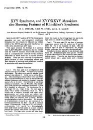
Butler, Reiss, France, and Briddon<br />
1 ii'II-<br />
b&<br />
- C _ .......4.... i4<br />
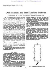
FIG. 1. Partial karyotype <strong>of</strong> a cultured amniotic fluid cell showing the Y chromosome in group G and an extra autosome at position <strong>13</strong>.<br />
d. _<br />
FIG. 2. The D(<strong>13</strong>-15) group from a similar cell treated with trypsin to reveal banding. The pattern is consistent<br />
with a diagnosis <strong>of</strong> trisomy <strong>13</strong>.<br />
which is in preparation. The banding patterns observed<br />
were consistent with a diagnosis <strong>of</strong> trisomy <strong>13</strong><br />
(Fig. 2).<br />
These results were subsequently confirmed using<br />
cultures <strong>of</strong> fetal skin.<br />
Management <strong>of</strong> Pregnancy<br />
The patient was informed immediately <strong>of</strong> the<br />
findings and she elected to have a termination by<br />
abdominal hysterotomy so that she could be sterilized<br />
by tubal ligation at the same time. This was<br />
performed at 20 weeks and the fetus in the intact<br />
gestation sac was removed with minimum damage<br />
for detailed pathological examination. The<br />
patient's postoperative progress was uneventful.<br />
A. Lit<br />
Ac,<br />
a<br />
Necropsy <strong>of</strong> Fetus<br />
The fetus was male and moderately hydropic<br />
(Fig. 3). The weight (442 g), and crown-rump<br />
length (16-5 cm) corresponded to a gestational age <strong>of</strong><br />
about 20 weeks. Over the vertex there was an area<br />
<strong>of</strong> apparent aplasia <strong>of</strong> the scalp measuring 1P9 x<br />
1 2 cm. Microscopically it was sharply differentiated<br />
from the surrounding normal immature<br />
skin and consisted <strong>of</strong> a thin layer <strong>of</strong> avascular myxomatous<br />
tissue the surface <strong>of</strong> which was covered by a<br />
single layer <strong>of</strong> cuboidal epithelium. No accessory<br />
skin organs were present. The ears were lowset but<br />
not otherwise abnormal and the upper lip was long<br />
with a clearly marked filtrum. The posterior half<br />
<strong>of</strong> the hard and s<strong>of</strong>t palate was cleft. There was a