Antenatal Diagnosis of Patau's Syndrome (Trisomy 13) including a ...
Antenatal Diagnosis of Patau's Syndrome (Trisomy 13) including a ...
Antenatal Diagnosis of Patau's Syndrome (Trisomy 13) including a ...

You also want an ePaper? Increase the reach of your titles
YUMPU automatically turns print PDFs into web optimized ePapers that Google loves.
370<br />
Downloaded from<br />
jmg.bmj.com on April 3, 20<strong>13</strong> - Published by group.bmj.com<br />
covered. Finally, in the group where the age <strong>of</strong> the<br />
mother was the important factor, the vast majority<br />
being over 40 years, 368 pregnancies were screened<br />
and 10 affected fetuses were detected, made up <strong>of</strong><br />
nine Down's syndrome (+ 21) and the present case<br />
<strong>of</strong> <strong>Patau's</strong> syndrome (+ <strong>13</strong>).<br />
In the high maternal age group the frequency <strong>of</strong><br />
anomaly <strong>of</strong> 2-7% is close to the figure predictable<br />
from the known frequencies <strong>of</strong> chromosome abnormalities,<br />
with Down's syndrome predominating<br />
as expected. Our experience, based on a 10-year<br />
survey in the North East Metropolitan Area <strong>of</strong><br />
London (population approximately 3-4 million),<br />
indicates that the frequency at birth <strong>of</strong> trisomy <strong>13</strong> is<br />
not greater than 1 in 15,000 after making allowances<br />
for those cases not studied because <strong>of</strong> lack <strong>of</strong> clinical<br />
recognition. Our discovery antenatally <strong>of</strong> a fetus<br />
with this condition is therefore somewhat fortuitous<br />
although, as in Down's syndrome, over half <strong>of</strong> all<br />
the cases are born to women in the 'over 35' age<br />
group.<br />
The social implications <strong>of</strong> the present case are<br />
worthy <strong>of</strong> emphasis. We were dealing with an unplanned<br />
pregnancy and therefore, presumably<br />
initially unwanted, though the mother was prepared<br />
to go to term if the chromosome pattern was<br />
normal. However, the result was adverse and she<br />
was spared the traumatic experience <strong>of</strong> giving birth<br />
to such a grossly abnormal child by having the<br />
pregnancy terminated at mid-term following our<br />
predictions. Tubal ligation was performed at the<br />
same time.<br />
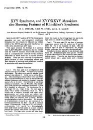
The spectrum <strong>of</strong> anatomical features <strong>of</strong> the fetus<br />
was similar to that found in most examples <strong>of</strong> trisomy<br />
<strong>13</strong> (Snodgrass et al, 1966). Examination <strong>of</strong><br />
an affected fetus <strong>of</strong> this maturity was <strong>of</strong> interest in<br />
that it threw some light on the development <strong>of</strong><br />
arrhinencephaly, a condition where the olfactory<br />
bulbs, tracts, and striae are absent. This is sometimes<br />
associated with developmental failure <strong>of</strong> the<br />
more central parts <strong>of</strong> the rhinencephalon such as the<br />
hippocampus, fimbria, indusium griseum, and<br />
fornices whilst the more severe forms involve agenesis<br />
<strong>of</strong> the corpus callosum or prosencephaly (Crome<br />
Butler, Reiss, France, and Briddon<br />
and Stern, 1972). All grades <strong>of</strong> arrhinencephaly<br />
have been found in cases <strong>of</strong> trisomy <strong>13</strong>. Microscopical<br />
examination <strong>of</strong> this fetus showed that<br />
migration <strong>of</strong> cells from the developing nervous<br />
system to the olfactory epithelium had occurred to<br />
form olfactory nerves <strong>of</strong> about normal size. In<br />
addition the mitral cells <strong>of</strong> the olfactory bulbs had<br />
developed, although in considerably reduced numbers,<br />
and their axons had formed olfactory tracts<br />
too small to be seen by the naked eye. It appears<br />
likely that in some cases <strong>of</strong> so-called arrhinencephaly,<br />
detailed examination would reveal hypoplasia <strong>of</strong> the<br />
olfactory bulbs and tracts as in this example.<br />
The histological features <strong>of</strong> the scalp were similar<br />
to those <strong>of</strong> the only three other cases which we have<br />
examined where the lesion was not infected. In all,<br />
the dermis showed poor development <strong>of</strong> collagen<br />
and the surface epithelium consisted <strong>of</strong> one to three<br />
layers <strong>of</strong> cells with complete absence <strong>of</strong> hair follicles<br />
and sweat glands. It is evident that this condition<br />
is a localized dysplasia <strong>of</strong> skin.<br />
We would like to thank Mr W. K. Sutton, Consultant<br />
Obstetrician, Southend-on-Sea for referring this patient<br />
to us for antenatal screening, Mrs Faith M. Byron for her<br />
technical assistance, and Miss Ivy K. Haverly for typing<br />
the manuscript.<br />
REFERENCES<br />
Butler, L. J. (1972). <strong>Antenatal</strong> detection <strong>of</strong> chromosomal and metabolic<br />
abnormalities. In Mental Retardation: Prenatal <strong>Diagnosis</strong><br />
and Infant Assessment, ed. by C. P. Douglas and K. S. Holt, pp. 1-<br />
16. Butterworth, London.<br />
Butler, L. J. and Reiss, H. E. (1970). <strong>Antenatal</strong> detection <strong>of</strong> chromosome<br />
abnormalities. Journal <strong>of</strong> Obstetrics and Gynaecology<br />
<strong>of</strong> the British Commonwealth, 77, 902-907.<br />
Crome, L. and Stem, J. (1972). Pathology <strong>of</strong> Mental Retardation,<br />
2nd ed., pp. <strong>13</strong>4. Churchill Livingstone, London.<br />
Ferguson-Smith, M. E., Ferguson-Smith, M. A., Nevin, N. C., and<br />
Stone, M. (1971). Chromosome analysis before birth and its<br />
value in genetic counselling. British MedicalJournal, 4, 69-74.<br />
Nadler, H. L. and Gerbie, A. B. (1971). Present status <strong>of</strong> amniocentesis<br />
in intra-uterine diagnosis <strong>of</strong> genetic defects. Obstetrics and<br />
Gyenecology, 38, 789-799.<br />
Seabright, M. (1971). A rapid banding technique from human<br />
chromosomes. Lancet, 2, 971-972.<br />
Snodgrass, G. J. A. I., Butler, L. J., France, N. E., Crome, L., and<br />
Russell, A. (1966). The 'D' (<strong>13</strong>-15) trisomy syndrome. An<br />
analysis <strong>of</strong> 7 examples. Archives <strong>of</strong> Disease in Childhood, 41, 250-<br />
261.