Lab 3: Cnidaria II -- Medusozoa & Anthozoa - Department of Biology
Lab 3: Cnidaria II -- Medusozoa & Anthozoa - Department of Biology
Lab 3: Cnidaria II -- Medusozoa & Anthozoa - Department of Biology

You also want an ePaper? Increase the reach of your titles
YUMPU automatically turns print PDFs into web optimized ePapers that Google loves.
ZOO 2203C / 6927 – LAB 3<br />
CNIDARIA: MEDUSOZOA <strong>II</strong> & ANTHOZOA<br />
Sections with bolded heading required, others optional for IZ, but required for AIZ.<br />
SCYPHOZOAN DIVERSITY<br />
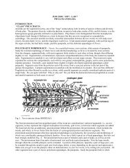
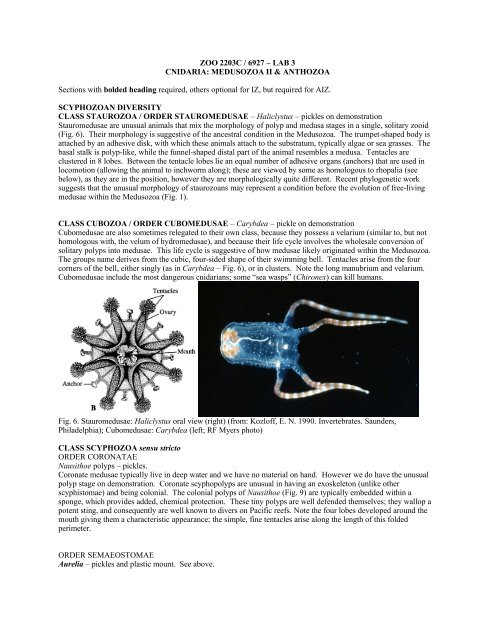
CLASS STAUROZOA / ORDER STAUROMEDUSAE – Haliclystus – pickles on demonstration<br />
Stauromedusae are unusual animals that mix the morphology <strong>of</strong> polyp and medusa stages in a single, solitary zooid<br />
(Fig. 6). Their morphology is suggestive <strong>of</strong> the ancestral condition in the <strong>Medusozoa</strong>. The trumpet-shaped body is<br />
attached by an adhesive disk, with which these animals attach to the substratum, typically algae or sea grasses. The<br />
basal stalk is polyp-like, while the funnel-shaped distal part <strong>of</strong> the animal resembles a medusa. Tentacles are<br />
clustered in 8 lobes. Between the tentacle lobes lie an equal number <strong>of</strong> adhesive organs (anchors) that are used in<br />
locomotion (allowing the animal to inchworm along); these are viewed by some as homologous to rhopalia (see<br />
below), as they are in the position, however they are morphologically quite different. Recent phylogenetic work<br />
suggests that the unusual morphology <strong>of</strong> staurozoans may represent a condition before the evolution <strong>of</strong> free-living<br />
medusae within the <strong>Medusozoa</strong> (Fig. 1).<br />
CLASS CUBOZOA / ORDER CUBOMEDUSAE – Carybdea – pickle on demonstration<br />
Cubomedusae are also sometimes relegated to their own class, because they possess a velarium (similar to, but not<br />
homologous with, the velum <strong>of</strong> hydromedusae), and because their life cycle involves the wholesale conversion <strong>of</strong><br />
solitary polyps into medusae. This life cycle is suggestive <strong>of</strong> how medusae likely originated within the <strong>Medusozoa</strong>.<br />
The groups name derives from the cubic, four-sided shape <strong>of</strong> their swimming bell. Tentacles arise from the four<br />
corners <strong>of</strong> the bell, either singly (as in Carybdea – Fig. 6), or in clusters. Note the long manubrium and velarium.<br />
Cubomedusae include the most dangerous cnidarians; some “sea wasps” (Chironex) can kill humans.<br />
Fig. 6. Stauromedusae: Haliclystus oral view (right) (from: Kozl<strong>of</strong>f, E. N. 1990. Invertebrates. Saunders,<br />
Philadelphia); Cubomedusae: Carybdea (left; RF Myers photo)<br />
CLASS SCYPHOZOA sensu stricto<br />
ORDER CORONATAE<br />
Nausithoe polyps – pickles.<br />
Coronate medusae typically live in deep water and we have no material on hand. However we do have the unusual<br />
polyp stage on demonstration. Coronate scyphopolyps are unusual in having an exoskeleton (unlike other<br />
scyphistomae) and being colonial. The colonial polyps <strong>of</strong> Nausithoe (Fig. 9) are typically embedded within a<br />
sponge, which provides added, chemical protection. These tiny polyps are well defended themselves; they wallop a<br />
potent sting, and consequently are well known to divers on Pacific reefs. Note the four lobes developed around the<br />
mouth giving them a characteristic appearance; the simple, fine tentacles arise along the length <strong>of</strong> this folded<br />
perimeter.<br />
ORDER SEMAEOSTOMAE<br />
Aurelia – pickles and plastic mount. See above.
Chrysaora – sea nettles – pickles, live when available<br />
Chrysaora is one <strong>of</strong> the common local scyphomedusae, with seasonal blooms on both coasts <strong>of</strong> Florida. Their<br />
abundance and nasty stings make them a well-known hazard to swimmers and net fishermen. Note that both the<br />
tentacles and oral arms are long – they serve as efficient fishing nets for these highly carnivorous animals.<br />
ORDER RHIZOSTOMAE<br />
Cassiopeia – pickles, live when available<br />
The upside down jellyfish is a common inhabitant <strong>of</strong> calm, shallow, tropical waters in Florida and elsewhere. It is an<br />
unusual medusa that typically rests upside down on the bottom (Fig. 9). Like other rhizostome (“root mouth”)<br />
medusae, it is microphagous – feeding on tiny organisms captured by the enlarged and highly branched oral arms, the<br />
tips <strong>of</strong> which open into large numbers <strong>of</strong> tiny mouths! It is also photosymbiotic and derives substantial nutrition<br />
from unicellular algal symbionts (zooxanthellae).<br />
Fig. 9. Coronatae: Nausithoe polyps, embedded in symbiotic sponge (left; GP photo); Cassiopeia (right; from<br />
Brusca, R.C. & G.J. Brusca. 2003. Invertebrates. 2nd Ed..)<br />
CLASS ANTHOZOA<br />
<strong>Anthozoa</strong> lack a medusa stage and have complex polyps, with a large gastrovascular cavity partially subdivided by<br />
numerous mesenteries and a pharynx (stomodaeum). This increased surface area allows anthozoan polyps to reach<br />
much larger sizes (to 1m!) than the simple medusozoan polyps. Two subclasses are currently recognized, the<br />
Hexacorallia, a heterogeneous group with highly variable polyp morphology and a tendency for six-fold symmetry,<br />
and the Octocorallia, a group in which polyp (but not colony) morphology is highly conserved and polyps always<br />
have 8 tentacles and mesenteries.<br />
SUBCLASS HEXACORALLIA<br />
HEXACORAL MORPHOLOGY: slides <strong>of</strong> transverse sections <strong>of</strong> Metridium, live Aiptasia.<br />
We will use actinian anemones to introduce the morphology <strong>of</strong> anthozoan polyps. Note that anemones take polyp<br />
complexity and size to its limits among cnidarians; some reach a meter in diameter or height, and have fleshy bodies<br />
with large numbers <strong>of</strong> mesenteries. Start your study by noting the external morphology <strong>of</strong> live Aiptasia. Most<br />
anemones are attached to hard bottom by a flat pedal disk and a cylindrical column, ending in wide oral disk. The<br />
mouth is at the center <strong>of</strong> the oral disk, while tentacles encircle (as in Aiptasia), or cover, the disk. Note the radiating<br />
lines on the oral disk <strong>of</strong> Aiptasia between the mouth and tentacles; these are the margins <strong>of</strong> the mesenteries that<br />
partially subdivide the gastrovascular cavity. Note also that the tentacles always arise in spaces between mesenteries,<br />
and the part <strong>of</strong> the gastrovascular cavity trapped between the mesenteries extends into the tentacle. The mouth <strong>of</strong><br />
hexacorals is usually compressed and slit-like, with one or two siphonoglyphs at its end(s). As a result hexacorals<br />
tend to depart from perfect radial symmetry to biradial symmetry. Siphonoglyphs are ciliated furrows that pump<br />
water into the gastrovascular cavity.<br />
Study the transverse and longitudinal sections <strong>of</strong> Metridium next, with Fig. 10 as your guide. We have several slides,<br />
and these differ substantially, so you may wish to look at more than one, as some features are clearer in some slides<br />
than others. Note that the oral-disk invaginates around the mouth, forming a pharynx that extends down into the<br />
column. The pharynx (or stomodaeum) is a characteristic <strong>of</strong> anthozoan polyps, absent in medusozoans. The<br />
transverse sections we have cut across the portion <strong>of</strong> the column with the pharynx (as also illustrated in the diagram<br />
below). You can clearly distinguish the outer epidermis, inner gastrodermis, and the sandwiched mesogloea on the
ody wall <strong>of</strong> the column. Note that circular muscle fibers run within the mesogloea, and permit contraction and thus<br />
elongation <strong>of</strong> the column. The body wall is also visible in the pharynx, but in reverse: here the innermost layer is<br />
epidermis, while the outermost layer is gastrodermis, as the pharynx is an invaginated structure. The gastrovascular<br />
cavity is subdivided by a series <strong>of</strong> radiating, vertical, sheet-like mesenteries. Note that the mesenteries come in pairs<br />
in actinians; however this is not the case in all anthozoans. The paired nature <strong>of</strong> actinian mesenteries may be<br />
evidence that actinians evolved from corals that lost their skeleton, such that in the past, calcareous, skeletal septa<br />
ran between such paired fleshy mesenteries, as they do in living corals (see later). Note the bulges (retractor muscle<br />
bundles) on the paired mesenteries are arranged to either face outward on both, or inward on both. Those<br />
mesenteries that reach and fuse with the pharynx are called complete; those that do not are termed incomplete. In<br />
small Metridium, as featured in most slides, there are 6 pairs <strong>of</strong> complete mesenteries – demonstrating the 6-fold<br />
symmetry <strong>of</strong> hexacorals. Note the numerous additional incomplete mesenteries <strong>of</strong> various sizes, again in pairs (these<br />
also tend to occur in multiples <strong>of</strong> 6). Mesenteries are added in a specific order during growth, the shorter ones being<br />
usually the most recently added. Mesenteries form as folds <strong>of</strong> the gastrodermis, with a mesogloeal core.<br />
Study a large incomplete mesentery on the slide (Fig. 10). Note the bulging retractor muscles (absent from small<br />
mesenteries), which can work as antagonists <strong>of</strong> the circular muscles <strong>of</strong> the column wall, with the gastrovascular<br />
cavity used as a hydrostatic skeleton, with the animal closing its mouth tightly. You may also see developing<br />
oocytes in more distal parts <strong>of</strong> the mesenteries, although the slides we have are largely from small, pre-reproductive<br />
juveniles. The innermost margin <strong>of</strong> a free mesentery is thickened into a mesenterial filament. This is like the thick<br />
cord sewn onto the edge <strong>of</strong> a curtain. The mesenterial filament is the results <strong>of</strong> three cord-like thickenings at the free<br />
end <strong>of</strong> the mesentery in hexacorals (<strong>of</strong>ten appearing as a trefoil in transverse sections – Fig. 10): a pair <strong>of</strong> ciliated<br />
bands that functions to circulate gastrovascular contents, and a cnido-glandular band that secretes digestive enzymes<br />
and also subdues swallowed prey with nematocysts. At the base <strong>of</strong> the anemone, where the mesenteries attach to the<br />
pedal disk, the mesenterial filament <strong>of</strong>ten does not terminate, but continues on, like a curtain cord that extend beyond<br />
and dangles <strong>of</strong>f the edge <strong>of</strong> a curtain. These free, nematocyst-laden mesenterial filaments are also called acontia.<br />
They can be used to subdue prey within the gastrovascular cavity, as well as in aggressive and defensive interactions<br />
when extruded from the body. Acontia are readily extruded by some hexacorals when they are disturbed, or when<br />
they interact aggressively with competing neighbors or predators. They can emerge through the mouth, be forced<br />
through the body wall, or come out through specialized pores in the body wall (=cinclides, see later).
Fig. 10. Anemone morphology: cut away <strong>of</strong> anemone (upper left); transverse section at level <strong>of</strong> actinopharynx<br />
(upper right); transverse section <strong>of</strong> incomplete mesentery (lower left); transverse section through mesenterial<br />
filament at end <strong>of</strong> free mesentery, with trefoil end (lower right) (from BIODIDAC)<br />
Once you are done with the slides, go back to the live anemone, Aiptasia, to examine it further. Share animals in<br />
groups <strong>of</strong> 4 to spare them. Start by studying the effectiveness <strong>of</strong> muscles vs. cilia by stimulating an extended animal<br />
until it contracts using its musculature; time this response. Now time how long it takes for the animal to re-expand.<br />
Which takes longer? Why? When you disturbed it, Aiptasia will likely discharge acontia in defense. Where do the<br />
acontia emerge from the body? What color are they? Take a piece <strong>of</strong> acontia and place it on a microscope slide,<br />
apply cover glass, and view under the compound scope to see the large nematocysts. By adding a drop <strong>of</strong> methylene<br />
blue, you can get the nematocysts to discharge. You may also want to look at a tentacle snipped from Aiptasia (they<br />
will readily regenerate it), to see the gastrovascular cavity running through it and the small (~8 μm) algal cells<br />
(zooxanthellae) that occur intracellularly within the gastrodermis and give Aiptasia its brown color. The same algae<br />
are the photosymbiotic partners <strong>of</strong> reef corals, s<strong>of</strong>t corals, and many other anthozoans. Aiptasia is abundant in our<br />
tanks as it buds asexually – it is actually a nuisance organism both in aquaria and in aquaculture settings (e.g. pearl<br />
farms) because <strong>of</strong> its habit to cover everything by its rapidly spreading clones.<br />
HEXACORAL DIVERSITY<br />
The Hexacorallia includes stony corals, black corals, and a diversity <strong>of</strong> anemones, and are differentiated from the<br />
other anthozoan group, the Octocorallia mainly on polyp morphology. While octocoral polyps are very conservative<br />
in their form and size, having 8 pinnately branched tentacles and 8 mesenteries, hexacoral polyps are highly variable,<br />
although they tend to have a 6-based symmetry in mesenteries and tentacles. The number <strong>of</strong> tentacles can vary from<br />
six to hundreds, as a result <strong>of</strong> both a variation in the number <strong>of</strong> mesenteries and in the number <strong>of</strong> tentacles per<br />
mesentery. Many hexacorals have but a single tentacle per mesentery and thus a ring <strong>of</strong> tentacles at the periphery <strong>of</strong><br />
the oral disk, while others have numerous tentacles per mesentery, covering the oral disk in a shag-carpet-like<br />
manner. Polyps range from 1m in diameter, and may be solitary or colonial. The Hexacorallia include<br />
two orders with well-developed skeletons: the Scleractinia (stony corals) with a calcareous skeleton and the<br />
Antipatharia (black corals) with an organic skeleton. They also include four orders that lack a solid skeleton,<br />
referred to broadly (sensu lato) as sea anemones: the Actiniaria, Corallimorpharia, Zoanthidea, and Ceriantharia.<br />
Among anemones the actinians are the most diverse, both in terms <strong>of</strong> species and body form. In contrast members <strong>of</strong><br />
other orders are more conservative in form and thus more recognizeable. Corallimorpharians are closely related to<br />
actinians; like actinians, but unlike other anemones, their mesenteries are arranged in pairs (a condition that may be<br />
evidence <strong>of</strong> the ultimate origin <strong>of</strong> these groups from skeletonized ancestors). Most tropical corallimorpharians occur<br />
in dense clonal aggregates, have very wide disks attached to <strong>of</strong>ten much narrower columns, and have short or even<br />
just bump-sized tentacles arranged in rows radiating from the mouth. Zoanthids are usually clonal or colonial<br />
anemones, with medium-sized (mostly 0.5-2cm) polyps, tentacles restricted to the periphery <strong>of</strong> the oral disk and<br />
alternating between an inner, erect and an outer, reflexed series. Cerianthids are burrowing, tube-dwelling anemones<br />
that usually live in sand or mud, with only their oral disk and tentacles emergent. They are readily differentiated<br />
from burrowing actinians in the field by the possession <strong>of</strong> tubes and arrangement <strong>of</strong> tentacles in two tiers: an outer<br />
ring <strong>of</strong> longer tentacles surrounding an inner cluster <strong>of</strong> shorter tentacles. Actinians, corallimorphs, and scleractinians<br />
are closely related, even potentially not monophyletic relative to each other, while the other hexacoral orders are<br />
quite divergent from these.<br />
Order Actiniaria: Calliactis, Condylactis, Epicystis Bunediopsis, Aiptasia, etc. – live.<br />
Actinians are by far the most diverse (1000+ species) group <strong>of</strong> sea anemones, and <strong>of</strong>ten considered to be sea
anemones in the narrow sense (sensu stricto). They are s<strong>of</strong>t-bodied, solitary anthozoans that generally lack a<br />
secreted skeleton, although a few can form a chitinous cuticle under their pedal disk. They resemble the three other<br />
groups <strong>of</strong> anemones: corallimorpharians, cerianthids, and zoanthids, and are most easily differentiated by the unique<br />
characters <strong>of</strong> these other, smaller, and more homogeneous groups (see above and below). Actiniarians have an oral<br />
disk that bears the mouth and tentacles, a column, and, in all but burrowing species, an adhesive, pedal disk that<br />
attaches to hard bottom. The tentacles range widely in shape, can be simple, branched, or more complex, and there<br />
may be more than one type <strong>of</strong> tentacle in an animal. Tentacles may be restricted to the margin <strong>of</strong> the oral disk, or<br />
cover most <strong>of</strong> it. The column may have verrucae, wart-like extensions that can be adhesive and help anchor the<br />
column to the substratum, or adhere sediment grains to the body. Although the nematocysts <strong>of</strong> most anemones do<br />
not affect humans, several species have nasty stings, and some readily feed on fishes foolish enough to brush against<br />
them. The sticky feeling to the tentacles <strong>of</strong> many anemones is due to the discharge <strong>of</strong> nematocysts (try this with<br />
some <strong>of</strong> the larger species – don’t worry, none <strong>of</strong> our pets are harmful stingers). Because <strong>of</strong> their stinging power<br />
anemones are favored as shelter by numerous animals, including copepods, shrimp, porcellanid crabs, and fish.<br />
Most larger anemones that live on coral reefs are photosymbiotic, harboring symbiotic din<strong>of</strong>lagellate algae<br />
(zooxanthellae) that contribute to their nutrition. Although actinians are solitary (with a single, recently discovered<br />
exception), many species are clonal and live in aggregations produced by a variety <strong>of</strong> modes <strong>of</strong> asexual reproduction.<br />
Examine the diverse anemones provided; note that some are in the large display aquaria in the back <strong>of</strong> the lab and<br />
downstairs. Which species do you think are zooxanthellate? How many circles <strong>of</strong> tentacles do each have?<br />
Calliactis is an unusual anemone in two senses. First, it commonly lives in symbiotic associations with hermit crabs,<br />
attached to their shells, giving the crab protection and benefiting from their messy eating habits. Second, it has<br />
unusual, pimple-shaped perforations on the sides <strong>of</strong> its column, called cinclides. These are specialized openings<br />
through which acontia are extruded when the animals are bothered. Calliactis is easily irritated; watch the<br />
demonstration <strong>of</strong> acontial discharge given by your TA. What color are its acontia?<br />
Order Corallimorpharia: live Discosoma, Ricordea<br />
Corallimorpharians include two rather different groups <strong>of</strong> anemones. One group (Discosomatidae & Ricordeidae) is<br />
comprised <strong>of</strong> large, zooxanthellate, warm-water species characterized by short, disc-shaped, flaring columns and<br />
wide oral disks, with radiating rows <strong>of</strong> short to barely bump-sized tentacles; these tend to live in large, clonal<br />
aggregates. This group lives on coral reefs and is represented by live specimens in the lab. The second group<br />
(Corallimorphidae) is characterized by azooxanthellate polyps sporting elongate tentacles with bulbous ends,<br />
arranged again, in radiating rows. These are known from temperate to tropical waters, including deep water, but are<br />
less common. Corallimorphs are so named because they resemble stony corals in their polyp anatomy, but lack a<br />
skeleton.<br />
Order Scleractinia: Acropora, Fungia, Manicina, Astrangia, etc. skeletons, plus live Oculina, Solenastrea,<br />
Euphyllia, Caulastrea.<br />
The Scleractinia are the “stony corals”. The word “coral” is applied more broadly to cnidarians with a solid<br />
carbonate skeleton, while “stony coral” refers to scleractinians. The vast majority <strong>of</strong> corals are scleractinians, with<br />
>1300 described species. Scleractinian corals are characterized by an aragonitic skeleton, with each polyp<br />
occupying a cup-like depression (calice) with radiating calcareous septa – skeletal elements that lie between fleshy<br />
mesenteries. Scleractinians can be either solitary, or more commonly, colonial. Scleractinians are the architects <strong>of</strong><br />
tropical coral reefs, and about half <strong>of</strong> the known species are reef builders. The rest live in a variety <strong>of</strong> habitats from<br />
the tropics to the poles, from shallow to deep water. While reef-dwelling species are mostly photosymbiotic, high<br />
latitude and deep water species lack symbiotic algae.<br />
Examine live scleractininans to see the range <strong>of</strong> polyp morphologies. Note that some species have large fleshy<br />
polyps that tend to be continuously extended (e.g. Euphyllia), while others have small, thin polyps, or have polyps<br />
that are largely withdrawn into the skeleton during the day (these tend to extend their tentacles at night (e.g.<br />
Caulastrea). While most corals have simple, finger-like or bulbous-tippes tentacles, others have branching tentacles<br />
(some Euphyllia). Note the brownish and greenish colors caused by an abundance <strong>of</strong> zooxanthellae. Look at the<br />
varied colony shapes formed by corals among the live and skeletal specimens provided, as well as the varied sizes<br />
and arrangements <strong>of</strong> polyps. Calices are <strong>of</strong>ten well defined, circular, with a wall delimiting them from the<br />
surrounding colonial coenenchyme and coenosteum (the skeletal surface underlaying the coenenchyme) (Fig. 1).<br />
New polyps can form either by the subdivision <strong>of</strong> existing polyps, or arise de novo from the coenosteal surface. Try<br />
to find examples <strong>of</strong> both kinds <strong>of</strong> new polyps in the skeletal material. In brain corals, new polyps tend to not lay<br />
down walls to separate them from other polyps in their row and thus remain in a single valley. The multiple polyps<br />
in such valleys are given away by their individual mouths, lying in series. Find the basic skeletal elements on the
coral skeleton: coenosteum, calices, walls, septa (radial elements inside the wall), costae (radial elements outside the<br />
wall, <strong>of</strong>ten contiguous with septa), and columella (in center <strong>of</strong> calices) (Fig. 1).<br />
Fig. 1. Scleractinian skeleton<br />
Order Zoanthidea: Palythoa pickles; live zoanthids in aquarium<br />
Zoanthids are a distinctive group <strong>of</strong> anemone-like anthozoans that have characteristic, relatively small polyps (3-20<br />
mm diameter), with short, finger-like tentacles restricted to the periphery <strong>of</strong> the oral disk and arranged in a<br />
distinctive, inner, erect and outer, reflexed cycle. This alteration <strong>of</strong> tentacles is clearly visible in expanded polyps<br />
and readily identifies live zoanthids (Fig. 2). Zoanthids have a single siphonoglyph, and alternating perfect (large)<br />
and imperfect (small) mesenteries. Polyps tend to be trumpet-shaped, with an expanded oral disk arising from a<br />
narrower column, although some colonial species have the column embedded in coenenchyme. They lack a secreted<br />
skeleton (except in one poorly known genus), but several genera incorporate sand grains in their mesogloea, which<br />
makes them very gritty. Other genera (e.g. Zoanthus) do not do this, and are s<strong>of</strong>t, and slippery. Zoanthids occur<br />
world-wide, but are especially common on coral reefs, where they can carpet large areas through clonal propagation.<br />
They are usually colonial, but a few species are solitary. Palythoa is one <strong>of</strong> the most common tropical zoanthids,<br />
characterized by a well developed colonial coenenchyme, heavily impregnated with sand. Reef-dwelling zoanthids<br />
always/almost always harbor zooxanthellae.<br />
Fig. 2. Zoanthid polyp (left): note how the tentacles are alternately held up and down, as typical for the order.<br />
Ceriantharian (right), not the two circles <strong>of</strong> tentacles; labial tentacles are marked with an arrow.<br />
Order Ceriantharia: live and pickled Cerianthiopsis<br />
Cerianthids are tube-dwelling anemones that differ so much from other hexacorallian anemones that some authors<br />
separate them into a different subclass. Unlike any other anthozoan, they live in leathery tubes, woven from<br />
discharged ptychocysts – specialized cnidae unique to these animals. They tend to live in and bury into s<strong>of</strong>t
substrata, and their tubes can extend into the sediment several times the length <strong>of</strong> the animals, <strong>of</strong>fering ample cover<br />
to withdraw into. And withdraw they can: cerianthids are unique among cnidarian polyps in being able to respond<br />
very quickly to disturbance, withdrawing with lightening speed. Onlike other anemones that have a sphincter muscle<br />
at the base <strong>of</strong> their oral disk that can close over withdrawn tentacles, ceriantharians lack this and are unable to<br />
withdraw their tentacles. They lack a pedal disk, all their mesenteries are complete, and they possess two tiers <strong>of</strong><br />
tentacles: an outer ring <strong>of</strong> longer tentacles surrounding an inner circle <strong>of</strong> labial tentacles. Examine the live<br />
Cerianthiopsis americanus provided, observing their habit, and tentacular crown. Gently touch the tentacles to see<br />
their rapid response. Note the elongate, vermiform shape <strong>of</strong> pickled or live animals that have been taken out <strong>of</strong> their<br />
tubes. Place a small piece <strong>of</strong> tube material under the scope to see how they are constructed.<br />
Order Antipatharia: dried or pickled<br />
The Antipatharia, or black corals, are a homogeneous group <strong>of</strong> colonial anthozoans that secrete an arborescent or<br />
whip-like, scleroproteinaceous skeleton. Although colonies superficially resemble gorgonians (see below), they can<br />
be readily distinguished in lacking the sclerite-filled rind that surrounds the axial skeleton <strong>of</strong> that group, in having the<br />
simplest polyps among hexacorals, with 6 simple (rather than 8 pinnate) tentacles (Fig. 3), and also by the thorny<br />
protuberances arising from the skeleton in many species. The polyps are obvious in the field, as they cannot<br />
withdraw into the coenenchyme, but are hard to recognize in dried specimens. Black corals range from the tropics to<br />
temperate latitudes, and are especially common in deep water <strong>of</strong>f reefs. The skeleton <strong>of</strong> large black corals is used to<br />
make jewelry, fetches a high price, and supports a deep diving industry in parts <strong>of</strong> the tropics.<br />
Fig. 3. Antipatharian branch showing simple, laterally compressed polyps with 6 tentacles (left); organization <strong>of</strong> a<br />
gorgonian octocoral (right; from BIODIDAC)<br />
SUBCLASS OCTOCORALLIA<br />
OCTOCORAL MORPHOLOGY- Model <strong>of</strong> Corallium, whole mount slides, piece <strong>of</strong> Leptogorgia, various living<br />
octocorals.<br />
We will look at octocoral morphology using gorgonians as a model. Gorgonians are the most common and diverse<br />
octocorals in our regions. They are a polyphyletic assemblage characterized by the possession <strong>of</strong> an axial tree- or<br />
fan-like skeleton made <strong>of</strong> scleroprotein and/or CaCO3. Look at the model <strong>of</strong> an octocoral (Corallium, the precious<br />
coral, a gorgonian with a calcareous axial skeleton) for a quick overview <strong>of</strong> octocoral organization. Note that polyp<br />
morphology, with 8 pinnate tentacles (pinnae are paired, lateral side branches), 8 mesenteries, 1 siphonoglyph, is<br />
rigidly conserved in this large subclass. In colonial octocorals there are masses <strong>of</strong> gastrovascular canals (solenia)<br />
that extend from the central gastrovascular cavity, permeate the coenenchyme, and allow direct food sharing among<br />
zooids.<br />
Study the whole mount slide <strong>of</strong> a gorgonian branch under low magnification to get a closeup view <strong>of</strong> polyp<br />
organization (Fig. 3). Note the pinnate tentacles. The long, thin, gut-like tube in the center <strong>of</strong> the polyps represents<br />
the space outlined by the inner margins <strong>of</strong> the mesenterial filaments that extend lengthwise through the polyp; these<br />
diverge and become curly below the extended polyp within a wide gastrovascular cavity. Note the solenia that<br />
connect these larger cavities and permeate the rind. The large central rod in the slide represents the axial skeleton <strong>of</strong><br />
gorgonians. In some slides it has a calcarous core, surrounded by a tough, organic, scleroproteinaceous skeleton,<br />
while other slides are <strong>of</strong> a species with a strictly scleroproteinaceous skeleton. Note that the rind also has abundant<br />
sclerites (=”spicules”), but these have been removed by acids during the preparation <strong>of</strong> these slides.
Take a small piece <strong>of</strong> a branch <strong>of</strong> Leptogorgia, our common local gorgonian. Note that in disturbed specimens the<br />
polyps can completely withdraw into the coenenchyme. In gorgonians the coenonchyme is usually so loaded with<br />
sclerites that give the rind a gritty toughness. Place a small branch segment into bleach to digest the rind, and thus<br />
expose the sclerites; mount the sclerites on a slide and examine.<br />
Study the expanded polyps <strong>of</strong> the various undisturbed, living octocorals in the aquaria. Note that the polyps <strong>of</strong> most<br />
octocorals can readily withdraw into the colonial coenenchyme, thus seeing extended ones is a treat – don’t disturb<br />
these animals. Note also that the polyps in some, or parts <strong>of</strong> some colonies, are indeed withdrawn, leaving little<br />
more evidence <strong>of</strong> their presence than a small dimple on the coenenchyme.<br />
OCTOCORAL DIVERSITY<br />
Unlike the relatively well-defined orders within the Hexacorallia, the classification <strong>of</strong> octocorals is less clear and is<br />
undergoing considerable flux at present. Octocorals have traditionally been classified into three orders:<br />
Coenothecalia (the blue coral, Heliopora), the Pennatulacea (sea pens and sea pansies), and the Alcyonacea, a large,<br />
heterogeneous group representing the rest <strong>of</strong> the octocorals. In the past people have also subdivided the Alcyonacea<br />
into multiple orders based on their level <strong>of</strong> colony organization. Recent molecular data suggest that both the<br />
Coenothecalia and Pennatulacea are specialized, relatively young <strong>of</strong>fshoots, and that the Alcyonacea is highly<br />
heterogeneous, with several traditional taxa poly- and paraphyletic. In this lab we will introduce you to some <strong>of</strong> the<br />
major groups <strong>of</strong> octocorals, without dwelling on their taxonomic relationships, for this reason we will not use<br />
taxonomic ranks to refer to these groups. Colony and skeletal organization have been the main considerations<br />
shaping alcyonacean taxonomy.<br />
Stolonifera: Carijoa – live & pickles.<br />
Stoloniferans are among the simplest octocorals; polyps arise by budding along stolons and there is little or no<br />
colonial coenenchyme. As a result colonies resemble those <strong>of</strong> hydroids, the stolons analogous to the hydrocaulus <strong>of</strong><br />
hydrozoan colonies. Stoloniferans can have creeping stolons that adhere to the substratum, or can grow into tree-like<br />
forms like Carijoa (these latter are <strong>of</strong>ten referred to as telestaceans). Note the simple polyps and connecting stolons.<br />
Stolonifera: Tubipora, the organ-pipe coral.<br />
Tubipora is unique among octocorals in having the sclerites fused into a more-or-less solid skeleton. Each polyp<br />
occupies one <strong>of</strong> the “organ-pipes” so produced, and thus is largely separate from its neighbors. Like other<br />
stoloniferans, Tubipora lacks a coenenchyme.<br />
“Alcyoniina”: s<strong>of</strong>t corals – pickled Lobophytum, Sarcophyton, Sinularia, live xeniids & nephtheids.<br />
Study the diverse s<strong>of</strong>t corals provided: live in our aquaria, and pickled up front. S<strong>of</strong>t corals are octocorals that<br />
develop a large, fleshy coenenchyme (colonial tissue between polyps). Polyps in many species can withdraw entirely<br />
into the coenenchyme. In addition to housing the polyps, the coenenchyme is loaded with sclerites, and solenia<br />
permeate it to provide nutrition throughout colony. The coenenchyme allows s<strong>of</strong>t corals to grow in a variety <strong>of</strong><br />
shapes: mats, massive colonies, to varied arborescent forms. Many, but not all s<strong>of</strong>t corals have specialized<br />
siphonozooids (Fig. 4), reduced polyps that lack tentacles, but have an efficient siphonoglyph; these inflate the<br />
colony by drawing water into the coelenteron. When polyps retract, siphonozoods are recognizable by their smaller<br />
opening than those <strong>of</strong> retracted gastrozooids, clearly visible on the surface <strong>of</strong> pickled Lobophytum or Sarcophyton.<br />
The siphonozooids also are the reproductive zooids in those s<strong>of</strong>t corals and gorgonians that have them, except sea<br />
pens. Study the variation in growth form <strong>of</strong> pickled and live s<strong>of</strong>t corals. Note also that the body <strong>of</strong> many species (all<br />
the pickled exemplars, for example) are packed solid with sclerites, and some <strong>of</strong> these sclerites are so large that they<br />
are easily discernible by the naked eye. S<strong>of</strong>t corals are abundant and locally dominant on Indo-Pacific reefs, from<br />
where all the pickled as well as live forms we have came from, but are effectively absent from Atlantic reefs. The so<br />
called Atlantic “s<strong>of</strong>t corals” are gorgonians. Many reef-associated s<strong>of</strong>t corals, including all those in the lab, are<br />
zooxanthellate. New research suggests that s<strong>of</strong>t corals are not a monophyletic group.
Fig. 4. Surface <strong>of</strong> s<strong>of</strong>t coral with siphonozooids (Sarcophyton; left); the sea pensie Renilla (right)<br />
Gorgonians (Scleraxonia, Holaxonia & Calcaxonia) – diverse pickles, live Leptogorgia and others.<br />
Study the diversity <strong>of</strong> pickled gorgonians and examine live Leptogorgia. Gorgonians are arborescent (tree-like)<br />
octocorals with an axial skeleton <strong>of</strong> gorgonin (a scleroprotein) and/or CaCO3. Gorgonians are diverse and<br />
traditionally divided into three orders (see above), largely based on the nature <strong>of</strong> their axial skeleton, although new<br />
research is showing that their relationships to each other and to other octocorals are more complex. Gorgonians have<br />
a well-developed coenenchyme, like s<strong>of</strong>t corals; they are effectively s<strong>of</strong>t corals on a stick. As in s<strong>of</strong>t corals, polyps<br />
can generally withdraw into the coenenchyme, and they share nutrients across abundant solenia (the model you<br />
studied earlier is <strong>of</strong> Corallum, a gorgonian with a calcitic axial skeleton). The rind <strong>of</strong> gorgonians is usually thinner<br />
than that <strong>of</strong> s<strong>of</strong>t corals and so full <strong>of</strong> sclerites as to be quite gritty. Study the diversity <strong>of</strong> growth forms among<br />
pickled gorgonians. Take a small piece <strong>of</strong> rind from some, place in a syracuse dish, cover with bleach, then wash<br />
with water, mount the sclerites on a slide, cover with cover slip, and examine them. Can you tell different species<br />
apart based on their sclerites?<br />
Coenothecalia: Heliopora – dry skeleton & photos <strong>of</strong> live colonies<br />
Heliopora coerulea, the only definite species <strong>of</strong> this group, is the only octocoral that is a “real” coral in the sense<br />
that it possesses a solid, CaCO3 skeleton (Tubipora’s skeleton is but weakly fused sclerites). The skeleton is built <strong>of</strong><br />
crystalline aragonite; the blue color derives from iron salts. Note the fine texture <strong>of</strong> the skeleton and the small<br />
openings from which the small, typical, octopolyps emerge, and into which they can withdraw. Polyps may be<br />
extended or withdrawn during the day. Check the photos <strong>of</strong> live Heliopora, to see the whitish polyps. Heliopora is<br />
zooxanthellate, and is abundant on Indo-Pacific reefs, its skeleton is a major component in many reef limestones. It<br />
is a “living fossil”: the genus has persisted largely unchanged since the Cretaceous, the age <strong>of</strong> dinosaurs.<br />
Pennatulacea: sea pens – pickled Renilla, Virgularia, Ptilosarcus, live Renilla if we are lucky<br />
Sea pens are the most complex <strong>of</strong> octocorals, they are a distinctive, readily recognizeable clade. Their<br />
distinctiveness have led to their treatment as a distinct order in the past, but recent work suggests that they are related<br />
to one particular family <strong>of</strong> gorgonians (Ellisellidae)! Sea pens have three types <strong>of</strong> zooids: the primary zooid that<br />
forms the axis <strong>of</strong> the colony, usually with a support rod running through it (absent in the aberrant Renilla) and a rootlike<br />
base that is anchored within the sediment. Other zooids are <strong>of</strong>ten organized in leaf-like branches that commonly<br />
project laterally from the colony axis (though again not in Renilla – which has them all on a single leafy surface).<br />
These are large, typical gastrozooids, and much smaller siphonozooids that draw water into the vast, colonial<br />
gastrovascular cavity. The gastrovascular cavity permeats the colony, and includes the large cavity running through<br />
the axis <strong>of</strong> the primary zooid. Sea pens live on s<strong>of</strong>t bottoms, anchored into sand or mud with the root-like basal<br />
extension <strong>of</strong> the primary polyp. Sea pens are common in a variety <strong>of</strong> environment from tropical reefs to cold waters,<br />
and are notable as perhaps the most diverse cnidarian group in the deep sea. Study the local sea pansy (Renilla) up<br />
close, and examine the other (more typical) genera on demonstration (Fig. 4).