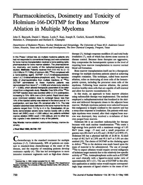
Pharmacokinetics, Dosimetry and Toxicity of Holmium-166-DOTMP ...
Pharmacokinetics, Dosimetry and Toxicity of Holmium-166-DOTMP ...
Pharmacokinetics, Dosimetry and Toxicity of Holmium-166-DOTMP ...

Create successful ePaper yourself
Turn your PDF publications into a flip-book with our unique Google optimized e-Paper software.
<strong>Pharmacokinetics</strong>, <strong>Dosimetry</strong> <strong>and</strong> <strong>Toxicity</strong> <strong>of</strong><br />
<strong>Holmium</strong>-<strong>166</strong>-<strong>DOTMP</strong> for Bone Marrow<br />
Ablation in Multiple Myeloma<br />
John E, Bayouth, Daniel J. Macey, Leela P. Kasi, Joseph R, Garlich, Kenneth McMillan,<br />
Meletios A. Dimopoulos <strong>and</strong> Richard E. Champlin<br />
Departments <strong>of</strong>Radiation Physics, Nuclear Medicine <strong>and</strong> Hematolo@ji, The University <strong>of</strong> Texas M.D. Anderson Cancer<br />
Center, Houston, Texas<strong>and</strong> Research <strong>and</strong> Developme,u, The Dow Chemical Company, Freepoi@,Texas<br />
In this Phase I dinicalthai,six multiplemyelomapatientswho<br />
therapy (5), biologic-response modifiers(6) <strong>and</strong> total body<br />
irradiation(7), each <strong>of</strong> which has shown some success in<br />
hadnotrespondedto conventional therapy<strong>and</strong>werescheduled disease control. Because these therapies are aggressive,<br />
for bonemarrowtransplantationrecelveda bone-seekingrad@ they compromise the hematopoietic system to the level <strong>of</strong><br />
pharmaceutical for bonemarrowablation.The pharmacoidnet requiring bone marrow transplantation to recover normal<br />
ics, dosimetry,<strong>and</strong> toxicity<strong>of</strong> this radiopharmaceutical were blood cell formation,<br />
studied. Methods: Patients recanted from 519 mCi to 2.1 Ci<br />
Bone marrow transplantation itself can be a therapeutic<br />
(19.2GBqto 77.7 GBa)<strong>of</strong>holmium-<strong>166</strong>(‘°°Ho) complexedwith<br />
a bone-seeldngagent <strong>DOTMP</strong> (1,4,7,10-tetraazacyclodode<br />
strategy for multiple myeloma patients aimed at achieving<br />
cane-i,4,7,10-tetramethylene-phosphonio acid).The reproduc complete remission. This technique, called bone marrow<br />
ibility<strong>of</strong> pharmacoidnetics frommultipleinjections<strong>of</strong> lseHo@ablation,<br />
relies on destroying all stem cells <strong>of</strong> the hemato<br />
<strong>DOTMP</strong> administeredto these myeloma patients was poietic system, including the precursor stem cells <strong>of</strong> the<br />
demonstratedfromblood(r@= 0.926)<strong>and</strong>whole-bodyretention myeloma. Following bone marrow ablation, the patient<br />
(r2= 0.983),whichallowedtherapeuticparametersto bedeter receives healthy stem cells that are capable <strong>of</strong> self-renewal<br />
minedfroma diagnosticstudy.Resulte:Over50%<strong>of</strong>the 1O6@ <strong>and</strong> allow for marrow reconstitution (8).<br />
<strong>DOTMP</strong>injecteddosewasexcretedwithin2-3 hr postinjection, In this study, an approach to bone marrow ablation<br />
increasingto 75%-85%overa 24-hrpeiiod.Rapidblooddear<br />
using radionucide therapywas implemented. The method<br />
ance minimizedradiationdoseto nontargetissue:less than<br />
employed a radiopharmaceuticalthat localized in the skel<br />
10% <strong>of</strong>the injected activity was retained in the blood pool at 1 hr<br />
postinjection,<strong>and</strong> lessthan 2% remainedafter5 hr. The total eton <strong>and</strong> delivered therapeuticdoses to the adjacent bone<br />
radiationabsorbeddosedeliveredtothebonemarrowforthesix marrow. Multiple myeloma patients were selected because<br />
patients ranged from 7.9 Gy to 41.4 Gy. Conclusion: Ail pa this malignancy is largely confined to the marrow <strong>and</strong> has<br />
tientsdemonstratedseverebonemarrowtoxicitywith a white a steep dose-response to irradiation.Six patients with mul<br />
bloodcell (WBC)count< I ,000CeIIS/IAJ, two patientsexhibited tiple myeloma were treated; after their originalbone mar<br />
marrowablation(WBC count
@<br />
As@. @: @j'@<br />
istered intravenouslyover a 1-mmperiod to each patient in four<br />
DlgnosUc<br />
Study<br />
Th•@apy I<br />
Th 2<br />
Th•mpy S R&IdUsIOA<br />
Dl separate fractions:a 30-mG (Lii GBq) diagnosticdose on one<br />
BoI'.<br />
(3•Sc, Tianaplvw<br />
Usirow day, followed by three daily therapy doses. The total amount <strong>of</strong><br />
Uwiow ,,<br />
administered‘@Ho was selectedto delivera prescribedredmar<br />
row dose <strong>of</strong>26 Gy at the firstdose level(three patients)<strong>and</strong>39 Gy<br />
attheseconddoselevel(threepatients).Serialbloodsamples<strong>and</strong><br />
@<br />
4*@@/ -f ‘<br />
.@<br />
.@ V<br />
//@<br />
all excreted urine were collected at several time intervals for 24 hr<br />
followingtheinjection<strong>of</strong>thediagnosticdose.Whole-bodygamma<br />
DaysFollowingFinalInjection<br />
camera images were acquired at approximately 1, 6 <strong>and</strong> 18hr after<br />
injection<strong>of</strong> the diagnosticdose, <strong>and</strong> 18hr after each <strong>of</strong> the three<br />
FiGURE1. limelineformarrowabletlonprotocol withlSOLI@o daily therapeutic injections. The harvested bone marrow was<br />
<strong>DOTMP</strong>indicatesthe administration times<strong>of</strong> onediagnostic<strong>and</strong> reinfusedafter the meanestimatedradiationdose rate withinthe<br />
threetherapeuticdoses<strong>of</strong> 1@Ho-<strong>DOTMP</strong> <strong>and</strong>radIOnUclIde image bone marrowcavitieshad fallento less than 1cGy/hr,typically5<br />
acquisitiontimes.Ifthediagnosticstudyshowedsufficientskeletal to 10daysafterthelastdose.<br />
uptake<strong>and</strong>negligiblelocai@ation innonskeletal tissues(asftdidin<br />
all patients),the patientreceWedthreetherapeuticInjections<strong>of</strong> Prs@on <strong>of</strong> 106Ho-OOTMP<br />
<strong>166</strong>Ho-<strong>DOTMP</strong>on consecutivedays.<br />
The radioisotope<strong>166</strong>}@o+3 was suppliedby the University<strong>of</strong><br />
Missouri Research Reactor (Columbia, MO). The isotope was<br />
@<br />
mium. In contrast, <strong>DOTMP</strong> aminophosphonic acid has<br />
been found to formkinetically inertcomplexes with samar<br />
ium <strong>and</strong> holmium (12). This property has allowed the use<br />
<strong>of</strong> <strong>DOTMP</strong> complexed with ‘@Ho for ablation studies in<br />
beagle dogs at a lig<strong>and</strong>-to-metalmolar ratio as low as 1.5:1<br />
produced by neutron capture reaction [1@Ho(n, y)—@‘@‘Ho] using<br />
1@Ho(NO3)3as the target material in a thermal neutron column<br />
(flux <strong>of</strong> 8 x 10¹³to 2.5 x 10¹⁴neutrons @-2 for irradiation<br />
periods <strong>of</strong> 84 to 155 hr). Radionucide purity was assessed by<br />
high-purity germanium (HPGe) spectrometzy to be less than one<br />
part in one million,where the impurityis ‘@Ho.After irradia<br />
(13).<br />
tion,thetargetwas dissolvedina sufficientamount<strong>of</strong> 0.1 N H@<br />
A major objective <strong>of</strong> this study was to evaluate the to keep the holmium concentration at or below 2.42 mM. After<br />
reproducibility <strong>of</strong> multiple injections <strong>of</strong> 1&@Ho@<strong>DOTMP</strong> ad sterile filtering, the vial was shipped overnight at ambient temper<br />
ministered to each patient by determining the rate at which ature to the M.D. Anderson Cancer Center. The quantity <strong>of</strong> ‘@Ho<br />
the injected activity was excreted from the body <strong>and</strong> the received was verified with a HPOe spectrometer <strong>and</strong> was subse<br />
percentage <strong>of</strong> the injected activity which was retained for<br />
each injection. Multiple dose injections rather than one<br />
single dose were necessitated by 1@Hoproduction limits<br />
tions. Additional objectives <strong>of</strong> this Phase I study were<br />
estimating the radiation dose to bone marrow <strong>and</strong> evaluat<br />
ingthe toxicity <strong>of</strong> ‘@Ho-<strong>DOTMP</strong> forbone marrowablation<br />
in a smallgroup<strong>of</strong>patients. Estimates <strong>of</strong> the absorbeddose<br />
quently used to cross-calibrate a reentrant-well air ionization<br />
chamber.<br />
The ‘@Ho-<strong>DOTMP</strong> complexwas preparedby adding5 ml <strong>of</strong><br />
1@Hoin 0.1 N H@to a lyophilized,sterile,pyrogen-freevacu<br />
ated vial containing1.5 equivalents<strong>of</strong> <strong>DOTMP</strong>(18 zmole)<strong>and</strong><br />
enough base to neutralize the hydrochloric acid. The resulting pH<br />
was >8 to ensure complexation. The degree <strong>of</strong> complexation was<br />
determined by simple cation <strong>and</strong> anion-exchange chromatography<br />
to the tumor sites were beyond the scope <strong>of</strong> this study, techniques.ThepHwas thenadjustedto between7 <strong>and</strong>8 by the<br />
particularlysince they would have required estimates <strong>of</strong> addition<strong>of</strong> sterile filteredphosphatebuffer.The finalradiophar<br />
the mass <strong>of</strong> uptake sites <strong>and</strong> no plans were made to biopsy maceutical was assayed for percentage <strong>of</strong> iseHo complexed using<br />
these sites.<br />
both cation <strong>and</strong> anion exchange chromatography techniques <strong>and</strong><br />
was consistently>99%complexed.An aliquot<strong>of</strong> the finaldose<br />
MATERIALS AND METHODS<br />
was testedforpyrogerncity<strong>and</strong>sterility.<br />
Patient Protocol<br />
Patientsunder 60years <strong>of</strong> agewith resistantmultiplemyeloma<br />
despite prior treatmentwith VAD chemotherapy(continuoUsin<br />
fusionvincristine<strong>and</strong> adriamycinwith pulses<strong>of</strong> dexamethasone)<br />
wereeligible.Patientswithextramedullarymyelomaas thedom<br />
inant site <strong>of</strong> disease were ineligible. Patients had to have a per<br />
formancestatuson the Zubrodscale <strong>of</strong> 0 (no symptoms)or 1<br />
(symptomsbut fullyambulatory).All patientsalso hadto have<br />
adequateorganfunctiondefinedas follows:creatininelevel 50mI/mm;bilirubinlevel L5<br />
mg/100ml, leftventricularejectionfraction 50%;FEY! or FVC<br />
70%<strong>and</strong>DLCO >50%<strong>of</strong> predicted.Therecouldbe no overt<br />
infection.Finally, all patients signed an informedconsent form<br />
<strong>and</strong>were registeredwith Data ManagementServices at The Urn<br />
versity <strong>of</strong> Texas MD. Anderson Cancer Center.<br />
A timelinefor bone marrowablationwith lseHo@<strong>DOTMP</strong> is<br />
shownin Figure1.Approximately1500cm@<strong>of</strong>bone marrowcells<br />
were harvestedby aspirationfromthe right<strong>and</strong>left iliaccrests<br />
priorto therapy.Thetotalradiopharmaceutical dosewas admin<br />
BoneMarrowAb@tionwith<strong>Holmium</strong>-<strong>166</strong>-<strong>DOTMP</strong> • Bayouthat al.<br />
Pharmacoidnetics <strong>of</strong> “Ho-<strong>DOTMP</strong> In Urln<br />
Pooled urine samples were collected for 0—2,2—4,4—6,6—8,<br />
8-12, 12-20, <strong>and</strong> 20-24-hr periods postinjectionto measure total<br />
body clearance <strong>of</strong> 1esHo. Urine samples were not collected from<br />
the firsttwo patients.Patients2—6were hydratedat 75 mI/hr<br />
duringthe week they received‘@Ho-<strong>DOTMP</strong> to accelerateclear<br />
ance <strong>of</strong> radioactivity from the renal system.<br />
Nal Probe Measurement<strong>of</strong> 1'Ho-<strong>DOTMP</strong> WhoI.-Body<br />
Retention<br />
Although urine was collected after the diagnostic injection <strong>of</strong> 30<br />
mCi (1.11 GBq), that was considered impractical for therapeutic<br />
injectionsbecause<strong>of</strong>the largeactivitiesadministered<strong>and</strong>the risk<br />
<strong>of</strong> contamination.Consequently,analternatemethodwas devel<br />
o_ tomeasurewhole-bodyretentionwithaNal probedetection<br />
system. The probe was a 2 x 2-inch Nal scintillation detector<br />
interfacedto an MCA(CanberraIndustries,ModelSeries20,<br />
Meriden, Cr). The Nal detector was positionedsuch that the<br />
central axis <strong>of</strong> the detector was 90 cm above the floor <strong>and</strong> the<br />
731
detector viewed the full length <strong>and</strong> width <strong>of</strong> the erect patient. The<br />
detector was shielded from extraneous sources such as residual<br />
urine in the bathroomor radioactivityin adjacenthospitalrooms<br />
with 5-cm thicklead bricks.The 50 keV <strong>and</strong> 80 keV photons<br />
emittedby <strong>166</strong>110were not used to measuretotalbody activity<br />
because they are more significantly affected by attenuation differ<br />
ences <strong>of</strong> bone <strong>and</strong> s<strong>of</strong>t tissue. Rather, the MCA allowed us to<br />
restrict measurement to include only the 1.38 MeV photons emit<br />
ted by the <strong>166</strong>110in the patient. The low-energy photons were<br />
ifiteredby placing4 mm <strong>of</strong>lead over the face <strong>of</strong>the detector. This<br />
permittedmeasurement<strong>of</strong> highintegralcount rates over the 1.38<br />
MeVphotonpeakwithoutoperatingthe detectionsystemunder<br />
signal overload (deadtime).<br />
Longitudinal dependence <strong>of</strong> the detector was evaluated by<br />
acquiringcounts with a @°Co point source positioned at 10-cm<br />
incrementsfrom 0 to 2 m. Latitudinaldependence was evaluated<br />
by acquiringcountswiththesamesourcepositioned90cm from<br />
the floor<strong>and</strong>displacedat 10-cmincrements<strong>of</strong>f the centralaxis,<br />
from0 to 1 m. The countratevariationwas within10%<strong>of</strong> the<br />
meanvalue for nearlyall source locationsalongthe length<strong>of</strong> the<br />
body <strong>and</strong> within 10%<strong>of</strong>the mean value for a patient up to ±70cm<br />
wide.<br />
To provideaninitial(t = 0) countrate,immediatelyfollowing<br />
the injection<strong>of</strong> 1@Ho-<strong>DOTMP</strong><strong>and</strong>beforethe patienturinated,<br />
thepatientstood2 metersfromthe face<strong>of</strong> the detectionsystem<br />
<strong>and</strong> photon counts were measured. The geometric mean <strong>of</strong> ante<br />
tior<strong>and</strong>posteriorcountswereusedtocompensatefortheeffect<strong>of</strong><br />
anysourcemovementinthetransverse(axial)plane<strong>of</strong> thepatient<br />
(14). To determine the percentage <strong>of</strong>the injected activity retained<br />
in the whole body, the photon count rate measured at each time<br />
pointpostinjectionwas correctedfor physicaldecay<strong>of</strong> 1@Ho<strong>and</strong><br />
normalizedto theinitialcountrate.<br />
Retention <strong>of</strong> 155Ho-<strong>DOTMP</strong> In the Whole Body from<br />
Multiple Injections<br />
Whole-body retention was measured for all diagnostic <strong>and</strong> ther<br />
apeutic injections using the Nal probe. The geometric mean <strong>of</strong> the<br />
anterior <strong>and</strong> posterior counts was calculated to yield the true<br />
countrateat eachtimepoint.<br />
Followingthe first injection,the counts recordedwith the<br />
probewere directlyproportionalto the activityretainedin the<br />
body at the time <strong>of</strong> the measurement. Count rates measured with<br />
theprobefollowingthesecondinjectionrepresentedthesumma<br />
tion <strong>of</strong> activity remainingfrom the first injection <strong>and</strong> the activity<br />
administeredin the second injection.Sinceeach patientreceived<br />
fourdailyinjections<strong>of</strong> 1@Ho,thedetectedcountsfromtheactiv<br />
ity remainingfrom previous injectionswas subtracted from the<br />
measured counts C@.The true count rate C,. at time t following<br />
theinitialinjectionwasdeterminedbycorrectingthecountsmea<br />
Bone Marrow RadIatIOn Dose Estimates<br />
Radiationdoses to the red marrowwere calculatedusing the<br />
MIRD formalism (15) <strong>and</strong> follow the variable model employed by<br />
Maxon et al. (16). The radiation dose to each target organ from all<br />
source organs (the S factors) was obtained using the MIRDOSE2<br />
computerprogramdistributedbyOakRidgeNationalLaboratory.<br />
Thebonemarrowdosimetrypresentedherefollowstheassump<br />
tionsuggestedby theMIRDCommittee(17)<strong>of</strong> anequalpartition<br />
(50/50) <strong>of</strong> skeletal activity in the trabecular <strong>and</strong> cortical bone <strong>and</strong><br />
neglectsthe dose to red marrow from activitycirculatingin the<br />
blood, which clears rapidly (18). The amount <strong>of</strong> activity required<br />
to delivertheprescribedradiationdose to thebonemarrow<strong>and</strong><br />
thewaitingperiodnecessaryto limittheradiationdoserateto the<br />
reinfusedmarrowto an acceptablelevel(1cGy/hr)for transplan<br />
tationweredeterminedas previouslydescribed(19).<br />
<strong>Toxicity</strong><br />
The primaryclinicalresponseparameterin this PhaseI trial<br />
was to achievebonemarrowablation,i.e., completeeradication<br />
<strong>of</strong> hematopoieticells(bloodcells)<strong>of</strong> thebonemarrow.Verifica<br />
tion<strong>of</strong> bonemarrowablationis complex,<strong>and</strong>theparameters<strong>and</strong><br />
techniques to confirmablation are not well established. In this<br />
protocol,bonemarrowablationwas evaluatedclinicallyby mea<br />
suringtheperipheralwhitebloodcell(WBC)count;ablationwas<br />
consideredto havebeen achievedwhen the WBCswere reduced<br />
to anadir<strong>of</strong> 100cellspermicroliter<strong>of</strong>blood.Inasecondmethod,<br />
bonemarrowaspiratesdrawnfromtheiliac<strong>of</strong> thelesHo@<strong>DOTMP</strong><br />
patientswere analyzedfor hematopoieticactivity.The aspirates<br />
drawn from the iliac crest <strong>of</strong> each patient were assessed for stem<br />
cell viability by visual examination. The plasma cell differential<br />
was determinedboth morphologicaily<strong>and</strong> from flowcytometiy<br />
Ablationwas consideredsuccessfulif the aspiratedemonstrated<br />
minimal activity. Tumor cells in the marrow aspirates were also<br />
counted<strong>and</strong>expressedas a percent<strong>of</strong> allthecells.<br />
RESULTS<br />
Urine<br />
The rate <strong>and</strong> magnitude<strong>of</strong> urinaryexcretion for Patients<br />
3—6are displayed as a log-linear plot in Figure 2. The<br />
cumulated urinary excretion data from a previous study <strong>of</strong><br />
patients receiving ‘53Sm-EDTMP for cancer metastatic to<br />
the bone (20) areincludedin Figure2 for comparison. Over<br />
50% <strong>of</strong> the ‘@‘Ho-<strong>DOTMP</strong> injected dose was excreted<br />
within 2—3hr postinjection, <strong>and</strong> over the period <strong>of</strong> 10 to 24<br />
hr, the activity appears to leave the body as a single expo<br />
sured from the previous injection C@(t—At) at time t — @t by<br />
nential. Whereas 50%<strong>of</strong> the ‘53Sm-EDTMP was excreted<br />
over a 24-hr period, nearly 75-85% <strong>of</strong> the 1@Ho<strong>DOTMP</strong><br />
was excreted over the same time. This difference may be<br />
attributableto the phosphonates, although animal studies<br />
applyinga decay factor e@' <strong>and</strong> subtractingthe result fromthe with ‘@Ho-<strong>DOTMP</strong> (12) <strong>and</strong> ‘@Ho-EDTMP (21) have in<br />
measured value at t. Mathematically, this is given by<br />
dicated that nearly50%<strong>of</strong> the injected dose localized in the<br />
C@(t)= C@,@(t) —C@(t— @t)* e<br />
Eq. 1<br />
skeleton. The relatively low uptake <strong>of</strong> 1@Ho-<strong>DOTMP</strong>ob<br />
served in these multiple myeloma patients may be the<br />
<strong>Pharmacokinetics</strong><strong>of</strong> 1'Ho-<strong>DOTMP</strong>InWholeBlood<br />
Serialbloodsampleswerecollectedin heparinizedtubesfrom<br />
eachpatientat 0.083,0i5, 0.5, 1, 2, 4, <strong>and</strong>24 hrpostinjection.<br />
Theactivityin a 1-rn!aliquot<strong>of</strong>whole bloodwas measuredinan<br />
automaticgammacountercalibratedwith two 1@Host<strong>and</strong>ards<br />
preparedfroma stocksolution.Each patientwas assumedto have<br />
result <strong>of</strong> disease involvement <strong>of</strong> the bone (22,23) <strong>and</strong> <strong>of</strong><br />
increased skeletal uptake <strong>of</strong> ‘53Sm-EDTMP by the blastic<br />
bone lesions (24). In support <strong>of</strong> the latter, the 19 153Sm<br />
EDTMP patients exhibited a larger variation in excretion<br />
than that observed in the ‘@‘Ho-<strong>DOTMP</strong> patients; this is<br />
perhaps attributableto the wide range in the number <strong>of</strong><br />
a blood pool <strong>of</strong> 5 liters.<br />
bone lesions noted in the ‘53Sm-EDTMP patients.<br />
732 The Journal<strong>of</strong> Nudear Medicine• Vol. 36 • No. 5 •May 1995
@<br />
100<br />
90<br />
U<br />
U<br />
SM<br />
0<br />
@ 60<br />
U<br />
(#3<br />
0<br />
U 40<br />
0 U<br />
.@b<br />
— 30<br />
‘4-<br />
0<br />
20<br />
0 5 10 15 20<br />
Time Post Injection (h)<br />
FiGURE2. Measuredcumuleted urk@wy excretion <strong>of</strong> ‘°°Ho<br />
<strong>DOTMP</strong> In patients receMng 30 mCI (1.11 GBa) as a diagnostic<br />
dose. Data from 1@Sm-EDTMP are shown for comparison. 1101mium-<strong>166</strong>-<strong>DOTMP</strong><br />
dearsslowiyfromthe bodyoverthe 6.-24-hr<br />
Validation<strong>of</strong> HalProbeMsasurementswithcumulated<br />
Urine Results<br />
Total body retention <strong>of</strong> ‘@Ho-<strong>DOTMP</strong>(measured with<br />
the Nal probe) should be equivalent to the residualactivity<br />
excreted in the urine, inasmuch as no other pathway for<br />
elimination <strong>of</strong> <strong>166</strong>jj@exists. Skeletal uptake was assumed<br />
to be equivalent to whole-body activity at 10 hr postinjec<br />
tion. This assumption was validated qualitatively by<br />
whole-body gamma camera images acquired at 4 hr postin<br />
jection (Fig. 3). These images show specific uptake in the<br />
skeleton, <strong>and</strong> images acquired from 18 to 66 hr postinjec<br />
tion indicated no detectable uptake <strong>of</strong> ‘@Ho-<strong>DOTMP</strong> in<br />
nonskeletal tissue.<br />
Whole Body Retention<br />
Whole-body retention <strong>of</strong> ‘@Ho-<strong>DOTMP</strong> for one patient,<br />
as measured by the Nal probe <strong>and</strong> corrected for physical<br />
decay <strong>of</strong> ‘@Ho, is shown in Figure 4A. The injected activ<br />
ity cleared the body in a two-compartment model: a rapid<br />
phase followed by a slow clearance. The rate <strong>and</strong> magni<br />
tude<strong>of</strong>whole-body clearancemeasuredwith the diagnostic<br />
study duplicated the response from the therapeutic injec<br />
tions at nearlyevery time point measured. The %IDvalues<br />
at 24 <strong>and</strong> 27 hr from the diagnostic dose are slightly below<br />
the values observed from therapy. This underestimation<br />
may have been due to statistical errorsin the small number<br />
<strong>of</strong> counts measured with the probe 24 hr postinjection for<br />
the diagnostic study, which was not a problem for the<br />
therapeutic studies. The %ID retained in the whole body<br />
following diagnostic injection was compared to the values<br />
derived from the therapeutic injections (Fig. 4B). These<br />
results show a direct correlation (r@= 0.983) between the<br />
BoneMarrowAblationwith<strong>Holmium</strong>-<strong>166</strong>-<strong>DOTMP</strong> • Bayouthat al.<br />
25<br />
@i<br />
@.<br />
Anterior<br />
. .<br />
Posterior<br />
FIGURE3. Whole-bodygammacameraimagesacquired4 hr<br />
afterthediagnosticinjection<strong>of</strong>3OmCi(1.11GBa)<strong>of</strong>1@Ho-<strong>DOTMP</strong><br />
administered to Patient 4. Localization is prIm&1Iyin the skeleton,<br />
withminimalu@akeviaualizedintheIddneys<strong>and</strong>bladder.<br />
diagnostic <strong>and</strong> therapeutic injections; almost every thera<br />
peutic value was within 5% <strong>of</strong> the diagnostic readings for<br />
all five patients studied. The correlation between diagnos<br />
tic <strong>and</strong> therapeuticinjectionswas not evaluated for Patient<br />
1 because hydration for this patient changed significantly<br />
during the study.<br />
An interpatient comparison <strong>of</strong> whole-body retention <strong>of</strong><br />
‘66Hois depicted in Figure 4C. Each curve represents a<br />
biexpoenetial fit <strong>of</strong> four injections for each patient; all data<br />
were corrected for physical decay <strong>of</strong> 1@Ho.The intrapa<br />
tient correlation observed in Figure 4A differs from the<br />
interpatient variability shown in Figure 4C. Interpatient<br />
variability is demonstrated both in the magnitude <strong>of</strong> skel<br />
etal uptake <strong>and</strong> the rate <strong>of</strong> biological clearance. These<br />
results clearly show the necessity for accurate measure<br />
ments <strong>of</strong> whole-body retention for each patient <strong>and</strong> dem<br />
onstrate that diagnostic studies <strong>of</strong> 1@Ho-<strong>DOTMP</strong>are pre<br />
dictive <strong>of</strong>whole-body retention for subsequent therapeutic<br />
injections.<br />
BlOOd<br />
The %ID<strong>of</strong> <strong>166</strong>t1j@retainedin whole blood following the<br />
diagnosticinjectionis shown for each patientin Figure5A.<br />
All patients exhibited biexponential clearance <strong>of</strong> activity: a<br />
rapid phase (avg. Tia 1.5 ±0.6 fun) followed by a<br />
slower phase (avg. Tia 64 ±23 TfliIl).Less than 10%<strong>of</strong><br />
the injected activity remainedin the blood pool 1 hr postin<br />
jection, <strong>and</strong> less than 2% remained after 5 hr. The clear<br />
ance <strong>of</strong> ‘@Ho-<strong>DOTMP</strong> fromwhole blood expressed as the<br />
*<br />
733
U<br />
U<br />
U 10<br />
@ U 00<br />
• 1@5 •<br />
5 10 20 25<br />
Time Post Injection (h)<br />
B 100<br />
@<br />
A<br />
,.‘ .@ 100<br />
00 90<br />
pehen'<br />
@<br />
@<br />
@<br />
@<br />
U<br />
0<br />
.U<br />
U<br />
U<br />
0<br />
@0<br />
U<br />
U<br />
U<br />
U<br />
80<br />
70 •<br />
@c<br />
60<br />
50 0+,,<br />
40 £O'4a<br />
:<br />
+<br />
80<br />
x •3<br />
0 44<br />
D@nos@c@udy<br />
a •5<br />
Therapyl<br />
0 a 46<br />
Therapy2<br />
60<br />
U<br />
0 #2<br />
Therapy3<br />
U<br />
a0x+0@,<br />
20<br />
0<br />
30 0 20 40 60 80<br />
Diagnostic Values <strong>of</strong> %ID<br />
C<br />
U<br />
@80<br />
U @60<br />
U<br />
0<br />
@U 40<br />
U<br />
U<br />
U<br />
— 20<br />
U<br />
I)<br />
U<br />
U<br />
100 p.@ 0<br />
BestFitolANFourInjectionsFor:<br />
— Patient #1<br />
...... Patent 12<br />
- - Patient 13<br />
Patient14<br />
. . Patient 15<br />
. . Patient #6<br />
0 5 10 15 20 25<br />
Time Post Injection (h)<br />
FiGURE4. Whole-bodyretention<strong>of</strong> 1@°Ho-<strong>DOTMP</strong> measuredwiththe Nalprobe.(A@Valuesareforeach <strong>of</strong> thefourInjections<strong>and</strong><br />
demonstratethe reprodudbftlty <strong>of</strong> mu@pIeinjections(Patient3). (B)Correlationanalysis<strong>of</strong> whole-bodyreterthonforthediagnostic<strong>and</strong><br />
therapeuticinjectionsinPatients2-6.Thereisa directcorrelationbetweenthe3O-m@i (1.11GB0)diagnosticinjection<strong>and</strong>thetherapeutic<br />
injections. (C)Whole-body retention<strong>of</strong>1@°Ho-<strong>DOTMP</strong> forPatients1-6 measuredwithNalprobe.Thesevaluesaretheb1-exponential fit <strong>of</strong><br />
thefourInjections.<br />
half-time for each phase was derived using an exponential<br />
stripping program (Table 1).<br />
Data obtained from blood samples indicate that the 1ev<br />
els <strong>of</strong> whole blood retention <strong>and</strong> clearance were qualita<br />
tively similarfor all four injections. The percentage <strong>of</strong> the<br />
injected activity measured in the blood is shown for Patient<br />
3 in Figure 5B. Small differences in blood activity between<br />
the four injectionswere observed until 24 hr postinjection,<br />
at which time the low activity yielded poor counting sta<br />
tistics. Comparison <strong>of</strong> the whole-body clearance in Figure<br />
5B demonstrates that the amount <strong>of</strong> activity in the blood<br />
pool was negligible relative to that retained in the whole<br />
body, <strong>and</strong> that the activity was cleared from the blood<br />
much more rapidly than from the whole body.<br />
Figure SC depicts the reproducibility <strong>of</strong> whole blood<br />
retention from multiple injections <strong>of</strong> ‘@Ho-<strong>DOTMP</strong>. For<br />
Patients 1—3, blood samples were collected 0.5, 1, 4, <strong>and</strong> 24<br />
hr postinjection for the diagnostic <strong>and</strong> all three therapeutic<br />
A<br />
* U<br />
0<br />
@0<br />
U<br />
U U<br />
at<br />
at<br />
U<br />
U<br />
U<br />
injections, allowing direct comparison. Analogous to<br />
whole-body retention measurements, the %ID <strong>of</strong> ‘@Hoin<br />
whole blood measured after therapeutic injections was<br />
found to be within 5% <strong>of</strong> the corresponding diagnostic<br />
values, with a correlation coefficient <strong>of</strong> r@= 0.926.<br />
Bone Marrow RadIatIOn Dose Estimates<br />
The values <strong>of</strong> the parameters required to calculate<br />
13(RM)<strong>and</strong>theresultsarelistedinTable2. Theaverage<br />
red marrow dose delivered from the diagnostic study was<br />
76 ±19cOy.Althoughmarrowdosesat thislevelfrom<br />
131I-labeledmonoclonal antibodies have produced Grades<br />
1-3 toxicity in Phase I clinical trials (25,26), experience<br />
with radio!abeled bone-seeking phosphonates such as<br />
‘53SmEDTMP (18@2O,24,27—29) has shown that myelotox<br />
icity at these marrow doses was reasonable. These <strong>166</strong>}@<br />
patients received increasing activity at each dose level,<br />
which resulted in increasing radiation dose to the bone<br />
0 0.5 1 1.5 2 2.5 3 3.5 4 4.5 5 0 5 10 15 20 25 30 0 5 10 15 20<br />
Time Post Injection (h) Time Post Injection (h) Diagnostic Values <strong>of</strong> %ID<br />
FIGURE5. Wholebloodretention<strong>of</strong>1@Ho-<strong>DOTMP</strong> inmumplemyelomapatients.(A)Clearancefollowingdiagnosticinjection<strong>of</strong>30 mCi<br />
(1.11 GBq)<strong>of</strong> 1e@Ho@<strong>DOTMP</strong> forallsixpatients.(B)Compahson<strong>of</strong>wholebody<strong>and</strong>bloodretention<strong>of</strong>Patient3 demonstrates theamount<br />
retalned,<strong>and</strong>therateOfclearanCe isSImIIarfOraN fourinjections. (C)Correlativeanalyals<strong>of</strong>bloodretentionforthediagnostic<strong>and</strong>therapeutic<br />
injections.<br />
734 TheJournal<strong>of</strong> NudearMedicine• Vol.36 • No.5 • May1995<br />
30
(mm)Phase<br />
BiolOgicalhalf-IIfs Inblood<br />
2Patientno.T1,@±uT1p@±@I IPhase<br />
2<br />
3<br />
4<br />
5<br />
12Mean 61.5<br />
1.6<br />
0.6<br />
1.9<br />
2.2<br />
1.30.2<br />
values1 .50.66423<br />
0.1<br />
02<br />
0.1<br />
0.2<br />
0.140<br />
55<br />
91<br />
73<br />
64<br />
615<br />
8<br />
10<br />
8<br />
10<br />
10'<br />
0<br />
Administered Effective %lDInthe<br />
Patient actMty<strong>of</strong> half-We skeletonat<br />
no. (cGy/mCI)1 Dose 1O6@(mCi) (hr) t = 0@Bone<br />
1.52<br />
1.63<br />
324<br />
2.35<br />
2.56<br />
2.0@VaIues<br />
TABLE I<br />
WholeBloodRetentiOn <strong>of</strong>1°@Ho-<strong>DOTMP</strong> Measured fromthe<br />
DiagnosticInjection<br />
marrow. In this study, the third patient was the first to<br />
receive a therapeutic dose calculated from the diagnostic<br />
data (after accounting for interpatient differences in<br />
skeletal uptake <strong>and</strong> biological clearance). As a conse<br />
quence <strong>of</strong> the unexpectedly low skeletal uptake <strong>and</strong> de<br />
termining the injected dose on a mCi/kg basis for the first<br />
two patients, they did not receive the prescribed radiation<br />
dose in Step I <strong>of</strong> the present protocol (26 Gy). Also, the<br />
first two patients demonstrated a variation in the cGy/mCi<br />
level to the bone marrow, which can be attributed to<br />
changes in the hydration rate for these patients during<br />
therapy. In step 2 (4—6)all three patients received a radi<br />
ation dose to the bone marrow near the prescribed dose<br />
<strong>of</strong> 39 Gy. The total radiation absorbed dose delivered to<br />
the bone marrowfor the six patients rangedfrom7.9 Gy to<br />
41.4 Gy.<br />
Figure 6 shows the radiation dose rate averaged over the<br />
50 100 150<br />
Hours Following Final Injection<br />
marrow<br />
dose @y@t<br />
(cGy)Relative<br />
DIagnostic 21.7 26.8 21<br />
Therapeutic 518.9 — —59 7862.7<br />
DIagnostic 29.2 26.8 16<br />
Therapeutic 647.5 — —59 10402.0<br />
DIagnOStiC 29.5 21.9 33<br />
Therapeutic 838.6 — —101 26673.4<br />
DIagnOStIC 31.7 19.9 26<br />
Therapeutic 1517.8 — —78 34802.5<br />
DIagnOStIC 35.9 16.7 33<br />
Therapeutic 1568.4 — —95 38572.6<br />
DIagnostic 30.5 18.3 23<br />
Therapeutic 2066.3 — —62 41382.0<br />
<strong>of</strong> %IDweremeasured>20 hrpostinjection<strong>and</strong>extrapolatedbackto t =0.<br />
tM@ doseestimatesarecalculatedasthemeanredmarrowdoseusingtheMIRDmethod.<br />
0<br />
U<br />
4-<br />
U<br />
(#3<br />
0<br />
0<br />
U<br />
0<br />
102<br />
101<br />
100<br />
Patient:<br />
0<br />
x #1<br />
0 •2<br />
+<br />
0 15<br />
* #6<br />
0 0 0 *QóX<br />
FiGURE6. Remalnkgcumulativedose rateinbonemarrowfol<br />
lowingadministration<strong>of</strong> 1°@Ho-<strong>DOTMP</strong>. Oncethe dose ratewlthki<br />
the marrowcavftywasbelow1cGyThr, the remainingdoseratewas<br />
conskleredsafefor re-Infusion<strong>of</strong> transplantmarrow.<br />
whole bone marrowfor all six ‘@Ho-<strong>DOTMP</strong> patients. The<br />
dose rate to the marrowdecreased exponentially as a func<br />
tion <strong>of</strong> the effective half-life <strong>of</strong> 1@Ho-<strong>DOTMP</strong>in the skel<br />
eton. Although the variation in biological clearance <strong>of</strong><br />
1@Ho-<strong>DOTMP</strong> from the skeleton <strong>and</strong> the resultant total<br />
marrow dose are large, the range inwaiting periods for a dose<br />
rate limit <strong>of</strong> 1 cGy/hrwas small; consequently, all patients<br />
were eligil,leto receive theirtransplantmarrowwithin 130hr<br />
following the final injection <strong>of</strong> @Ho-<strong>DOTMP</strong>. This short<br />
TABLE 2<br />
RadiationDose Estimatesto the Marrowfrom Whole-body RetentionMeasurementswith the Nal Probe<br />
BoneMarrowAblationw@i<strong>Holmium</strong>-<strong>166</strong>-<strong>DOTMP</strong> • Bayouthet al. 735<br />
200
@<br />
0<br />
. .<br />
TABLE 3<br />
Analysis<strong>of</strong> BoneMarrowAblationin MultipleMyaloma<br />
Patients ReceMng 1esHo-<strong>DOTMP</strong><br />
<strong>of</strong> the transplant<strong>and</strong> continuing until the WBC count was<br />
>5,000 cells/pJ. The WBC response <strong>of</strong> Patient 4 was sim<br />
ilar to the responses <strong>of</strong> Patients 1—3, even though the red<br />
Days<br />
ablation marrow dose estimate for all three patients in the second<br />
Patient nadir WBCs observedin step <strong>of</strong> the protocol (38 ±3 Gy) was significantlygreater<br />
no.WBC as@wate10.50No20.60No30.50No40.70No50.116Yes60.113Yes<br />
(x 10@C@IlS/pJ)# 300 CeIIS/IAJMarrow<br />
than the estimates for the first step (15—26Gy). Patients 5<br />
<strong>and</strong> 6 demonstrated a more complete WBC response.<br />
DISCUSSIONANDCONCLUSION<br />
waiting period is an advantage <strong>of</strong> <strong>166</strong>}4@, which nearly halves<br />
the dose rate deliveredto the bone marrowevery 24 hr.<br />
The pharmacokinetics <strong>of</strong> 1@Ho-<strong>DOTMP</strong>in the blood,<br />
urine <strong>and</strong> whole body <strong>of</strong> six multiple myeloma patients was<br />
investigated. The amount <strong>of</strong> activity retained in the skele<br />
ton <strong>of</strong> these patients, 15%—30%<strong>of</strong> the injected dose, was<br />
lower than expected based on experience with a similar<br />
bone-seekingradiopharmaceutical(153Sm-EDTMP). Also,<br />
<strong>Toxicity</strong><br />
unlike the result in ‘535m-EDTMPstudies, the biological<br />
The degree <strong>of</strong> bone marrow eradication for all patients half-life <strong>of</strong> 1@Ho-<strong>DOTMP</strong> was not infinite. The shortest<br />
appears in Table 3. All patients demonstrated severe bone biological half-time for clearance <strong>of</strong> ‘@Ho-<strong>DOTMP</strong>from<br />
marrow toxicity with a WBC count
in trabecularbone to be seven times greaterthanthatfound 9. Cathoun3M, CessnaIT, CourseyBM, HoppesDD, SchimaFJ, Unter<br />
in cortical bone. The MIRD formalism utilizing “5†fac<br />
tors (17) assumes a uniform distribution <strong>of</strong> activity in each<br />
wegerMP. St<strong>and</strong>ardization<strong>of</strong> holinium-<strong>166</strong>by the CIEMAT/NISTliquid<br />
scintillation efficiency-tracing method. Radioactivity <strong>and</strong> Radiochemi@tiy<br />
19922:38-45.<br />
source organ <strong>and</strong> that each patient is configured as Refer 10. BrowneE, FirestoneRB. ToNe<strong>of</strong>nzdioacsiwiweopes.New York: Wiley;<br />
ence Man. A more rigorous model would be useful to<br />
quantify regional radiationdose distributionsdelivered to<br />
bone marrow <strong>and</strong> prescribe a therapeutic dose that de<br />
pends on total skeletal mass. This type <strong>of</strong> model would be<br />
necessary to assess stromal damage. Fibrosis is a consid<br />
1986.<br />
11. VolkertWA, Simon3, KetringAR, HolmesPA, LattimerLC, CorwinLA.<br />
Radiolabekd @honk acidchelates: potential therapeutic agentsfor<br />
treatment <strong>of</strong> skeletal metastasis. Thugs <strong>of</strong>the Future 1989;14:799-811.<br />
12. GarlichJR,McMiHanK, MastersonTF, Simon1,WilsonDA.Chemistiy<strong>of</strong><br />
novel macrocyclic aminophosphonic acids chelates <strong>of</strong> rare earth radionu<br />
clides<strong>and</strong>theirinvivobiodistribution [Abstractj. INuciMed1993;34:244.<br />
eration because <strong>of</strong> the nonuniform pattern <strong>of</strong> absorbed 13. ParksNJ, Kawakami1, Avila M, Ct al. Bone marrow transplantationin<br />
dogsafterndioablationwith a Ho.<strong>166</strong>aminophosphonicacidbone-seeking<br />
dose delivered to the skeletal system from ‘@“Ho-<strong>DOTMP</strong>.<br />
agent.Blood 1993;82:318—325.<br />
Fibrosis is likely above absorbed doses <strong>of</strong> about 90 Ciy. 14. Larsson5k Gammacameraemissiontomography:Development<strong>and</strong>prop<br />
Prompt recovery <strong>of</strong> blood counts after re-infusion <strong>of</strong> the erties <strong>of</strong> a multisectionalemission computed tomographysystem. Ada<br />
purged marrow in every patient, however, indirectly mdi<br />
cated that damageto the stromawas not a significantprob<br />
Radial 198@(sepp@:363.<br />
15. LoevingerR, BermanM. A revisedschemafor calculatingthe absorbed<br />
dose from biologicallydistributedradionucides. MIRDpamphlet No. 1.<br />
1cmfor this radionucide therapy approach.<br />
New York: Society <strong>of</strong> Nuclear Medicine; 1975.<br />
To date, nine patientswho received ‘@Ho-<strong>DOTMP</strong>, two<br />
at the 50 Gy level, indicateda clinical response as assessed<br />
16. MaxonHR, DeutschEA, Thomas SR. et al. Rhethum-186(Sn)-HEDPfor<br />
treatment <strong>of</strong> multiplemetastatic foci in bone: human biodistoibution<strong>and</strong><br />
dosimetric studies. Radiology 1988;<strong>166</strong>:501-507.<br />
by a drop in myeloma marker protein. One <strong>of</strong> these pa 17. Snyder VS. Ford MR, Warner 06, Watson SB. MlRDpwnphletNo. 11.S<br />
tients has had a complete response <strong>and</strong> the proteinmarkers<br />
indicate a greaterthan 90%drop8 mo aftertreatment.The<br />
abLwthed dose per cumulated activity for selected audionuclides <strong>and</strong> or'<br />
gans. New York: Society <strong>of</strong> Nuclear Medicine; 1975.<br />
18. TurnerJH, MartindaleAA, SorbyP, Ctal. Samarium-153-EDTMPtherapy<br />
second patient had a partial transient fail in protein <strong>and</strong> <strong>of</strong> disseminated skeletal metastasis. EurJ Nuci Med 1989;15:784-795.<br />
subsequently progressed<br />
marrow transplantation.<br />
to successful autologous bone<br />
19. BayouthJE, Macey DJ. Dosünetiy considerations<strong>of</strong> bone-seekingradio<br />
nuclides for bone marrow ablation. Med Phys 1993;20:1089-1096.<br />
20. Bayouth JE, Macey DJ, Kasi LP, Fossella FV. Dcainoetiy <strong>and</strong> toxicity <strong>of</strong><br />
@ lS3@@yi@<br />
for bone due to skeletal metastases. I<br />
ACKNOWLEDGMENTS<br />
Theauthorsthankthefollowingindividualsfortheircontribu<br />
tions to this research effort: Raymond Alexanian, MD; Donald<br />
Podol<strong>of</strong>f,MD;Jim Simon,PhD; Pat Williams,RN; MiguelDiaz;<br />
SeyedImam,PhD;FarokhDesai;VijuBhadkamkar,MS;<strong>and</strong>E.<br />
Joe Grant, MS. We also thank Alan Kettering, PhD <strong>and</strong> Lynn<br />
Ayers, MS. <strong>of</strong> The University <strong>of</strong> Missouri Research Reactor<br />
Facility.ThisworkwasfinanciallysupportedbyTheDowChem<br />
ical Company.<br />
REFERENCES<br />
1. McLaugjilin P, Alexanian R. Myeloma protein kinetics follo@ng chemo<br />
therapy. Bkx4 1982;6l@851-855.<br />
2. Schnipper LE, McCaffrey RP. Multiple myeloma <strong>and</strong> plasma cell dyscra<br />
sias. In: Cancer Manual, 8th edition. Boston: American Cancer So&ty<br />
1990:353-359.<br />
3. Lee OR, BithellIC, FoersterJ, Athens3W, <strong>and</strong> LukensJN. IVUZtmIYe'S<br />
clinicalhematology,9thedition,Baltimore:Lea& Febiger;1993.<br />
4. Cavo M, Gobbi M, Tura S. PeptiChemiain multiple myeloma. Hematolog<br />
ica 1981;66:208—215.<br />
5. AlexanianR, BarlogieB. New treatmentstrategiesfor mul@p1cmyeloma.<br />
An, I Hematol199035:194-198.<br />
6. Oken MM. New agentsfor the treatment <strong>of</strong> multiple mycloma <strong>and</strong> non<br />
Hodgkin'slymphoma.Cancer 1992;70:946-948.<br />
7. Rostom AY, O'Cathail SM, Folkes A. Systemic irradiation in multiple<br />
myeloma:a report on nineteencases. BrJ Haematol 198458:423-431.<br />
8. Till JE, Mcculloch EA. A direct measurement<strong>of</strong>the radiation sensitivity <strong>of</strong><br />
normal mouse bone marrow cells. Radiat Res 1961;14:213—216.<br />
Bone MarrowAblation wfth <strong>Holmium</strong>-<strong>166</strong>-<strong>DOTMP</strong>• Bayouth et al.<br />
NuciMed 199435:63-69.<br />
21. Appelbaum FR, Brown PA, S<strong>and</strong>maier BM, et al. Specific marrow ablation<br />
before marrow transplantation using an aininOphOsphOniCacid conjugate<br />
‘@Ho-EDTh1P. Blood 1992;80:1608-1613.<br />
22.Woolfenden JM,PittMi, DuneBOM,MoonTE. Comparison <strong>of</strong>bone<br />
scintigraphyin multiplemyeloma.Radiology19801M:723-fl8.<br />
23. Valentin-Opran A, Charhon SA, Meunier PJ, Edouard CM, Arlot ME.<br />
Quantitativehistology<strong>of</strong> myeloma-inducedbone changes.BrI Haernatol<br />
198252:601—610.<br />
24. Farhanghi M, Holmes RA, Volkert WA, Logan W, Singh A. Samarium<br />
153-EDThIP:pharmacokinetic, toxicity <strong>and</strong> pain responseusingan escalat<br />
lag dose schedule in treatment <strong>of</strong> metastatic bone cancer. I Nuci Med<br />
199233:1451—1458.<br />
25. Larson SM, RaubitschekA, ReynoldsJC, Ctal. Comparison<strong>of</strong> bone mar<br />
row dosimetly<strong>and</strong> toxic effect<strong>of</strong> hiaji dose ‘311-labeled monoclonalanti<br />
bodiesadministeredto man.NuclMedBioI 1989;16:153-158.<br />
26. RosenblumMG, MaceyD, Podol<strong>of</strong>fD, CtaL A Phase I pharmacokinetic,<br />
toxicity<strong>and</strong>dosimetiystudy<strong>of</strong>'311.labdedIMMU-4F(ab')@inpatientswith<br />
advanced colorectal carcinoma. Antibody Jmmwsoconj Radiopharm 1993;<br />
6:239—255.<br />
27. Turner JH, thringbold P0, Hethcrington EL, Sorby P, Martindale AA. A<br />
Phase I clinical study <strong>of</strong> samarium-153-ethyknediaminetetramethylene<br />
phosphonatetherapy for disseminatedskeletal metastases.I Clin Oncol<br />
1989;7:1926—1931.<br />
28. Collins C, Eamy IF, Donaldson 0, et al. Samarium-153-EDTMP in bone<br />
metastases <strong>of</strong> hormone refractosy prostate carcinoma: a Phase I/lI trial. I<br />
NuciMed 1993;M:1839-1844.<br />
29. EamyJF, Collins C, Stabin M, Ctal. Samarium-153-EDTMPbiodistribution<br />
<strong>and</strong> dosimetmy estimation. INuciMed 199334:1031-1036.<br />
30.AppelbaumFR,S<strong>and</strong>maicr B, BrownPA,etaLMyclosuppression <strong>and</strong><br />
mechanism<strong>of</strong>recovery followingadministration<strong>of</strong> samarium-153-EDThIP.<br />
Antibod Im@nw;oc@.mj Radiop#zann 1988;1:263—270.<br />
737