Cleft palate and glue ear - Archives of Disease in Childhood
Cleft palate and glue ear - Archives of Disease in Childhood
Cleft palate and glue ear - Archives of Disease in Childhood

You also want an ePaper? Increase the reach of your titles
YUMPU automatically turns print PDFs into web optimized ePapers that Google loves.
Downloaded from<br />
adc.bmj.com on April 4, 2013 - Published by group.bmj.com<br />
<strong>Archives</strong> <strong>of</strong> <strong>Disease</strong> <strong>in</strong> <strong>Childhood</strong>, 1988, 63, 176-179<br />
<strong>Cleft</strong> <strong>palate</strong> <strong>and</strong> <strong>glue</strong> <strong>ear</strong><br />
H R GRANT,* R E QUINEY,* D M MERCER,t AND S LODGE*<br />
* University College Hospital, London, <strong>and</strong> tQueen Victoria Hospital, East Gr<strong>in</strong>stead (Participat<strong>in</strong>g<br />
hospitals: University College Hospital, London, The Hospitals for Si Children, Great Ormond Street <strong>and</strong><br />
Queen Elizabeth Hospital, Hackney, <strong>and</strong> Queen Victoria Hospital, East Gr<strong>in</strong>stead)<br />
SUMMARY The <strong>in</strong>variable presence <strong>of</strong> otitis media with effusion <strong>in</strong> children with cleft <strong>palate</strong><br />
aged from 2 to 20 months is confirmed <strong>in</strong> a prospective trial, <strong>and</strong> the diagnosis was made <strong>in</strong> each<br />
case by myr<strong>in</strong>gotomy. Treatment plann<strong>in</strong>g should take account <strong>of</strong> this, <strong>and</strong> long term ventilation<br />
<strong>of</strong> at least one <strong>ear</strong> seems to be m<strong>and</strong>atory <strong>in</strong> order to correct bilateral congenital deafness. Close<br />
cooperation between otologist <strong>and</strong> plastic surgeon is essential for diagnosis <strong>and</strong> treatment <strong>of</strong> otitis<br />
media <strong>in</strong> these patients.<br />
The association between h<strong>ear</strong><strong>in</strong>g loss <strong>and</strong> a cleft<br />
<strong>palate</strong> is well established. 1-4 The h<strong>ear</strong><strong>in</strong>g loss<br />
observed <strong>in</strong> adults with cleft <strong>palate</strong> is usually<br />
conductive <strong>and</strong> related to scarr<strong>in</strong>g, adhesions, <strong>and</strong><br />
perforations <strong>of</strong> the tympanic membrane. It is<br />
suggested that these symptoms are late sequelae <strong>of</strong><br />
-untreated or unresolved middle <strong>ear</strong> effusions.5 6<br />
Much recent res<strong>ear</strong>ch centres on otological problems<br />
<strong>in</strong> children <strong>and</strong> <strong>in</strong>fants with cleft <strong>palate</strong>.<br />
Various <strong>in</strong>cidences <strong>of</strong> otitis media (OME) have been<br />
recorded,7 8 however, the precise <strong>in</strong>cidence is not<br />
established. This is largely due to methodological<br />
differences <strong>in</strong> the studies concerned, particularly the<br />
use <strong>of</strong> different techniques to assess middle <strong>ear</strong><br />
function. The effects <strong>of</strong> OME on h<strong>ear</strong><strong>in</strong>g, speech,<br />
<strong>and</strong> <strong>in</strong>tellectual development have also been the<br />
subject <strong>of</strong> cl<strong>in</strong>ical res<strong>ear</strong>ch.9 10 Thus at the present<br />
time it seems important to establish the <strong>in</strong>cidence <strong>of</strong><br />
OME <strong>in</strong> children with cleft <strong>palate</strong>, <strong>and</strong> it is<br />
important to decide whether <strong>ear</strong>ly <strong>and</strong> active treatment<br />
<strong>of</strong> middle <strong>ear</strong> effusions will prevent or m<strong>in</strong>imise<br />
the long term otological <strong>and</strong> developmental<br />
sequelae.<br />
In 1984 a multicentre prospective trial was established<br />
at four hospitals with both plastic surgical <strong>and</strong><br />
<strong>ear</strong>, nose, <strong>and</strong> throat units: University College<br />
Hospital, London, The Hospitals for Sick Children<br />
(Great Ormond Street, <strong>and</strong> Queen Elizabeth Hospital,<br />
Hackney), <strong>and</strong> the Queen Victoria Hospital,<br />
East Gr<strong>in</strong>stead. In this trial myr<strong>in</strong>gotomy was<br />
performed for all children with cleft <strong>palate</strong> irrespective<br />
<strong>of</strong> previous otological f<strong>in</strong>d<strong>in</strong>gs. When OME was<br />
confirmed one <strong>ear</strong> only was ventilated, thus each<br />
child provided a ventilated <strong>and</strong> a non-ventilated<br />
(control) <strong>ear</strong>. The ma<strong>in</strong> aim <strong>of</strong> the trial was to<br />
answer these questions: (1) What is the <strong>in</strong>cidence <strong>of</strong><br />
OME <strong>in</strong> cleft <strong>palate</strong> children? (2) What is the effect<br />
<strong>of</strong> <strong>palate</strong> repair on subsequent eustachian tube<br />
function? (3) Are there any advantages <strong>in</strong> <strong>ear</strong>ly long<br />
term ventilation <strong>of</strong> the middle <strong>ear</strong>? This report<br />
concentrates on the first <strong>of</strong> these questions only.<br />
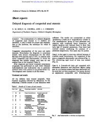
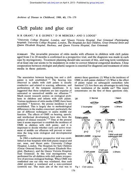
Lip<br />
0 Normal<br />
2 Incomplete 3 Complete<br />
Primary <strong>palate</strong><br />
0 Normal<br />
Secondary polate<br />
2 S<strong>of</strong>t <strong>palate</strong><br />
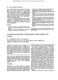
Fig 1 <strong>Cleft</strong> defects.<br />
176<br />
1 Notch<br />
O Normal<br />
1 Notch<br />
4 Others<br />
2 Complete 3 Other<br />
1 Bifid uvula <strong>and</strong><br />
submucous cleft<br />
3 S<strong>of</strong>t <strong>and</strong><br />
hard <strong>palate</strong> 4 Others
Patients <strong>and</strong> methods<br />
Downloaded from<br />
adc.bmj.com on April 4, 2013 - Published by group.bmj.com<br />
Cooperative otological <strong>and</strong> plastic surgery was performed<br />
at each participat<strong>in</strong>g hospital to m<strong>in</strong>imise<br />
admissions <strong>and</strong> anaesthetics. Investigations <strong>in</strong>cluded<br />
a formal assessment <strong>of</strong> cleft defect at presentation<br />
(fig 1), otoscopic <strong>in</strong>spection <strong>of</strong> tympanic membranes<br />
<strong>and</strong> impedance measurement, otoscopic exam<strong>in</strong>ation<br />
with a microscope, <strong>and</strong> bilateral myr<strong>in</strong>gotomy under<br />
anaesthetic. All observations were documented <strong>and</strong><br />
a database established us<strong>in</strong>g a dBASE 11 data<br />
management system <strong>and</strong> an Apple llE computer.<br />
With parental consent children with cleft <strong>palate</strong><br />
who were treated <strong>in</strong> the plastic surgery departments<br />
<strong>of</strong> the above hospitals were registered <strong>and</strong> entered<br />
<strong>in</strong>to the trial, some at birth, others just before<br />
plastic surgery.<br />
The child attended the <strong>ear</strong>, nose, <strong>and</strong> throat<br />
outpatient cl<strong>in</strong>ic the day before surgery, for otoscopy<br />
<strong>and</strong> tympanometry. (The type <strong>of</strong> impedance<br />
meter varied: at the Queen Victoria Hospital an<br />
American AR85 was available, <strong>and</strong> at the other<br />
three hospitals a Graystad GS1 28 was used.) The<br />
next day, under general anaesthetic, both <strong>ear</strong>s were<br />
<strong>Cleft</strong> <strong>palate</strong> <strong>and</strong> <strong>glue</strong> <strong>ear</strong> 177<br />
<strong>in</strong>spected with an operat<strong>in</strong>g microscope, <strong>and</strong><br />
myr<strong>in</strong>gotomy <strong>and</strong> aspiration <strong>of</strong> middle <strong>ear</strong> contents<br />
by microsuction was performed. With OME confirmed<br />
by aspiration a long term ventilation tube<br />
(the protocol recommended a Goode 'T' tube) was<br />
<strong>in</strong>serted <strong>in</strong> one <strong>ear</strong> at r<strong>and</strong>om. For children readmitted<br />
for a second exam<strong>in</strong>ation with a microscope<br />
the above procedure was repeated for the<br />
non-ventilated (control) <strong>ear</strong>, <strong>and</strong> patency <strong>of</strong> the <strong>in</strong><br />
situ ventilation tube confirmed.<br />
Otological follow up was ma<strong>in</strong>ta<strong>in</strong>ed to assess the<br />
patency <strong>of</strong> the ventilation tube, the state <strong>of</strong> the <strong>ear</strong><br />
undergo<strong>in</strong>g myr<strong>in</strong>gotomy alone, <strong>and</strong> to monitor<br />
h<strong>ear</strong><strong>in</strong>g. Early liaison with a speech therapist<br />
ensured subsequent speech <strong>and</strong> language assessment.<br />
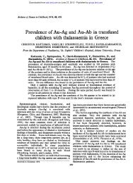
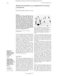
The trial protocol is outl<strong>in</strong>ed <strong>in</strong> fig 2.<br />
Results<br />
By July 1986, 112 children had been registered <strong>in</strong> the<br />
study. Fifty five children had undergone exam<strong>in</strong>ation<br />
with a microscope <strong>and</strong> bilateral myr<strong>in</strong>gotomy:<br />
15 at the time <strong>of</strong> lip repair, 34 at <strong>palate</strong> repair, <strong>and</strong><br />
six as a separate procedure (see fig 2). Six children<br />
had a second exam<strong>in</strong>ation with myr<strong>in</strong>gotomy <strong>of</strong> the<br />
Fig 2 Trial proool. * With late registration bilateral myr<strong>in</strong>gotomy <strong>and</strong> ventilation <strong>of</strong> one <strong>ear</strong> were performed<br />
at <strong>palate</strong> repair.
178 Grant, Qu<strong>in</strong>ey, Mercer, <strong>and</strong> Lodge<br />
Table Age at myr<strong>in</strong>gotomy: 61 operations <strong>in</strong> 55 children<br />
Age <strong>in</strong> months - 2 3 4 5 6 7 8 9 10 11 12 13 14 15 16 17 18 19 20<br />
Nos <strong>of</strong> children - 1 12 6 - - 2 - 3 5 1 8 5 3 7 5 1 - 1 1<br />
'control' (non-ventilated) <strong>ear</strong>. Thus observations<br />
were recorded for 116 <strong>ear</strong>s (bilaterally at 55 operations<br />
<strong>and</strong> unilaterally at six operations). The table<br />
shows the age at myr<strong>in</strong>gotomy. Age at surgical<br />
repair varied between hospitals <strong>and</strong> surgeons, but<br />
on average lip repair was at 3-3 months (range 2-4<br />
months) <strong>and</strong> <strong>palate</strong> repair at 13 months (range 7-17<br />
months).<br />
The diagnostic rate <strong>of</strong> tympanometry <strong>in</strong> 51 <strong>ear</strong>s<br />
(55 children) was: normal peaked curve, four<br />
(7.8%) <strong>and</strong> flat (type B) curve, 47 (92.2%). Wax<br />
occluded the external auditory canal <strong>in</strong> 42 out the<br />
116 <strong>ear</strong>s, which prevented reliable observation. The<br />
small number <strong>of</strong> <strong>ear</strong>s effectively tested by impedance<br />
measurement reflects practical difficulties<br />
associated with this technique. Tympanometry<br />
accurately predicted f<strong>in</strong>d<strong>in</strong>gs at myr<strong>in</strong>gotomy for 46<br />
out <strong>of</strong> 51 <strong>ear</strong>s.<br />
On exam<strong>in</strong>ation with an operat<strong>in</strong>g microscope<br />
otoscopic f<strong>in</strong>d<strong>in</strong>gs on 116 <strong>ear</strong>s <strong>in</strong> 55 children (two<br />
observations were made <strong>in</strong> six <strong>ear</strong>s) were: normal<br />
tympanic membrane, five <strong>ear</strong>s (4.3%) <strong>and</strong> diagnostic<br />
app<strong>ear</strong>ance <strong>of</strong> OME, 107 <strong>ear</strong>s (92.2%). Some<br />
children with apparently normal tympanic membranes<br />
had effusions at myr<strong>in</strong>gotomy; for two<br />
patients (four <strong>ear</strong>s) the app<strong>ear</strong>ance <strong>of</strong> the tympanic<br />
membranes was not documented. When confirmed<br />
by myr<strong>in</strong>gotomy there was a high <strong>in</strong>cidence <strong>of</strong> OME<br />
<strong>in</strong> 113 <strong>of</strong> the 116 <strong>ear</strong>s (97.4%). Three middle <strong>ear</strong>s<br />
conta<strong>in</strong>ed no fluid.<br />
The type <strong>of</strong> fluid aspirated at myr<strong>in</strong>gotomy <strong>in</strong> the<br />
113 <strong>ear</strong>s with OME was: serous, <strong>in</strong> 11 <strong>ear</strong>s (9.7%);<br />
mucoid, <strong>in</strong> 23 <strong>ear</strong>s (20.4%); '<strong>glue</strong>', <strong>in</strong> 57 <strong>ear</strong>s<br />
(50.4%); <strong>and</strong> it was not recorded <strong>in</strong> 22 <strong>ear</strong>s (19.5%).<br />
The distribution was similar to that seen <strong>in</strong> children<br />
without a cleft <strong>palate</strong> who have OME.<br />
Discussion<br />
Downloaded from<br />
adc.bmj.com on April 4, 2013 - Published by group.bmj.com<br />
In this typical sample <strong>of</strong> children with cleft <strong>palate</strong><br />
myr<strong>in</strong>gotomies were performed irrespective <strong>of</strong> previous<br />
otological f<strong>in</strong>d<strong>in</strong>gs, <strong>and</strong> were not limited to<br />
those children with symptoms. The age <strong>of</strong> objective<br />
diagnosis <strong>of</strong> OME by myr<strong>in</strong>gotomy, <strong>and</strong> the 97%<br />
<strong>in</strong>cidence reported here, merit careful consideration.<br />
Furthermore, had any <strong>of</strong> the three 'dry' <strong>ear</strong>s<br />
conta<strong>in</strong>ed serous fluid capable <strong>of</strong> displacement<br />
under general anaesthetic the <strong>in</strong>cidence <strong>of</strong> OME<br />
would have been even higher.<br />
Too-Chung has suggested the middle <strong>ear</strong> is<br />
aerated from birth until 4 months <strong>of</strong> age <strong>in</strong> children<br />
with cleft <strong>palate</strong>." This claim is not supported by<br />
our evidence. In this study no myr<strong>in</strong>gotomies were<br />
performed before 2 months <strong>of</strong> age, <strong>and</strong> therefore<br />
the state <strong>of</strong> the middle <strong>ear</strong> at birth cannot be<br />
<strong>in</strong>ferred. OME was confirmed, however, by myr<strong>in</strong>gotomy<br />
from 2 months onwards; this supports <strong>ear</strong>lier<br />
f<strong>in</strong>d<strong>in</strong>gs that OME is present <strong>in</strong> very young children<br />
with cleft <strong>palate</strong>, <strong>and</strong> as it occurs throughout the<br />
observed age range spontaneous resolution seems<br />
unlikely.8 In addition, Too-Chung's observations<br />
relied on tympanometry, which may not be the most<br />
appropriate technique for assessment <strong>of</strong> middle <strong>ear</strong><br />
function <strong>in</strong> very small children.<br />
In this study the observation rate for tympanometry<br />
was disappo<strong>in</strong>t<strong>in</strong>g. For 55 children with<br />
cleft <strong>palate</strong>, only 51 <strong>ear</strong>s were effectively tested with<br />
an impedance meter. In our experience wax occlud<strong>in</strong>g<br />
the external auditory canal, a narrow pliable <strong>ear</strong><br />
canal, or an uncooperative child all mitigate aga<strong>in</strong>st<br />
successful assessment. In addition impedance<br />
meters <strong>and</strong> technical help were not always available<br />
at short notice. Thus despite a 90% diagnostic<br />
reliability many children simply could not be tested.<br />
By contrast the observation rate for exam<strong>in</strong>ation<br />
with an operat<strong>in</strong>g microscope was 100%.<br />
Many authorities have placed great emphasis on<br />
impedance test<strong>in</strong>g to confirm or exclude OME <strong>in</strong><br />
children with cleft <strong>palate</strong> <strong>and</strong> a wide range <strong>of</strong><br />
<strong>in</strong>cidences is reported. Unfortunately many <strong>of</strong> these<br />
series are retrospective <strong>and</strong> the reliability <strong>of</strong> this<br />
technique cannot be confirmed. Although a dedicated<br />
impedance meter <strong>and</strong> technical help <strong>in</strong> each<br />
plastic surgery unit would have improved the<br />
observation rate achieved <strong>in</strong> this study, for the<br />
reasons listed above, we conclude that accurate<br />
diagnosis <strong>of</strong> OME must rely on otoscopy followed<br />
by myr<strong>in</strong>gotomy <strong>in</strong> all children with cleft <strong>palate</strong>.<br />
The advantages <strong>and</strong> disadvantages <strong>of</strong> <strong>in</strong>sert<strong>in</strong>g<br />
grommets to treat OME <strong>in</strong> normal children is<br />
currently a matter <strong>of</strong> heated debate. 12 13 The precise<br />
aetiology <strong>of</strong> OME rema<strong>in</strong>s uncerta<strong>in</strong> although<br />
eustachian tube function is strongly implicated <strong>in</strong><br />
children with cleft <strong>palate</strong>.14 Medical treatment <strong>of</strong><br />
persist<strong>in</strong>g OME is disappo<strong>in</strong>t<strong>in</strong>g. In consequence<br />
reliable restoration <strong>of</strong> h<strong>ear</strong><strong>in</strong>g by surgical <strong>in</strong>tervention<br />
<strong>and</strong> <strong>in</strong>sertion <strong>of</strong> grommets is favoured <strong>and</strong><br />
accounts for this procedure be<strong>in</strong>g the most common<br />
paediatric operation performed <strong>in</strong> both the United<br />
K<strong>in</strong>gdom <strong>and</strong> North America.
Downloaded from<br />
adc.bmj.com on April 4, 2013 - Published by group.bmj.com<br />
In children with a cleft <strong>palate</strong> the cl<strong>in</strong>ical picture<br />
<strong>of</strong> OME from birth or shortly before is very different<br />
from that found <strong>in</strong> children without a cleft <strong>palate</strong><br />
where the peak <strong>in</strong>cidence is between 3 <strong>and</strong> 6 y<strong>ear</strong>s <strong>of</strong><br />
age. For the former group the onset <strong>of</strong> OME <strong>and</strong><br />
associated h<strong>ear</strong><strong>in</strong>g loss preceeds language acquisition<br />
<strong>and</strong> <strong>ear</strong>ly speech development. Furthermore<br />
the condition seems to persist throughout <strong>in</strong>fancy<br />
<strong>and</strong> <strong>ear</strong>ly childhood. Thus <strong>in</strong> addition to risks <strong>of</strong><br />
long term otological sequelae <strong>of</strong> OME, the untreated<br />
child with cleft <strong>palate</strong> may also suffer<br />
language, <strong>in</strong>tellectual, <strong>and</strong> emotional disability as a<br />
consequence <strong>of</strong> both <strong>ear</strong>ly h<strong>ear</strong><strong>in</strong>g loss <strong>and</strong> orig<strong>in</strong>al<br />
deformity.9 Adenoidectomy can play no part <strong>in</strong><br />
treatment <strong>of</strong> children with cleft <strong>palate</strong> for f<strong>ear</strong> <strong>of</strong><br />
further impair<strong>in</strong>g velopharyngeal competence or<br />
exaggerat<strong>in</strong>g rh<strong>in</strong>olalia aperta, <strong>and</strong> it is also hazardous<br />
<strong>in</strong> very young children. Active treatment <strong>of</strong><br />
deafness must therefore <strong>in</strong>clude ventilation <strong>of</strong> the<br />
middle <strong>ear</strong> on one or both sides, but should logically<br />
only follow accurate diagnosis.<br />
If parents accept the need for surgical diagnosis<br />
<strong>and</strong> treatment <strong>of</strong> OME this should not require<br />
separate anaesthesia because it can be l<strong>in</strong>ked to<br />
surgical repair <strong>of</strong> the lip or <strong>palate</strong>, or both, if close<br />
cooperation between surgeons is achieved. Symptomatic<br />
OME necessitat<strong>in</strong>g myr<strong>in</strong>gotomy before or<br />
after plastic surgery may need a further anaesthetic,<br />
but <strong>in</strong> our experience the number <strong>of</strong> children<br />
requir<strong>in</strong>g this will be small.<br />
The Shepard grommet may not be the most<br />
suitable device for ma<strong>in</strong>ta<strong>in</strong><strong>in</strong>g middle <strong>ear</strong> aeration<br />
<strong>in</strong> children with cleft <strong>palate</strong> because it will be<br />
extruded on average at 6.6 months,15 <strong>and</strong> recurrent<br />
OME will necessitate repeated anaesthetics. The<br />
Goode T tube, on the other h<strong>and</strong>, has been shown<br />
to rema<strong>in</strong> <strong>in</strong> situ for substantially longer periods<br />
without caus<strong>in</strong>g additional tympanic membrane or<br />
middle <strong>ear</strong> complications.16 Long term ventilation<br />
with this type <strong>of</strong> tube should reduce the <strong>in</strong>cidence <strong>of</strong><br />
the late sequelae <strong>of</strong> OME <strong>in</strong>clud<strong>in</strong>g adhesive otitis,<br />
ossicular damage, <strong>and</strong> cholesteatoma, all <strong>of</strong> which<br />
are particularly evident <strong>in</strong> untreated children with<br />
cleft <strong>palate</strong>.3 It is hoped that long term follow up <strong>in</strong><br />
this trial will yield additional <strong>in</strong>formation on this<br />
matter.<br />
<strong>Cleft</strong> <strong>palate</strong> <strong>and</strong> <strong>glue</strong> <strong>ear</strong> 179<br />
The members <strong>of</strong> the <strong>Cleft</strong> Palate Study are: plastic surgeons: JE<br />
Bowen, IW Broomhead, TD Cochrane, BM Jones, BD Morgan,<br />
CC Walker; otologists: CM Bailey, JM Graham, CM Milton, RJ<br />
Sergeant; <strong>and</strong> an audiologist: SC Bellman. The authors k<strong>in</strong>dly<br />
thank them for their enthusiastic participation, <strong>and</strong> their advice <strong>in</strong><br />
prepar<strong>in</strong>g this report. Figures were prepared by the Department <strong>of</strong><br />
Cl<strong>in</strong>ical Illustration, Queen Victoria Hospital, East Gr<strong>in</strong>stead.<br />
References<br />
Yules RB. H<strong>ear</strong><strong>in</strong>g <strong>in</strong> cleft <strong>palate</strong> patients. Arch Otolaryngol<br />
1970;91:319-23.<br />
2 Bluestone CD. Eustachian tube obstruction <strong>in</strong> the <strong>in</strong>fant with<br />
cleft <strong>palate</strong>. Ann Otol Rh<strong>in</strong>ol Laryngol 1971;80(suppl 2):1-30.<br />
3Bennett M. The older cleft <strong>palate</strong> patient. Laryngoscope<br />
1972;86:1217-25.<br />
4Paradise JL. Middle <strong>ear</strong> problems associated with cleft <strong>palate</strong>.<br />
An <strong>in</strong>ternationally-oriented review. <strong>Cleft</strong> Palate J 1975;12:<br />
17-20.<br />
5Tos M. Relationship between secretory otitis media <strong>in</strong> childhood<br />
<strong>and</strong> chronic otitis <strong>and</strong> its sequelae <strong>in</strong> adults. J Laryngol Otol<br />
1981 ;95:1011-22.<br />
6 Brooks DN. Possible long-term consequences <strong>of</strong> middle <strong>ear</strong><br />
effusion. Ann Otol Rh<strong>in</strong>ol Laryngol 1980;89(suppl 68):246-8.<br />
7Stool SE, R<strong>and</strong>all P. Unexpected <strong>ear</strong> disease <strong>in</strong> <strong>in</strong>fants with cleft<br />
<strong>palate</strong>. <strong>Cleft</strong> Palate J 1967;4:99-103.<br />
Paradise JL, Bluestone CD, Felder H. The universality <strong>of</strong> otitis<br />
media <strong>in</strong> 50 <strong>in</strong>fants with cleft <strong>palate</strong>. Pediatrics 1969;44:35-42.<br />
9 Lamb M, Wilson F, Leeper H. A comparison <strong>of</strong> selected cleft<br />
<strong>palate</strong> children <strong>and</strong> their sibl<strong>in</strong>gs on the variables <strong>of</strong> <strong>in</strong>telligence,<br />
h<strong>ear</strong><strong>in</strong>g loss, <strong>and</strong> visual-perceptual-motor abilities. <strong>Cleft</strong> Palate J<br />
1972;9:218-28.<br />
10 Hubbard TW, Paradise JL, McWilliams BJ, Elster BA,<br />
Taylor FH. Consequences <strong>of</strong> unremitt<strong>in</strong>g middle-<strong>ear</strong> disease <strong>in</strong><br />
<strong>ear</strong>ly life. Otological, audiological <strong>and</strong> developmental f<strong>in</strong>d<strong>in</strong>gs<br />
<strong>in</strong> children with cleft <strong>palate</strong>. N Engl J Med 1985;312:1529-34.<br />
Too-Chung MA. The assessment <strong>of</strong> middle <strong>ear</strong> function <strong>and</strong><br />
h<strong>ear</strong><strong>in</strong>g by tympanometry <strong>in</strong> children before <strong>and</strong> after <strong>ear</strong>ly cleft<br />
<strong>palate</strong> repair. Br J Plast Surg 1983;36:295-9.<br />
2 Black NA. Surgery for <strong>glue</strong> <strong>ear</strong>-a modern epidemic. Lancet<br />
1984;i:835-7.<br />
13 Maran ADG, Wilson JA. Glue <strong>ear</strong> <strong>and</strong> speech development. Br<br />
Med J 1986;293:713.<br />
14 Bluestone CD, Wittel RA, Paradise JL. Roentgenographic<br />
evaluation <strong>of</strong> eustachian tube function <strong>in</strong> <strong>in</strong>fants with cleft <strong>and</strong><br />
normal <strong>palate</strong>s. <strong>Cleft</strong> Palate J 1972;9:93-100.<br />
'5 Mawson SR, Fagan P. Tympanic effusions <strong>in</strong> children.<br />
J Laryngol Otol 1972;86:105-19.<br />
16 Rothera MP, Gratn HR. Long-term ventilation <strong>of</strong> the middle<br />
<strong>ear</strong> us<strong>in</strong>g the Goode T-tube. J Laryngol Otol 1985;99:335-7.<br />
Correspondence to Mr HR Grant, <strong>Cleft</strong> Palate Study, University<br />
College Hospital, Gower Street, London WC1E 6AU.<br />
Received 16 September 1987
Downloaded from<br />
adc.bmj.com on April 4, 2013 - Published by group.bmj.com<br />
Email alert<strong>in</strong>g<br />
service<br />
Notes<br />
<strong>Cleft</strong> <strong>palate</strong> <strong>and</strong> <strong>glue</strong> <strong>ear</strong>.<br />
H R Grant, R E Qu<strong>in</strong>ey, D M Mercer, et al.<br />
Arch Dis Child 1988 63: 176-179<br />
doi: 10.1136/adc.63.2.176<br />
Updated <strong>in</strong>formation <strong>and</strong> services can be found at:<br />
http://adc.bmj.com/content/63/2/176<br />
These <strong>in</strong>clude:<br />
To request permissions go to:<br />
http://group.bmj.com/group/rights-licens<strong>in</strong>g/permissions<br />
To order repr<strong>in</strong>ts go to:<br />
http://journals.bmj.com/cgi/repr<strong>in</strong>tform<br />
To subscribe to BMJ go to:<br />
http://group.bmj.com/subscribe/<br />
Receive free email alerts when new articles cite this<br />
article. Sign up <strong>in</strong> the box at the top right corner <strong>of</strong><br />
the onl<strong>in</strong>e article.