Delayed diagnosis of congenital anal stenosis - Archives of Disease ...
Delayed diagnosis of congenital anal stenosis - Archives of Disease ...
Delayed diagnosis of congenital anal stenosis - Archives of Disease ...

Create successful ePaper yourself
Turn your PDF publications into a flip-book with our unique Google optimized e-Paper software.
<strong>Archives</strong> <strong>of</strong> <strong>Disease</strong> in Childhood, 1979, 54, 68-79<br />
Short reports<br />
<strong>Delayed</strong> <strong>diagnosis</strong> <strong>of</strong> <strong>congenital</strong> <strong>anal</strong> <strong>stenosis</strong><br />
E. M. KIELY, R. CHOPRA, AND J. J. CORKERY<br />
Department <strong>of</strong>Paediatric Surgery, Children's Hospital, Birmingham<br />
SUMMARY The consequences <strong>of</strong> a delayed <strong>diagnosis</strong><br />
<strong>of</strong> <strong>congenital</strong> <strong>anal</strong> <strong>stenosis</strong> in 11 children are<br />
described. A plea is made for proper <strong>anal</strong> examination<br />
in the newborn, the technique for which is<br />
described.<br />
Congenital abnormalities <strong>of</strong> the anus are easily<br />
detected. Nevertheless the <strong>diagnosis</strong> is sometimes<br />
missed, with serious consequences for the child.<br />
Between 1970 and the end <strong>of</strong> 1977, 11 children with<br />
minor <strong>congenital</strong> <strong>anal</strong> abnormalities, in whom the<br />
<strong>diagnosis</strong> was initially missed, were seen on one<br />
surgical firm at this hospital (Table 1).<br />
After <strong>diagnosis</strong> and initial treatment intravenous<br />
urography was performed on each child; bilateral<br />
hydronephrosis and hydroureter due to posterior<br />
urethral valves were present in one patient (Case 11).<br />
The urograms were normal in all the others.<br />
Treatment and results<br />
Downloaded from<br />
adc.bmj.com on July 5, 2013 - Published by group.bmj.com<br />
All the children were treated surgically. Nine<br />
required anoplasty followed by dilatations. In the<br />
remaining 2 limited courses <strong>of</strong> <strong>anal</strong> dilatation were<br />
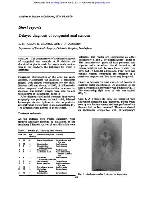
Table 1 Details <strong>of</strong> 11 cases <strong>of</strong> <strong>anal</strong> <strong>stenosis</strong><br />
Case Sex Age Presenting complaint Anomaly<br />
(months)<br />
I M 42 Faecal incontinence Covered anus and<br />
skin tags<br />
2 M 5 Constipation Anal <strong>stenosis</strong><br />
3 F 5 Constipation; Anal <strong>stenosis</strong><br />
abdominal mass<br />
4 F 6 Constipation Anal <strong>stenosis</strong><br />
5 F 7 Abdominal mass Anal membrane;<br />
<strong>stenosis</strong> and skin<br />
tags<br />
6 F 5 Abdominal Anal <strong>stenosis</strong><br />
distension; diarrhoea<br />
7 F 4 Constipation Covered anus<br />
8 F 4 Rectal bleeding Anal <strong>stenosis</strong> and<br />
skin tags<br />
9 M 1 Oseophageal atresia Anal <strong>stenosis</strong><br />
10 M 4I Abdominal Covered anus<br />
distension<br />
11 M 1 Constipation Covered anus<br />
sufficient. The results are summarised as either<br />
'satisfactory' (Table 2) or 'unsatisfactory' (Table 3).<br />
The 'unsatisfactory' group all have persistent constipation<br />
with occasional faecal impaction; all<br />
require laxatives and, between them to date, they<br />
have had 17 hospital admissions. Four have had<br />
contrast enemas confirming the presence <strong>of</strong> a<br />
persistent megarectum. Two cases may be quoted.<br />
Case 1. A boy aged 3 i years was referred because <strong>of</strong><br />
overflow rectal incontinence. On inspection <strong>of</strong> the<br />
anus a <strong>congenital</strong> abnormality was obvious (Fig. 1).<br />
The obstructing rigid hood <strong>of</strong> skin was excised<br />
(Fig. 2).<br />
Case 6. A 5-month-old baby girl presented with<br />
abdominal distension and diarrhoea. Before being<br />
seen by us a barium enema had been performed but<br />
the anus had not been examined. The enema showed<br />
an appearance compatible with Hirschsprung's<br />
Fig. 1 Anal abnormality is obvious on inspection.<br />
68
disease (Fig. 3) and it was for this reason that a<br />
surgical opinion was sought. The anus looked normal<br />
but on attempting digital examination a tight <strong>stenosis</strong><br />
at the dentate line was detected. Rectal biopsy was<br />
normal.<br />
Table 2 'Satisfactory' group<br />
Case Age at presentation Functional result Duration <strong>of</strong><br />
(months) treatment needed<br />
(months)<br />
7 4 Normal 2<br />
8 4 Normal 4<br />
9 1 Normal 3<br />
10 ij Normal 7<br />
11 1 Normal 3<br />
Fig. 2 Obstructing skin has been excised.<br />
Fig. 3 Barium enema (Case 6). Appearance Is<br />
compatible with Hirschsprung's disease.<br />
Downloaded from<br />
adc.bmj.com on July 5, 2013 - Published by group.bmj.com<br />
<strong>Delayed</strong> <strong>diagnosis</strong> <strong>of</strong> <strong>congenital</strong> <strong>anal</strong> <strong>stenosis</strong> 69<br />
Table 3 'Unsatisfactory' group<br />
Case Age at presentation Functional result Duration <strong>of</strong><br />
(months) treatment to date<br />
(months)<br />
1 42 Unsatisfactory 12<br />
2 5 Unsatisfactory 54<br />
3 5 Unsatisfactory II<br />
4 6 Unsatisfactory 18<br />
5 7 Unsatisfactory 21<br />
6 5 Unsatisfactory 12 then lost to<br />
follow-up<br />
Discussion<br />
Although classed by us as a minor abnormality, <strong>anal</strong><br />
<strong>stenosis</strong>, if neglected, becomes a major one requiring<br />
prolonged surgical treatment and supervision. The<br />
longer the <strong>stenosis</strong> is untreated the more severely<br />
affected the rectum becomes so that in the worst<br />
cases the rectum becomes grossly distended, insensitive,<br />
and apparently aperistaltic (Fig. 4). This<br />
constitutes a severe disability, the treatment is<br />
prolonged and major surgery may be needed. Our<br />
results (Tables 2 and 3) and the experience <strong>of</strong> others<br />
(Partridge and Gough, 1961; Stephens and Smith,<br />
1971; Taylor et al., 1973) show that the sooner the<br />
<strong>diagnosis</strong> is made and effective treatment instituted,<br />
the better the prognosis.<br />
In 3 <strong>of</strong> these children simple inspection <strong>of</strong> the anus<br />
was sufficient to reveal an abnormality requiring<br />
closer scrutiny. However, the anus can look perfectly<br />
Fig. 4 Barium enema in Case 5 showing huge<br />
megarectum full <strong>of</strong>faeces.
Downloaded from<br />
adc.bmj.com on July 5, 2013 - Published by group.bmj.com<br />
70 Kiely, Chopra, and Corkery<br />
normal and yet be severely stenosed. The normal<br />
passage <strong>of</strong> meconium and stools is not a reliable<br />
guide to the state <strong>of</strong> the anus, as a stenosed anus will<br />
<strong>of</strong>ten allow meconium and the s<strong>of</strong>t stool <strong>of</strong> the newborn<br />
to escape. Similarly a rectal thermometer can<br />
usually be easily introduced into the rectum in these<br />
cases.<br />
The technique <strong>of</strong> <strong>anal</strong> examination in the newborn<br />
is easy. The anus should first be inspected and then<br />
palpated. The little finger, well lubricated, should be<br />
used. The finger is inserted into the anus, pad first,<br />
very slowly and very gently. The normal anus will<br />
stretch as this is done and in most cases a little finger<br />
can enter the rectum. However, the examiner's<br />
finger may be too big or, as in the case <strong>of</strong> a very<br />
small baby, it may obviously be unwise to over dilate<br />
and injure the anus. With practice all degrees <strong>of</strong> <strong>anal</strong><br />
<strong>stenosis</strong> can be excluded without the finger tip<br />
entering the rectum. During the palpation two points<br />
in particular should be noted. Firstly, the absolute<br />
size <strong>of</strong> the anus. More important than this is the<br />
suppleness or otherwise <strong>of</strong> the c<strong>anal</strong>. A rigid <strong>anal</strong><br />
c<strong>anal</strong> is an abnormal one.<br />
The anus is easily amenable to examination in the<br />
newborn. If there is any doubt that there is even a<br />
slight element <strong>of</strong> <strong>stenosis</strong> an expert surgical opinion<br />
should be urgently obtained.<br />
References<br />
Partridge, J. P., and Gough, M. H. (1961). Congenital<br />
abnormalities <strong>of</strong> the anus and rectum. British Journal <strong>of</strong><br />
Surgery, 49, 37-50.<br />
Stephens, F. D., and Smith, E. D. (1971). Anorectal Malformations<br />
in Children. Year Book Medical Publishers Inc.:<br />
Chicago.<br />
Taylor, I., Duthie, H. L., and Zachary, R. B. (1973). Anal<br />
continence following surgery for imperforate anus. Journal<br />
<strong>of</strong> Pediatric Surgery, 8, 497-503.<br />
Correspondence to E. M. Kiely, FRCS, The Children's<br />
Hospital, Ladywood Middleway, Ladywood, Birmingham<br />
B16 8ET.<br />
Acute anuric renal failure in an infant with systemic candidiasis<br />
J. Z. HECKMATT, S. R. MEADOW, AND C. K. ANDERSON<br />
Department <strong>of</strong> Paediatrics, University <strong>of</strong> Leeds, Seacr<strong>of</strong>t Hospital, and the University Department <strong>of</strong><br />
Pathology, General Infirmary, Leeds<br />
SUMMARY We report a newborn baby who presented<br />
with acute anuricrenal failure resulting from systemic<br />
candidiasis. The predisposing factors and diagnostic<br />
features are examined.<br />
Although the newborn baby is susceptible to<br />
infection and mucocutaneous thrush is common,<br />
systemic candidiasis is rare. When it does occur in<br />
babies it usually causes osteomyelitis, arthritis, and<br />
meningitis. Renal failure is not a recognised feature.<br />
Case history<br />
A boy was born in August 1977 at29 weeks' gestation,<br />
birthweight 1.430 kg. Respiratory distress syndrome<br />
developed and he required ventilation for 23 days. A<br />
blood culture taken at 2 days grew Staphylococcus<br />
aureus and he was given ampicillin and cloxacillin.<br />
Subsequently he received courses <strong>of</strong> gentamicin and<br />
carbenicillin.<br />
He was fed intravenously from 3 to 22 days <strong>of</strong> age<br />
via silastic cannulae. These were placed in various<br />
veins, including the left long saphenous. At 21 days<br />
the long saphenous site became infected and blood<br />
cultures grew Candida albicans. The cannula was<br />
therefore removed. Five days later 3 ml pus was<br />
drained from the abscess; Gram's stain showed yeast<br />
cells and mycelial elements; cultures produced a<br />
pr<strong>of</strong>use growth <strong>of</strong> C. albicans. He was given benzylpenicillin,<br />
streptomycin, and lincomycin from 25 to<br />
30 days and the abscess healed. Thereafter he made<br />
good progress and for 5 weeks he fed normally and<br />
gained weight. Five blood cultures during this<br />
period were sterile.<br />
When he was 64 days old he suddenly became<br />
anuric and was transferred to Leeds. There was no<br />
urine in the bladder on catheterisation. An intravenous<br />
urogram showed nephrograms <strong>of</strong>2 normal size<br />
kidneys which failed to excrete dye into the renal<br />
pelvis. A cystogram was normal; there was no<br />
urethral obstruction and no vesico ureteric reflux.<br />
After a week <strong>of</strong> anuria blood tests showed: Hb<br />
6.4 g/dl, WBC 14.8 x 10⁹/1 (14800/mms) (75%
Downloaded from<br />
adc.bmj.com on July 5, 2013 - Published by group.bmj.com<br />
Email alerting<br />
service<br />
Notes<br />
<strong>Delayed</strong> <strong>diagnosis</strong> <strong>of</strong> <strong>congenital</strong><br />
<strong>anal</strong> <strong>stenosis</strong><br />
E. M. Kiely, R. Chopra and J. J. Corkery<br />
Arch Dis Child 1979 54: 68-70<br />
doi: 10.1136/adc.54.1.68<br />
Updated information and services can be found at:<br />
http://adc.bmj.com/content/54/1/68<br />
These include:<br />
To request permissions go to:<br />
http://group.bmj.com/group/rights-licensing/permissions<br />
To order reprints go to:<br />
http://journals.bmj.com/cgi/reprintform<br />
To subscribe to BMJ go to:<br />
http://group.bmj.com/subscribe/<br />
Receive free email alerts when new articles cite this<br />
article. Sign up in the box at the top right corner <strong>of</strong><br />
the online article.