Sagaria cilentana gen. et sp. nov.-A New Angiosperm Fructification ...
Sagaria cilentana gen. et sp. nov.-A New Angiosperm Fructification ...
Sagaria cilentana gen. et sp. nov.-A New Angiosperm Fructification ...

Create successful ePaper yourself
Turn your PDF publications into a flip-book with our unique Google optimized e-Paper software.
286<br />
Derivation of Name: <strong>Sagaria</strong> is named in honor of the collector of<br />
the <strong>sp</strong>ecimen; Giovanni <strong>Sagaria</strong>.<br />
Type <strong>sp</strong>ecies: <strong>Sagaria</strong> <strong>cilentana</strong> <strong>sp</strong>. <strong>nov</strong>.<br />
Generic Diagnosis. Angio<strong>sp</strong>erm, axis with leaves and fruits.<br />
Leaves lobate, p<strong>et</strong>iolate. Distal tips of foliar lobes mucronate.<br />
Inflorescence cymose. Fruits borne by an elongate peduncle, on an<br />
expanded cup-shaped recepticle, up to 1.1 cm long, and 0.6 cm<br />
wide and composed of at least three follicles with a maximum<br />
width of 3 mm and partially fused in their basal three-quarters.<br />
Distal tips of the follicles mucronate.<br />
Derivation of <strong>sp</strong>ecific epith<strong>et</strong>. From the area, Cilento, in which the<br />
<strong>sp</strong>ecimen was found.<br />
Holotype. Specimen 301a housed at Museum of Paleobotany and<br />
Ethnobotany, at Orto Botanico University of Naples. (Fig. 3 A) [part]<br />
and <strong>sp</strong>ecimen 301b housed at the Paleontology Museum of<br />
Magliano V<strong>et</strong>ere (Cilento National Park), Provence of Salerno<br />
(Fig. 3B) [counterpart].<br />
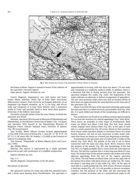
Type locality. Monti Alburni locality located approximately<br />
1.5 km SSW of the town of P<strong>et</strong>ina [Fig. 1; circa 40 31’ 18’’ N 15 22<br />
05’’ E; I. G. M. she<strong>et</strong> 198, I SE–Aul<strong>et</strong>ta; 1:25.000], in the Provence of<br />
Salerno in Campania, Italy.<br />
Type Stratum. ‘‘Plattenkalk’’ at Monte Alburni (Bravi and Garassino,<br />
1998).<br />
Age. Middle Albian.<br />
Material. The <strong>sp</strong>ecies is represented by a single <strong>sp</strong>ecimen<br />
preserved as part and counterpart of a coalified compression.<br />
<strong>Sagaria</strong> <strong>cilentana</strong> <strong>sp</strong>. <strong>nov</strong>.<br />
Figs. 3–5<br />
Specific diagnosis. Characteristics as for the <strong>gen</strong>us.<br />
Description of <strong>sp</strong>ecimen<br />
The <strong>sp</strong>ecimen consists of a main axis with four attached leaves<br />
and a distal cyme bearing three fructifications. The <strong>sp</strong>ecimen is<br />
S. Bravi <strong>et</strong> al. / Cr<strong>et</strong>aceous Research 31 (2010) 285–290<br />
Fig. 1. Map showing the location of the plattenkalk of Monte Alburni in Campania.<br />
approximately 8 cm long, with the main axis about 1.75 mm wide<br />
and remaining at a relatively uniform width. In addition, there is<br />
a d<strong>et</strong>ached leaf that is clearly derived from the <strong>sp</strong>ecimen. The<br />
<strong>sp</strong>ecimen di<strong>sp</strong>lays five nodes (Fig. 3A,B). The di<strong>sp</strong>osition of the<br />
nodes indicates an alternate or helical arrangement of appendices.<br />
The two most proximal nodes show axes that are broken off but at<br />
their bases are approximately the same diam<strong>et</strong>er as the main axis of<br />
the <strong>sp</strong>ecimen (Fig. 3A).<br />
About 4 cm from the base of the <strong>sp</strong>ecimen and lying underneath<br />
the main axis there is a lobed structure, presumably a leaf (Fig. 3A,B,<br />
4G). It is quite folded and d<strong>et</strong>ails of the morphology are difficult to<br />
discern.<br />
Two small leaves are found in an axillary position approximately<br />
0.5 cm from the insertion of a lateral appendage (Figs. 3A,B, 4D,H).<br />
These leaves are probably in early stages of development. Both<br />
leaves are lobate and about 2 mm long. One leaf, more clearly<br />
visible, di<strong>sp</strong>lays three distinct lobes (Fig. 4H); at the apex of each<br />
lobe is a small, pointed tip (Fig. 4H arrow). Based on their position,<br />
these leaves could represent stipules or immature leaves of a small<br />
branch, however, the lack of such structures at other nodes indicates<br />
that these are leaves on a branch just beginning to develop.<br />
In proximity to the <strong>sp</strong>ecimen, part of a lobed leaf is visible on the<br />
matrix (Fig. 3A,B, 4D,E). The p<strong>et</strong>iole of this leaf is not in direct<br />
connection with the main axis (Figs. fig3A, 4D,H); however, from<br />
the position of the p<strong>et</strong>iole (Fig. 4H), and morphology of the leaf<br />
(Fig. 4D), it is highly probable that it comes from the same plant as<br />
the axis. The leaf has a p<strong>et</strong>iole that is 4 mm long and about 0.5 mm<br />
wide. The broken base of the p<strong>et</strong>iole matches the broken end of the<br />
adjacent appendage from the axis of the <strong>Sagaria</strong> <strong>sp</strong>ecimen<br />
(Fig. 4D,H). The leaf shows an obtuse base, and on the right side of<br />
the lamina three lobes with deep sinuses are visible (Fig. 4E). Lobes<br />
are about 2 mm deep and 1.5 mm wide (Fig. 4D,E). At the apex of<br />
each lobe is a small pointed tip (Fig. 4D,E arrow). Primary venation<br />
is faint but medial (Fig. 4D). It is not possible to discern secondary<br />
venation, but the position of the lobes and the mucronate tip<br />
suggest a median secondary vein at a perpendicular angle to the