J <strong>Indian</strong> Acad <strong>Forensic</strong> Med. Jan- March 2012, Vol. 34, No. 1 ISSN 0971-0973 haemorrhage occurring because <strong>of</strong> rupture is massive and can be life threatening, unless diagnosed and treated promptly. [5] Obstructed labour due to cephalo-pelvic disproportion and malpresentations, continued to be a major causative factor <strong>of</strong> rupture uterus. Sinha and Roy also recently reported an incidence <strong>of</strong> 24.4% scar rupture, while Kulkarni and Kendre reported 56.12% scar rupture in their series on rupture uterus in rural India. There are increasing number <strong>of</strong> cases <strong>of</strong> scar rupture is due to an increasing use <strong>of</strong> caesarean section in place <strong>of</strong> difficult vaginal delivery. Although better alternatives in terms <strong>of</strong> fetal outcome and decreased maternal morbidity, improved these caesarean sections should not be accompanied by an increase in the rate <strong>of</strong> scar rupture. All patients with previous caesarean scars should be made aware <strong>of</strong> the importance <strong>of</strong> ante-natal care in all subsequent pregnancies. They also require careful pre-natal supervision, proper selection <strong>of</strong> cases for vaginal delivery, early hospital admission, and closesupervision in labour. [6, 7] The high risk mother with contracted pelvic, previous history <strong>of</strong> caesarean section, hysterotomy or myomectomy, uncorrected transverse lie, grand multiparity are likely to rupture should have mandatory hospital delivery. Ultrasonography (USG) may be helpful in diagnosing such anomalies before rupture, which will help in decreasing the morbidity and mortality associated with rapid and massive hemoperitoneum occurring because <strong>of</strong> rupture <strong>of</strong> uterus. [8] Treatment usually involved is removal <strong>of</strong> ruptured horn. As it leaves a scar on upper part <strong>of</strong> the uterus, it is important to avoid pregnancy for at least one year by barrier or hormonal contraceptives. In addition, future pregnancy requires proper monitoring, early hospitalization, and elective caesarean section at term. As to the choice <strong>of</strong> surgery, conservation <strong>of</strong> the uterus by re- suturing the rent should be attempted wherever possible. With availability <strong>of</strong> higher antibiotics and better non-steroidal anti- inflammatory drugs, good results have been obtained. However, in cases with severe haemorrhage and shock requiring Hysterectomy, operative time and exposure to anaesthesia are vital factors, and a quick sub total hysterectomy should be resorted too. [4] Conclusion: Most cases <strong>of</strong> rupture uterus are preventable with good ante-natal and intrapartum care, and proper identification <strong>of</strong> high- 84 risk cases. High risk cases should have mandatory hospital delivery. Fatal out comes in all cases are either due to mishandling the case or improper/incomplete treatment was provided to the deceased. The treating doctor should keep in the mind such complications especially in the case <strong>of</strong> previous operation /elderly primi / primi with foeto-pelvic disproportion. References: 1. Nagarkatti RS, Ambiye VR, Vaidya PR. Rupture uterus: changing trends in aetiology and management. Journal <strong>of</strong> Post Graduate <strong>Medicine</strong>, India, 1991; 37-3, 136-139. 2. D. C .Dutta. Textbook <strong>of</strong> Obstetrics; Injuries to the birth canal; 4th edition, 1998; pg 454-466. 3. Mane S, Chaudhry R, Nandanwar Y. An unusual presentation <strong>of</strong> pregnancy in bicornuate uterus. Journal <strong>of</strong> Obstetrics & Gynaecology, India 1994; 44-1, 154-155. 4. S Kore, A Pandole, R Akolekar, N Vaidya, VR Ambiye. Rupture <strong>of</strong> left horn <strong>of</strong> bicornuate uterus at twenty weeks <strong>of</strong> gestation. Journal <strong>of</strong> Post Graduate <strong>Medicine</strong>, India, 2000; 46-1, 39-40. 5. Chang JC, Lin YC. Rupture <strong>of</strong> rudimentary horn pregnancy. Acta Obstet Gynecol Scand 1992; 71:235-238. 6. Sinha J, Roy S. A retrospective study <strong>of</strong> rupture uterus at Patna Medical College Hospital during five years period 1978-1982. J Obstet Gynec India: 1986; 36:241-245. 7. Kulkarni NP, Kendre BV. Rupture uterus -experience at a rural medical college. J. Obstet Gynec India 1990; 40:, 75-79. 8. Achiron R, Tadmor O, Kamar R, Aboulafia Y, Diamant Y. Prerupture ultrasound diagnosis <strong>of</strong> interstitial and rudimentary uterine horn pregnancy in second trimester. A report <strong>of</strong> two cases. J Reprod Med 1992; 37:89-92. Photographs <strong>of</strong> Case 1A 1B
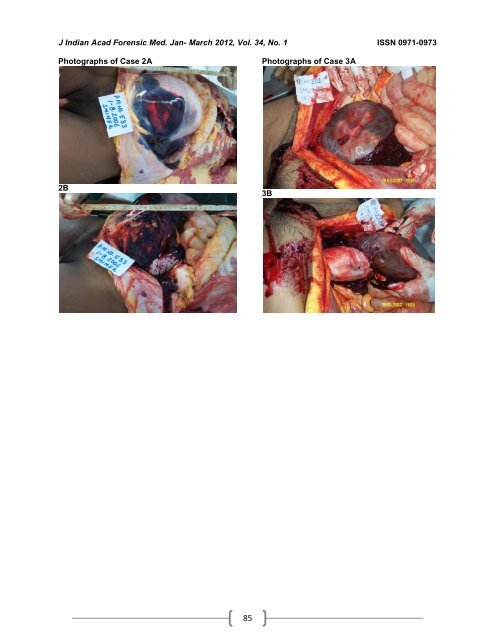
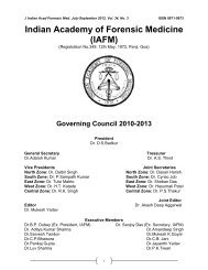
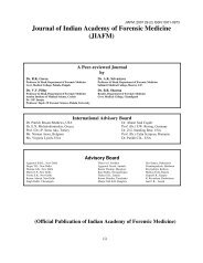
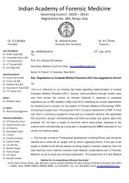
J <strong>Indian</strong> Acad <strong>Forensic</strong> Med. Jan- March 2012, Vol. 34, No. 1 ISSN 0971-0973 Photographs <strong>of</strong> Case 2A 2B 85 Photographs <strong>of</strong> Case 3A 3B