Indian Academy of Forensic Medicine (IAFM) - Official website of IAFM
Indian Academy of Forensic Medicine (IAFM) - Official website of IAFM
Indian Academy of Forensic Medicine (IAFM) - Official website of IAFM

You also want an ePaper? Increase the reach of your titles
YUMPU automatically turns print PDFs into web optimized ePapers that Google loves.
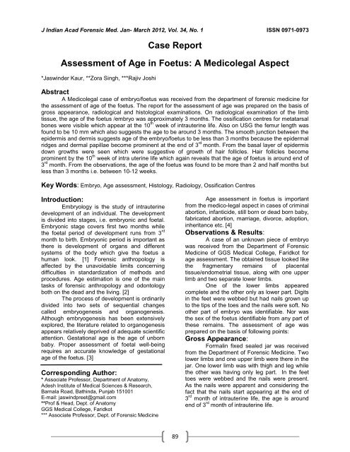
J <strong>Indian</strong> Acad <strong>Forensic</strong> Med. Jan- March 2012, Vol. 34, No. 1 ISSN 0971-0973<br />
Case Report<br />
Assessment <strong>of</strong> Age in Foetus: A Medicolegal Aspect<br />
*Jaswinder Kaur, **Zora Singh, ***Rajiv Joshi<br />
Abstract<br />
A Medicolegal case <strong>of</strong> embryo/foetus was received from the department <strong>of</strong> forensic medicine for<br />
the assessment <strong>of</strong> age <strong>of</strong> the foetus. The report for the assessment <strong>of</strong> age was prepared on the basis <strong>of</strong><br />
gross appearance, radiological and histological examinations. On radiological examination <strong>of</strong> the limb<br />
tissue, the age <strong>of</strong> the foetus /embryo was approximately 3 months. The ossification centres for metatarsal<br />
bones were visible which appear at the 10 th week <strong>of</strong> intrauterine life. Also on USG the femur length was<br />
found to be 10 mm which also suggests the age to be around 3 months. The smooth junction between the<br />
epidermis and dermis suggests age <strong>of</strong> the embryo/foetus to be less than 3 months because the epidermal<br />
ridges and dermal papillae become prominent at the end <strong>of</strong> 3 rd month. From the basal layer <strong>of</strong> epidermis<br />
down growths were seen which were suggestive <strong>of</strong> growth <strong>of</strong> hair follicles. Hair follicles become<br />
prominent by the 10 th week <strong>of</strong> intra uterine life which again reveals that the age <strong>of</strong> foetus is around end <strong>of</strong><br />
3 rd month. From the observations, the age <strong>of</strong> the foetus was found to be more than 2 and half months but<br />
less than 3 months i.e. between 10-12 weeks.<br />
Key Words: Embryo, Age assessment, Histology, Radiology, Ossification Centres<br />
Introduction:<br />
Embryology is the study <strong>of</strong> intrauterine<br />
development <strong>of</strong> an individual. The development<br />
is divided into stages, i.e. embryonic and foetal.<br />
Embryonic stage covers first two months while<br />
the foetal period <strong>of</strong> development runs from 3 rd<br />
month to birth. Embryonic period is important as<br />
there is development <strong>of</strong> organs and different<br />
systems <strong>of</strong> the body which give the foetus a<br />
human look. [1] <strong>Forensic</strong> anthropology is<br />
affected by the unavoidable limits concerning<br />
difficulties in standardization <strong>of</strong> methods and<br />
procedures. Age estimation is one <strong>of</strong> the main<br />
tasks <strong>of</strong> forensic anthropology and odontology<br />
both on the dead and the living. [2]<br />
The process <strong>of</strong> development is ordinarily<br />
divided into two sets <strong>of</strong> sequential changes<br />
called embryogenesis and organogenesis.<br />
Although embryogenesis has been extensively<br />
explored, the literature related to organogenesis<br />
appears relatively deprived <strong>of</strong> adequate scientific<br />
attention. Gestational age is the age <strong>of</strong> unborn<br />
baby. Proper assessment <strong>of</strong> foetal well-being<br />
requires an accurate knowledge <strong>of</strong> gestational<br />
age <strong>of</strong> the foetus. [3]<br />
Corresponding Author:<br />
* Associate Pr<strong>of</strong>essor, Department <strong>of</strong> Anatomy,<br />
Adesh Institute <strong>of</strong> Medical Sciences & Research,<br />
Barnala Road, Bathinda, Punjab 151001<br />
E-mail: jaswindpreet@gmail.com<br />
**Pr<strong>of</strong> & Head, Dept. <strong>of</strong> Anatomy<br />
GGS Medical College, Faridkot<br />
*** Associate Pr<strong>of</strong>essor, Dept. <strong>of</strong> <strong>Forensic</strong> <strong>Medicine</strong><br />
89<br />
Age assessment in foetus is important<br />
from the medico-legal aspect in cases <strong>of</strong> criminal<br />
abortion, infanticide, still born or dead born baby,<br />
fabricated abortion, marriage, divorce, adoption,<br />
inheritance etc. [4]<br />
Observations & Results:<br />
A case <strong>of</strong> an unknown piece <strong>of</strong> embryo<br />
was received from the Department <strong>of</strong> <strong>Forensic</strong><br />
<strong>Medicine</strong> <strong>of</strong> GGS Medical College, Faridkot for<br />
age assessment. The obtained tissue looked like<br />
the fragmentary remains <strong>of</strong> placental<br />
tissue/endometrial tissue, along with one upper<br />
limb and two separate lower limbs.<br />
One <strong>of</strong> the lower limbs appeared<br />
complete and the other only as lower part. Digits<br />
in the feet were webbed but had nails grown up<br />
to the tips <strong>of</strong> the toes and the nails were s<strong>of</strong>t. No<br />
other part <strong>of</strong> embryo was identifiable. Nor was<br />
the sex <strong>of</strong> the foetus identifiable from any part <strong>of</strong><br />
these remains. The assessment <strong>of</strong> age was<br />
prepared on the basis <strong>of</strong> following points:<br />
Gross Appearance:<br />
Formalin fixed sealed jar was received<br />
from the Department <strong>of</strong> <strong>Forensic</strong> <strong>Medicine</strong>. Two<br />
lower limbs and one upper limb were there in the<br />
jar. One lower limb was with thigh and leg while<br />
the other was having only leg part. In the feet<br />
toes were webbed and the nails were present.<br />
As the nails were apparent and considering the<br />
fact that the nails start appearing at the end <strong>of</strong><br />
3 rd month <strong>of</strong> intrauterine life, the age is around<br />
end <strong>of</strong> 3 rd month <strong>of</strong> intrauterine life.