Download - Angelo State University
Download - Angelo State University
Download - Angelo State University

Create successful ePaper yourself
Turn your PDF publications into a flip-book with our unique Google optimized e-Paper software.
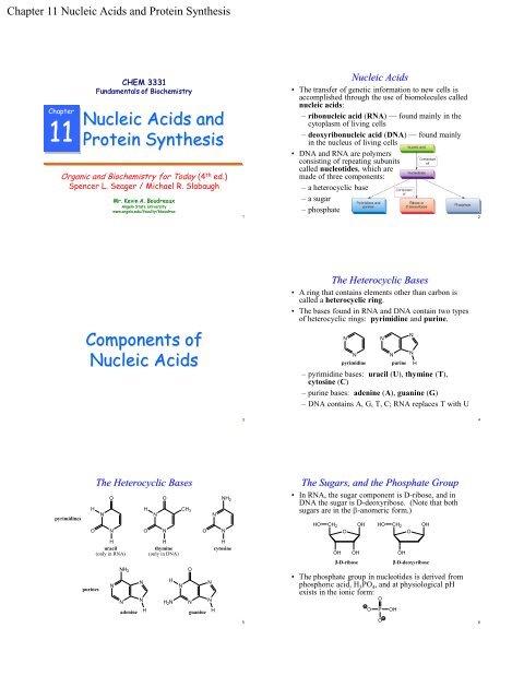
Chapter 11 Nucleic Acids and Protein Synthesis<br />
Chapter<br />
11<br />
CHEM 3331<br />
Fundamentals of Biochemistry<br />
Nucleic Acids and<br />
Protein Synthesis<br />
Organic and Biochemistry for Today (4 th ed.)<br />
Spencer L. Seager / Michael R. Slabaugh<br />
pyrimidines<br />
Mr. Kevin A. Boudreaux<br />
<strong>Angelo</strong> <strong>State</strong> <strong>University</strong><br />
www.angelo.edu/faculty/kboudrea<br />
Components of<br />
Nucleic Acids<br />
H<br />
O<br />
purines<br />
The Heterocyclic Bases<br />
N<br />
O<br />
N<br />
H<br />
uracil<br />
(only in RNA)<br />
N<br />
NH 2<br />
N<br />
adenine<br />
N<br />
N<br />
H<br />
O<br />
H<br />
N<br />
O<br />
N<br />
H<br />
thymine<br />
(only in DNA)<br />
H<br />
H 2N<br />
N<br />
CH 3<br />
O<br />
N<br />
O<br />
guanine<br />
N<br />
N<br />
N<br />
NH 2<br />
N<br />
H<br />
cytosine<br />
H<br />
1<br />
3<br />
5<br />
Nucleic Acids<br />
• The transfer of genetic information to new cells is<br />
accomplished through the use of biomolecules called<br />
nucleic acids:<br />
– ribonucleic acid (RNA) — found mainly in the<br />
cytoplasm of living cells<br />
– deoxyribonucleic acid (DNA) — found mainly<br />
in the nucleus of living cells<br />
• DNA and RNA are polymers<br />
consisting of repeating subunits<br />
called nucleotides, which are<br />
made of three components:<br />
– a heterocyclic base<br />
– a sugar<br />
– phosphate<br />
The Heterocyclic Bases<br />
• A ring that contains elements other than carbon is<br />
called a heterocyclic ring.<br />
• The bases found in RNA and DNA contain two types<br />
of heterocyclic rings: pyrimidine and purine.<br />
N<br />
N<br />
pyrimidine<br />
N<br />
– pyrimidine bases: uracil (U), thymine (T),<br />
cytosine (C)<br />
– purine bases: adenine (A), guanine (G)<br />
– DNA contains A, G, T, C; RNA replaces T with U<br />
N<br />
N<br />
N<br />
purine H<br />
The Sugars, and the Phosphate Group<br />
• In RNA, the sugar component is D-ribose, and in<br />
DNA the sugar is D-deoxyribose. (Note that both<br />
sugars are in the -anomeric form.)<br />
HO<br />
CH 2<br />
O<br />
OH OH<br />
-D-ribose<br />
OH<br />
• The phosphate group in nucleotides is derived from<br />
phosphoric acid, H3PO4, and at physiological pH<br />
exists in the ionic form:<br />
O<br />
HO<br />
O<br />
P<br />
O<br />
CH 2<br />
OH<br />
OH<br />
O<br />
OH<br />
-D-deoxyribose<br />
2<br />
4<br />
6
Chapter 11 Nucleic Acids and Protein Synthesis<br />
O<br />
Putting the Pieces Together: Nucleotides<br />
• Nucleotides are formed from the combination of a<br />
sugar with a phosphate group at the 5′ position and a<br />
heterocyclic base at the 1′ position. (′ is used to<br />
indicate the carbon number in the sugar to<br />
distinguish them from the atoms in the bases.)<br />
O<br />
P<br />
O<br />
OH<br />
NH2 adenine<br />
H O<br />
N<br />
CH 2<br />
N<br />
O<br />
N<br />
N<br />
OH OH<br />
-D-ribose<br />
Abbreviated as ACGT<br />
H<br />
OH<br />
O<br />
O<br />
P<br />
O<br />
N<br />
NH 2<br />
N<br />
5'<br />
O CH2 O<br />
4'<br />
3'<br />
OH OH<br />
N<br />
N<br />
adenosine 5'-monophosphate<br />
(AMP)<br />
The Structure<br />
of DNA<br />
2'<br />
1'<br />
+ 2H 2O<br />
7<br />
9<br />
11<br />
O<br />
General Nucleotide Structure<br />
• The general structure of a nucleotide is illustrated<br />
below:<br />
O<br />
P<br />
O<br />
O<br />
CH 2<br />
OH<br />
O<br />
a nucleotide<br />
OH<br />
base<br />
A<br />
T (U in RNA)<br />
G<br />
C<br />
OH in RNA (ribose)<br />
H in DNA (deoxyribose)<br />
The Primary Structure of DNA<br />
• DNA is one of the largest molecules known,<br />
containing between 1 and 100 million nucleotide<br />
units.<br />
• The nucleotides in DNA are linked by phosphate<br />
groups that connect the 5′ carbon of one nucleotide<br />
to the 3′ carbon of the next.<br />
– Because these connections occur on two oxygen<br />
atoms of the phosphate group, they are called<br />
phosphodiester bonds.<br />
• The nucleic acid backbone then is a sequence of<br />
sugar-phosphate groups, which differ only in the<br />
sequence of bases attached to the sugars along the<br />
backbone (the primary structure of DNA):<br />
The Secondary Structure of DNA<br />
• The bases hydrogen bond to each other in a specific<br />
way: A hydrogen bonds to T, and G hydrogen<br />
bonds to C, forming a set of complementary base<br />
pairs:<br />
H<br />
N<br />
N<br />
H H<br />
N<br />
A<br />
N<br />
N<br />
O<br />
H<br />
CH 3<br />
N N<br />
O<br />
T<br />
H<br />
H<br />
N<br />
N<br />
N<br />
H<br />
C<br />
O<br />
H<br />
H<br />
H<br />
O<br />
N<br />
N<br />
H<br />
N<br />
N<br />
G<br />
N<br />
H<br />
8<br />
10<br />
12
Chapter 11 Nucleic Acids and Protein Synthesis<br />
The Secondary Structure of DNA<br />
• This allows two separate strands of sugarphosphate<br />
backbones to run alongside<br />
each other, held together by the hydrogen<br />
bonds between the complementary base<br />
pairs:<br />
S<br />
S<br />
P S P S P S P<br />
A T C G<br />
T A G C<br />
P S P S P S P<br />
The Double Helix<br />
• The two intertwined polynucleotide chains run in<br />
opposite (antiparallel) directions, with the 5′ end of<br />
one chain on the same side as the 3′ end of the other.<br />
– The base sequence of a DNA strand is always<br />
written from the 5′ end to the 3′ end.<br />
• The sugar-phosphate backbone runs along the<br />
outside of the helix, with the bases pointing inwards,<br />
where they form hydrogen bonds to each other.<br />
• The two strands of DNA are complementary to<br />
each other, because of the specific pairing of G to C<br />
and A to T.<br />
A<br />
G<br />
G<br />
T<br />
C<br />
G<br />
A<br />
G<br />
C<br />
C<br />
T<br />
C<br />
C<br />
A<br />
G<br />
C<br />
T<br />
C<br />
G<br />
G<br />
13<br />
15<br />
The Double Helix<br />
• The ―ladder-like‖ structure<br />
folds in on itself to form a<br />
double helix, with the<br />
bases on the inside and the<br />
sugar-phosphate backbone<br />
on the outside:<br />
The Double Helix<br />
17 18<br />
14<br />
16
Chapter 11 Nucleic Acids and Protein Synthesis<br />
The Discovery of the DNA Structure<br />
• DNA was discovered in 1869 by the Swiss physician<br />
Friedrich Miescher in the pus of discarded surgical<br />
bandages; he named it ―nuclein‖ because it was<br />
located in the nucleus of the cell.<br />
• In 1878, Albrecht Kossel isolated the pure nucleic<br />
acid, and later isolated the five nitrogenous bases.<br />
• Many scientists believed that nucleic acids were far<br />
too simple to be the agent that carried genetic<br />
information from one generation to the next, and that<br />
the genetic material would turn out to be a protein.<br />
• In 1943, Oswald Avery, Colin MacLeod, and<br />
Maclyn McCarty identified DNA as the carrier of<br />
genetic information.<br />
• The race was on to determine the structure of DNA,<br />
and how it was able to transmit genetic information.<br />
The Discovery of the DNA Structure<br />
We wish to suggest a structure for the<br />
salt of deoxyribose nucleic acid<br />
(DNA). This structure has novel<br />
features which are of considerable<br />
biological interest. ...<br />
It has not escaped our notice that the<br />
specific pairing we have postulated<br />
immediately suggests a possible<br />
copying mechanism for the genetic<br />
material.<br />
―Molecular Structure of Nucleic Acids: A Structure for<br />
Deoxyribose Nucleic Acid,‖ Nature (April 25, 1953)<br />
DNA Replication<br />
19<br />
21<br />
23<br />
The Discovery of the DNA Structure<br />
• In 1952, Rosalind Franklin obtained an<br />
X-ray crystal structure (―Photo 51‖) of<br />
a sample of DNA which contained<br />
structural features which lead James D.<br />
Watson and Francis H. C. Crick to<br />
deduce the double helix structure of<br />
DNA (Nobel Prize in Medicine, 1962).<br />
A Structure for Deoxyribose<br />
Nucleic Acid<br />
J. D. Watson and F. H. C. Crick,<br />
Nature 171, 737-738 (1953)<br />
Molecular Configuration in<br />
Sodium Thymonucleate<br />
R. Franklin, and R. G. Gosling,<br />
Nature 171, 740-741 (1953)<br />
Examples: The Structure of DNA<br />
• One strand of a DNA molecule has the base<br />
sequence CCATTG. What is the base sequence for<br />
the complementary strand?<br />
Chromosomes<br />
• A normal human<br />
cell contains 46<br />
chromosomes, each<br />
of which contains a<br />
molecule of DNA<br />
coiled tightly<br />
around a group of<br />
small basic proteins<br />
called histones.<br />
20<br />
22<br />
24
Chapter 11 Nucleic Acids and Protein Synthesis<br />
Replication<br />
• Replication is the process by which an exact copy<br />
of DNA is produced.<br />
– Two strands of DNA separate, and each one<br />
serves as the template for the construction of its<br />
own complement, generating new DNA strands<br />
that are exact replicas of the original molecule.<br />
– The two daughter DNA molecules have exactly<br />
the same base sequences of the parent DNA.<br />
– Each daughter contains one strand of the parent<br />
and one new strand that is complementary to the<br />
parent strand. This type of replication is called<br />
semiconservative replication.<br />
Steps in DNA Replication<br />
• Step 1: Unwinding of the<br />
double helix.<br />
– The enzyme helicase<br />
catalyzes the separation<br />
and unwinding of the<br />
nucleic acid strands at a<br />
specific point called a<br />
replication fork.<br />
– The hydrogen bonds<br />
between the base pairs<br />
are broken, and the<br />
bases are exposed.<br />
– An RNA primer<br />
attaches to the DNA at<br />
the point where<br />
replication begins.<br />
leading<br />
strand<br />
lagging<br />
strand<br />
25<br />
27<br />
Movie:<br />
DNA 29<br />
Replication<br />
Genes<br />
• Individual sections of DNA molecules make up the<br />
genes, which are the fundamental units of heredity<br />
that direct the synthesis of proteins.<br />
– Viruses contain a few to several hundred genes.<br />
– Escherichia coli (E. coli) contains ~1000 genes.<br />
– Humans cells contain ~25,000 genes.<br />
Replication<br />
Steps in DNA Replication<br />
• Step 2: Synthesis of DNA segments.<br />
– DNA replication takes place from the 3′ end<br />
towards the 5′ end of the exposed strands (the<br />
template).<br />
– Because the strands are antiparallel, the synthesis<br />
of new nucleic acid strands proceeds:<br />
• toward the replication fork on one strand (the<br />
leading strand)<br />
• away from the replication fork on the other<br />
strand (the lagging strand).<br />
– Nucleotides complementary to the ones on the<br />
exposed strands are attached to the growing<br />
chain, and are linked together by the enzyme<br />
DNA polymerase to form a new daughter strand.<br />
26<br />
28<br />
30
Chapter 11 Nucleic Acids and Protein Synthesis<br />
Steps in DNA Replication<br />
• Step 2: Synthesis of DNA segments.<br />
– As the replication fork moves down the DNA<br />
backbone, the leading strand grows smoothly<br />
towards the 5′ end.<br />
– Since the lagging strand was growing away from<br />
the first fork, new segments grow from the new<br />
location of the replication fork, until they meet<br />
the areas where the RNA primers are located.<br />
– This daughter strand is thus synthesized as a<br />
series of fragments that are bound together in<br />
Step 3. The gaps or breaks between segments in<br />
this daughter strand are called nicks, and the<br />
DNA fragments separated by the nicks are called<br />
Okazaki fragments (after Reiji Okazaki).<br />
Steps in DNA Replication — Summary<br />
• Step 1: The DNA is unwound by helicase and a replication fork forms.<br />
• Step 2: With the help of the enzyme DNA polymerase, DNA is<br />
replicated smoothly along the leading strand which grows towards the<br />
replication fork. DNA segments (Okazaki fragments) are synthesized<br />
by DNA polymerase along the lagging strand as the replication fork<br />
moves.<br />
• Step 3: The Okazaki fragments are joined by DNA ligase, resulting in<br />
two new DNA molecules.<br />
Polymerase Chain Reaction (PCR)<br />
• An important laboratory technique called the<br />
polymerase chain reaction (PCR) mimics the<br />
natural process of replication.<br />
– A small quantity of target DNA, a buffered<br />
solution of DNA polymerase, the cofactor MgCl 2,<br />
the four nucleotide building blocks, and primers<br />
are added to a test tube.<br />
• The primers are short polynucleotides that bind<br />
to the DNA strands and serve as starting points<br />
for new chain growth.<br />
– The mixture goes through several three-step<br />
replication cycles:<br />
• Heat (94-96°C) is used for one to several<br />
minutes to unravel DNA into single strands.<br />
31<br />
33<br />
[Kary Mullins<br />
and Michael<br />
Smith, Nobel<br />
Prize, 1993]<br />
35<br />
Steps in DNA Replication<br />
• Step 3: Closing the nicks.<br />
– The daughter strand along the leading strand is<br />
synthesized smoothly, without any nicks.<br />
– The Okazaki fragments along the lagging strand<br />
are joined by an enzyme called DNA ligase,<br />
which removes the RNA primer and replaces it<br />
with the correct nucleotides.<br />
– The result is two DNA double-helix molecules of<br />
DNA that are identical to the original DNA<br />
molecule, each of which contains one old strand<br />
from the parent DNA and one new daughter<br />
strand (semiconservative replication).<br />
DNA Replication in More Than One Place<br />
• In eukaryotes, DNA replication occurs simultaneously<br />
at many replication forks along the original molecule.<br />
The zones where replication occur eventually combine<br />
to form complete strands. This allows long molecules<br />
to be replicated quickly.<br />
– The largest chromosome of the fruit fly (Drosophila) would take more<br />
than 16 days to replicate in one segment. The actual process takes less<br />
then three minutes because it occurs at more than 6000 replication forks.<br />
Polymerase Chain Reaction (PCR)<br />
• The tube is cooled to 50-65°C for one to<br />
several minutes to allow primers to hydrogenbond<br />
to the separated strands of target DNA.<br />
• The tube is heated to 72°C for one to several<br />
minutes while DNA polymerase synthesizes<br />
new strands.<br />
– Each cycle doubles the amount of DNA;<br />
following 30 cycles, a theoretical amplification<br />
factor of 1 billion is attained.<br />
32<br />
34<br />
36
Chapter 11 Nucleic Acids and Protein Synthesis<br />
Polymerase Chain Reaction (PCR)<br />
• PCR is a standard research technique that:<br />
– detects all manner of mutations associated with<br />
genetic disease.<br />
– is used to detect presence of unwanted DNA<br />
(bacterial or viral infection).<br />
– is a fast and simple to lengthy procedures<br />
involving sample cultures that can take weeks.<br />
– can be used on degraded DNA samples:<br />
• forensic analysis, DNA fingerprinting.<br />
• Recovery of DNA from extinct mammals,<br />
Egyptian mummies, and ancient insects<br />
trapped in amber to be amplified and analyzed.<br />
Ribonucleic Acid<br />
(RNA)<br />
Secondary Structure of RNA<br />
• Most RNA molecules are single-stranded, although<br />
many contain regions of double-helical structure<br />
where they form loops. (A::U, G:::C)<br />
37 38<br />
39<br />
41<br />
RNA<br />
• RNA is a long unbranched polymer consisting of<br />
nucleotides joined by 3′ to 5′ phosphodiester bonds.<br />
• RNA strands consist of from 73 to many thousands<br />
of nucleotides.<br />
• Whereas DNA is only found in the nucleus, RNA is<br />
found throughout cells: in the nucleus, in the<br />
cytoplasm, and in the mitochondria.<br />
• Differences in RNA and DNA primary structures:<br />
– In RNA the sugar is ribose instead of deoxyribose.<br />
– In RNA, the base uracil (U) is used instead of<br />
thymine (T).<br />
O<br />
O<br />
HO CH2 O<br />
OH OH<br />
-D-ribose<br />
OH<br />
HO CH2 O<br />
OH<br />
OH<br />
-D-deoxyribose<br />
H<br />
N<br />
O<br />
N<br />
H<br />
uracil<br />
(only in RNA)<br />
H<br />
N<br />
O<br />
N<br />
H<br />
thymine<br />
(only in DNA)<br />
Kinds of RNA — Messenger RNA (mRNA)<br />
• There are three kinds of RNA: messenger RNA<br />
(mRNA), ribosomal RNA (rRNA), and transfer<br />
RNA (tRNA).<br />
• Messenger RNA (mRNA) — functions as a carrier<br />
of genetic information from the DNA in the cell<br />
nucleus to the site of protein synthesis in the<br />
cytoplasm.<br />
– The bases of mRNA are in a complementary<br />
sequence to the base sequence of one of the<br />
strands of nuclear DNA.<br />
– mRNA has a short lifetime (usually less than one<br />
hour); it is synthesized as it is needed, then<br />
rapidly degraded to the constituent nucleotides.<br />
CH 3<br />
40<br />
42
Chapter 11 Nucleic Acids and Protein Synthesis<br />
Kinds of RNA — Ribosomal RNA (rRNA)<br />
• Ribosomal RNA (rRNA) — the main component<br />
of ribosomes that are the site of protein synthesis.<br />
– rRNA accounts for 80-85% of the total RNA of<br />
the cell.<br />
– rRNA accounts for 65% of a ribosome’s structure<br />
(the remaining 35% is protein).<br />
Kinds of RNA — Transfer RNA (tRNA)<br />
• Transfer RNA (tRNA) — cont.<br />
– tRNA has regions of hydrogen bonding between<br />
complementary base pairs, separated by loops<br />
where there is no hydrogen bonding.<br />
– Two regions of tRNA have important functions:<br />
• the anticodon is a three-base sequence which<br />
allows tRNA to bind to mRNA during protein<br />
synthesis. (It is complementary to one of the<br />
codons in mRNA.)<br />
• the 3′ end of the molecule binds to an amino<br />
acid with an ester bond and transports it to the<br />
site of protein synthesis. An enzyme matches<br />
the tRNA molecule to the correct amino acid,<br />
―activating‖ it for protein synthesis.<br />
The Flow of Genetic<br />
Information<br />
43<br />
45<br />
47<br />
Kinds of RNA — Transfer RNA (tRNA)<br />
• Transfer RNA (tRNA) — delivers individual<br />
amino acids to the site of protein synthesis.<br />
– tRNA is specific to one type of amino acid; cells<br />
contain at least one specific type of tRNA for<br />
each of the 20 common amino acids.<br />
– tRNA is the smallest of the nucleic acids, with<br />
73-93 nucleotides per chain.<br />
Kinds of RNA — Transfer RNA (tRNA)<br />
transfer RNA<br />
activated<br />
tRNA<br />
44<br />
activated<br />
tRNA<br />
(schematic) 46<br />
The Central Dogma of Molecular Biology<br />
• The central dogma of molecular biology states that<br />
genetic information contained in the DNA is<br />
transferred to RNA molecules and then expressed in<br />
the structure of synthesized proteins.<br />
• Genes are segments of DNA that contain the<br />
information needed for the synthesis of proteins.<br />
• Each protein in the body corresponds to a DNA<br />
gene.<br />
48
Chapter 11 Nucleic Acids and Protein Synthesis<br />
Transcription, Translation, and Information Flow<br />
• There are two steps in the flow of genetic information:<br />
– transcription — in eukaryotes, the DNA<br />
containing the stored information is in the nucleus<br />
of the cell, and protein synthesis occurs in the<br />
cytoplasm. The information stored in the DNA<br />
must be carried out of the nucleus by mRNA.<br />
– translation — mRNA serves as a template on<br />
which amino acids are assembled in the sequence<br />
necessary to produce the correct protein. The code<br />
carried by mRNA is translated into an amino acid<br />
sequence by tRNA.<br />
• The communicative relationship between mRNA<br />
nucleotides and amino acids in a protein is called the<br />
genetic code.<br />
Transcription: RNA Synthesis<br />
• Under the influence of the enzyme RNA<br />
polymerase, the DNA double helix unwinds at a<br />
point near the gene that is being transcribed (the<br />
initiation sequence). Only one strand of the DNA<br />
is transcribed.<br />
• Ribonucleotides are linked along the DNA strand in<br />
a sequence determined by the base pairing of the<br />
DNA and ribonucleotide bases (A::U, G:::C).<br />
• mRNA synthesis occurs in the 3′ to 5′ direction<br />
along the DNA strand (in the 5′ to 3′ direction along<br />
the RNA strand) until the termination sequence is<br />
reached.<br />
• The newly-synthesized mRNA strand moves away<br />
from the DNA, which rewinds into the double helix.<br />
• Synthesis of tRNA and rRNA is similar to this.<br />
Examples: The Synthesis of RNA<br />
• Write the sequence for the mRNA that could be<br />
synthesized using the following DNA base sequence<br />
as a template:<br />
5′ G-C-A-A-C-T-T-G 3′<br />
49 50<br />
51<br />
53<br />
Transcription: RNA Synthesis<br />
Movie:<br />
Transcription<br />
Introns and Exons<br />
• In prokaryotes, each gene is a continuous segment<br />
along a DNA molecule. Transcription of the gene<br />
produces mRNA that is translated into a protein<br />
almost immediately, because there is no nuclear<br />
membrane separating the DNA from the cytoplasm.<br />
• In eukaryotes, the gene segments of DNA that code<br />
for proteins (exons) are interrupted by segments that<br />
do not carry an amino acid code (introns).<br />
– Both exon and intron segments are transcribed,<br />
producing heterogenous nuclear RNA<br />
(hnRNA).<br />
– A series of enzymes cut out the intron segments<br />
and splice the exon segments together to produce<br />
mRNA.<br />
52<br />
54
Chapter 11 Nucleic Acids and Protein Synthesis<br />
Introns and Exons<br />
The Genetic Code<br />
Characteristics of the Genetic Code<br />
• The genetic code applies almost universally: with<br />
minor exceptions, the same amino acid is<br />
represented by the same codon(s) in all species.<br />
• Most amino acids are represented by more than one<br />
codon (a feature known as degeneracy).<br />
– Only methionine and tryptophan are represented by a single codon.<br />
– Leucine, serine, and arginine are represented by six codons.<br />
– No codon codes for more than one amino acid.<br />
• Only 61 of the 64 possible triplets represent amino<br />
acids. The other three are used as signals for chain<br />
termination (a ―stop‖ signal).<br />
• The AUG codon (which also codes for methionine)<br />
functions as a ―start‖ signal, but only when it occurs<br />
as the first codon in a sequence.<br />
55<br />
The Genetic Code<br />
• Once the 3D structure of DNA was known, it was<br />
clear that the sequence of the bases along the<br />
backbone in some way directed the order in which<br />
amino acids were stacked to make proteins.<br />
• In 1961, Marshall Nirenberg and his coworkers<br />
began to unravel the connection between the base<br />
sequence in DNA and the amino acid sequence in<br />
proteins.<br />
• The genetic code uses a sequence of three bases (a<br />
triplet code) to specify each amino acid. (A triplet<br />
code gives 4 3 =64 possible combinations, which is<br />
more than enough to specify the 20 amino acids.)<br />
• Each base triplet sequence that represents a code<br />
word on mRNA molecules is called a codon.<br />
57 58<br />
59<br />
The Genetic Code<br />
56<br />
60
Chapter 11 Nucleic Acids and Protein Synthesis<br />
Translation and Protein Synthesis<br />
Step 1: Initiation of the polypeptide chain.<br />
• mRNA and a small ribosomal subunit join; the<br />
initiating codon (AUG) is aligned with P (peptidyl)<br />
site of the subunit.<br />
• tRNA brings in methionine (eukaryotes) or<br />
N-formylmethionine (prokaryotes).<br />
• The resulting complex binds to the large ribosomal<br />
subunit to form a unit called the initiation complex.<br />
Ribosome<br />
red = large subunit<br />
blue = small subunit<br />
Translation and Protein Synthesis<br />
Step 2: Elongation of the chain, cont.<br />
• The ―empty‖ tRNA released, and the whole<br />
ribosome moves one codon along the mRNA<br />
towards the 3’ end (translocation).<br />
• Another tRNA attaches to the A site, and the<br />
elongation process is repeated.<br />
Movie:<br />
Translation<br />
Translation and Protein Synthesis<br />
• Several ribosomes can move along a single strand of<br />
mRNA, producing several identical proteins<br />
simultaneously. These complexes are called<br />
polyribosomes or polysomes.<br />
• The growing polypeptide chain emerging from the<br />
end of the ribosome spontaneously folds into the<br />
characteristic 3D shape of that protein.<br />
61<br />
63<br />
Translation and Protein Synthesis<br />
Step 2: Elongation of the chain.<br />
• The next incoming tRNA with an anticodon that is<br />
complementary to the mRNA codon bonds at the A<br />
(aminoacyl) site on the mRNA.<br />
• A peptide bond is formed between the amino acid<br />
segments, (catalyzed by peptidyl transferase), which<br />
releases the amino acid chain from the P site.<br />
Translation and Protein Synthesis<br />
Step 3: Termination of polypeptide synthesis.<br />
• Elongation continues until the ribosome complex<br />
reaches a stop codon (UAA, UAG, or UGA).<br />
• A termination factor protein binds to the stop codon,<br />
and separates the protein from the final tRNA.<br />
• The ribosome can then synthesize another protein<br />
molecule.<br />
65 66<br />
62<br />
64
Chapter 11 Nucleic Acids and Protein Synthesis<br />
Mutations,<br />
Recombinant DNA,<br />
and<br />
Genetic Engineering<br />
Mutations<br />
• Mutations may be beneficial to an organism by<br />
making it more capable of surviving in its<br />
environment, ultimately (over millions of years of<br />
accumulating changes) leading to the evolution of<br />
new species.<br />
• Since much of an organisms DNA does not code for<br />
anything, mutations in these regions are neutral.<br />
• Other mutations can be harmful, either producing<br />
genetic diseases or other debilitating conditions.<br />
Restriction Enzymes<br />
• Restriction enzymes, found in a wide variety of<br />
bacterial cells, catalyze the cleaving of DNA<br />
molecules. These enzymes are normally part of a<br />
mechanism that protects certain bacteria from<br />
invasion by foreign DNA such as that in viruses. In<br />
these bacteria, some of the bases in their DNA have<br />
methyl groups attached:<br />
O<br />
N<br />
NH 2<br />
N<br />
CH<br />
5 3<br />
H<br />
5-methylcytosine<br />
CH3 1<br />
N<br />
• The methylated DNA of these bacteria is left<br />
untouched by the restriction enzymes, for foreign<br />
DNA that lacks these bases undergoes rapid<br />
cleavage, and is rendered nonfunctional.<br />
O<br />
N<br />
H2N N<br />
N<br />
H<br />
1-methylguanine<br />
67<br />
69<br />
71<br />
Mutations<br />
• Mutations are any changes resulting in an incorrect<br />
base sequence on DNA.<br />
• Even though the base-pairing mechanism provides a<br />
nearly perfect way of copying DNA, on average one<br />
out of every 10 10 bases are copied incorrectly.<br />
– This leads to a change in the amino acid sequence<br />
in a protein, or causes the protein not to be made<br />
at all.<br />
• Mutations occur naturally during replication. They<br />
can also be induced by environmental factors:<br />
– ionizing radiation (X-rays, UV, gamma rays).<br />
– mutagens, which are chemical agents.<br />
Recombinant DNA<br />
• Recombinant DNA is produced when segments of<br />
DNA from one organism are introduced into the<br />
genetic material of another organism.<br />
• ―Genetic engineering‖ of E. coli to include the gene<br />
for the production of human insulin enables large<br />
quantities of insulin to be made available for the<br />
treatment of diabetes.<br />
Restriction Enzymes<br />
• Restriction enzymes act at sites on DNA called<br />
palindromes, where two strands have the same<br />
sequence but run in opposite directions:<br />
point of attack<br />
• Restriction enzymes are used to break DNA up into<br />
fragments of known size and nucleotide sequence,<br />
which can then be spliced together with DNA<br />
ligases.<br />
68<br />
70<br />
72
Chapter 11 Nucleic Acids and Protein Synthesis<br />
Plasmids<br />
• The introduction of a new DNA segment (gene) into<br />
a bacterial cell requires a DNA carrier called a<br />
vector, which is often a circular piece of doublestranded<br />
DNA called a plasmid.<br />
– Plasmids range from 2000 to several hundred<br />
thousand nucleotides, and are found in the<br />
cytoplasm of bacterial cells.<br />
– Plasmids function as accessories to chromosomes<br />
by carrying genes for the inactivation of<br />
antibiotics and the production of toxins. They are<br />
also able to replicate independently of<br />
chromosomal DNA.<br />
The Formation of Recombinant DNA<br />
• The breaks in the strands are joined using DNA<br />
ligase, and the plasmid becomes a circular piece of<br />
double-stranded, recombinant DNA.<br />
The End<br />
73<br />
75<br />
77<br />
The Formation of Recombinant DNA<br />
• A plasmid is isolated from a bacterium, and a<br />
restriction enzyme is added, which cleaves it at a<br />
specific site:<br />
• When the circular DNA is cut, two ―sticky ends‖ are<br />
produced, which have unpaired bases.<br />
• The ―sticky ends‖ are provided with complementary<br />
sections for pairing from a human chromosome to<br />
which the same restriction enzyme has been used:<br />
The Formation of Recombinant DNA<br />
• When the bacteria reproduce, they replicate all of the<br />
genes, including the new recombinant DNA<br />
plasmids.<br />
• Because bacteria multiply quickly, there are soon a<br />
large number of bacteria containing the modified<br />
plasmid, which are capable of manufacturing the<br />
desired protein.<br />
74<br />
76