
Create successful ePaper yourself
Turn your PDF publications into a flip-book with our unique Google optimized e-Paper software.
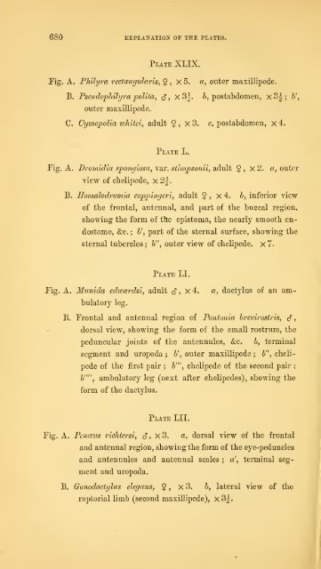
680 explanatiox of the plates.<br />
Plate XLIX.<br />
Fig. A. Philyra rectangvlaris, 5 , X 5. a, outer maxilHpede.<br />
B. PseudoplnJyra poUta, c5", x3|. &, postabdomen, X 3| ; b\<br />
outer maxillipede.<br />
C. Cymopolia luhitet, adult $<br />
Plate L.<br />
, x 3. c, postabdomen, x 4.<br />
Pig. A. Dromidia spongiosa, var. stimpsonii, adult §<br />
view of cbelipede, x 2^.<br />
B. Homalodromia coppingeri, adult 5<br />
, X iJ. «, outer<br />
, X 4. 6, inferior view<br />
of the frontal, antennal, and part of the buccal region,<br />
showing the form of the epistoma, the nearly smooth en-<br />
dostome, &c. ; 6', part of the sternal surface, showing the<br />
sternal tubercles ; h", outer view of chelipede. x 7.<br />
Plate LI.<br />
Fig. A. Munida ediuardsi, adult (5" , x 4. a, dactylus of an am-<br />
bulatory leg.<br />
B. Frontal and antennal region of Pontonia brevirostris, c?<br />
dorsal view, showing the form of the small rostrum, the<br />
peduncular joints of the antennules, &c. b, terminal<br />
segment and uropoda ; b', outer maxillipede ; b", cheli-<br />
pede of the first pair ; b'", chelipede of the second pair ;<br />
6"", ambulatory leg (next after chelipedes), showing the<br />
form of the dactylus.<br />
Plate LII.<br />
Fig. A. Penceus ricJitersi, c?, x3. a, dorsal view of the frontal<br />
and antennal region, showing the form of the eye-peduncles<br />
and antennules and antennal scales ; a', terminal seg-<br />
ment and uropoda.<br />
B. Gonodactijlus elegans, $<br />
, X 3. b, lateral view of the<br />
raptorial limb (second maxillipede), x3|.<br />
,