Cleavage and gastrulation in the shrimp Penaeus (Litopenaeus ...
Cleavage and gastrulation in the shrimp Penaeus (Litopenaeus ...
Cleavage and gastrulation in the shrimp Penaeus (Litopenaeus ...

Create successful ePaper yourself
Turn your PDF publications into a flip-book with our unique Google optimized e-Paper software.
458<br />
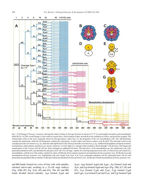
Fig. 1. Cell l<strong>in</strong>eage of <strong>Penaeus</strong> vannamei, show<strong>in</strong>g <strong>the</strong> relative tim<strong>in</strong>g of cleavage divisions <strong>in</strong> m<strong>in</strong> ps at 27 8C (A) <strong>and</strong> naupliar mesoderm <strong>and</strong> mesendoderm<br />
detail (B–E). (A) The overall l<strong>in</strong>eage is color coded as to germ layer, with ectoderm <strong>in</strong> blue, mesoderm <strong>in</strong> red, endoderm <strong>in</strong> yellow, <strong>and</strong> germ l<strong>in</strong>e <strong>in</strong> purple. The<br />
nomenclature refers to <strong>the</strong> most commonly observed cleavage pattern, where DIIrZX, <strong>the</strong> progenitor of <strong>the</strong> two mesendoderm cells. The AB blastomere<br />
conta<strong>in</strong>s <strong>the</strong> animal pole, while <strong>the</strong> CD blastomere conta<strong>in</strong>s <strong>the</strong> vegetal pole. A <strong>and</strong> C make cross-furrow contact, as do B <strong>and</strong> D. The subscript I refers to <strong>the</strong><br />
cell adjacent to <strong>the</strong> cross-furrow (e.g. A I), while <strong>the</strong> subscript II refers to <strong>the</strong> cell away from <strong>the</strong> cross furrow (e.g. A II). Additional designations are based on relative<br />
axial positions, ei<strong>the</strong>r anterior–posterior (a, p), dorsal–ventral (d, v) or left–right (l, r). L<strong>in</strong>eage of <strong>the</strong> ectoderm is shown through 7 cell divisions. The l<strong>in</strong>eage <strong>and</strong><br />
nomenclature of <strong>the</strong> 9 crown cells K1–K9 are shown through two additional divisions. The derivatives of <strong>the</strong> mesendoderm are: <strong>the</strong> endodermal yolk cells,<br />
primordial endoderm cell E, mesoteloblast M, <strong>and</strong> germ cell G. (B) 122-cell stage, vegetal view show<strong>in</strong>g replicated crown cells. (C) Dorsal (X d) <strong>and</strong> ventral (X v)<br />
mesendoblasts, with <strong>the</strong> rest of <strong>the</strong> embryo omitted for clarity. (D) Result of first (anterior–posterior) mesendoderm division, posterior view. (E) Result of second<br />
mesendoderm division, posterior view. Endodermal yolk cells (Xdpd, Xdal, Xdar, Xval, Xvar), primordial endoderm cell (E), primordial germ cell (G), <strong>and</strong><br />
primordial mesoteloblast (M) are labeled. (F) Result of third mesendoderm division, posterior view. (G) Result of fourth mesendoderm division, posterior view.<br />
<strong>and</strong> BD b<strong>and</strong>s formed two rows of four cells with sp<strong>in</strong>dles<br />
oriented end-to-end, result<strong>in</strong>g <strong>in</strong> a 32-cell stage embryo<br />
(Fig. 2(M)–(P), Fig. 3(A), (D) <strong>and</strong> (G)). The AC <strong>and</strong> BD<br />
b<strong>and</strong>s divided dorsal–ventrally. AIIa formed AIIad <strong>and</strong><br />
P.L. Hertzler / Arthropod Structure & Development 34 (2005) 455–469<br />
AIIav, AIIp formed AIIpd <strong>and</strong> AIIpv, AIa formed AIad <strong>and</strong><br />
AIav, <strong>and</strong> AIp formed AIpd <strong>and</strong> AIpv (Fig. 3(B), (C), (E) <strong>and</strong><br />
(F)). CIIa formed CIIad <strong>and</strong> CIIav, CIIp formed CIIpd<br />
<strong>and</strong> CIIpv, CIa formed CIad <strong>and</strong> CIav, <strong>and</strong> CIp formed CIpd