Mirizzi syndrome - medIND
Mirizzi syndrome - medIND
Mirizzi syndrome - medIND

Create successful ePaper yourself
Turn your PDF publications into a flip-book with our unique Google optimized e-Paper software.
CASE REPORT<br />
<strong>Mirizzi</strong> <strong>syndrome</strong><br />
Col PV Rama Mohan*, Wg Cdr M Kumar + , Maj R Pacharu #<br />
MJAFI 2011;67:280–281<br />
INTRODUCTION<br />
<strong>Mirizzi</strong> <strong>syndrome</strong> (MS) is a rare form of obstructive jaundice<br />
that occurs as an infrequent complication of gallstones in about<br />
0.1–0.7% of patients who have gallstones. 1 <strong>Mirizzi</strong> was the first<br />
to describe this phenomenon as “functional hepatic <strong>syndrome</strong>”<br />
in 1948. 2 It is a benign condition resulting from a chronically impacted<br />
stone or stones in the neck of gall bladder or cystic duct,<br />
which over time induces sufficient pericholecystic inflammation<br />
to narrow and obstruct the adjacent common hepatic duct.<br />
We present a case of MS type II who presented with<br />
obstructive jaundice.<br />
CASE REPORT<br />
A 50-year-old male patient, a smoker and consumer of alcohol,<br />
presented with features of painless progressive jaundice, anorexia,<br />
weight loss, and passing clay coloured stool for two<br />
months duration. Clinical examination revealed icterus, firm,<br />
non-tender hepatomegaly with rounded edge and a palpable<br />
nodule in it 8 cm below the right subcostal margin. Gall bladder<br />
was not palpable.<br />
EVALUATION<br />
Liver Function Tests<br />
Serum bilirubin 25.3 mg/dL; conjugated 17.6 mg/dL; ALP 184 IU/<br />
dL (15–112). USG abdomen showed mild central IHBR dilatation,<br />
GB contracted with sludge in its lumen and a 29 mm calculus<br />
at the neck. CECT abdomen showed asymmetric wall thickening<br />
of GB, without any fat stranding or liver infiltration. A 29 mm<br />
calculus seen in cystic duct with extrinsic compression of common<br />
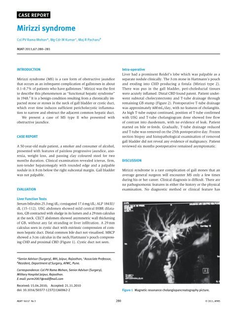
hepatic duct. Distal common bile duct not visualised. MRCP<br />
showed a 3 cm calculus in the neck/Hartmann’s pouch compressing<br />
CHD and proximal CBD (Figure 1). Cystic duct not seen.<br />
*Senior Advisor (Surgery), MH, Jaipur, Rajasthan, + Associate Professor,<br />
# Resident, Department of Surgery, AFMC, Pune.<br />
Correspondence: Col PV Rama Mohan, Senior Advisor (Surgery),<br />
Military Hospital Jaipur, Rajasthan.<br />
E-mail: pvrm2007@rediffmail.com<br />
Received: 15.04.2010; Accepted: 21.11.2010<br />
doi: 10.1016/S0377-1237(11)60062-2<br />
Intra-operative<br />
Liver had a prominent Reidel’s lobe which was palpable as a<br />
separate nodule clinically. The 3 cm stone in Hartmann’s pouch<br />
and eroding into CHD producing a fistula (<strong>Mirizzi</strong> type 2).<br />
There was pus in the gall bladder, peri-choleductal tissues<br />
were acutely inflamed. Distal CBD found patent. Patient underwent<br />
subtotal cholecystectomy and T-tube drainage through<br />
remaining GB stump (Figure 2). Postoperative T-tube drainage<br />
was approximately 600 mL/day, with no features of cholangitis.<br />
As high T-tube output continued, position of T-tube confirmed<br />
with USG and T-tube cholangiogram done showed free flow<br />
of contrast into duodenum, with no evidence of leak. Patient<br />
started on bile re-feeds. Gradually, T-tube drainage reduced<br />
and T-tube was removed on the 25th postoperative day. Frozen<br />
section biopsy and histopathological examination of removed<br />
gall bladder did not reveal any evidence of malignancy. Patient<br />
reviewed six months postoperative remained asymptomatic.<br />
DISCUSSION<br />
<strong>Mirizzi</strong> <strong>syndrome</strong> is a rare complication of gall stones that an<br />
average general surgeon will encounter MS only a few times<br />
during his or her career. Clinical diagnosis is difficult. There are<br />
no pathognomonic features in either the history or the physical<br />
examination. No diagnostic method or clinical feature has<br />
Figure 1 Magnetic resonance cholangiopancreatography picture.<br />
MJAFI Vol 67 No 3 280 © 2011, AFMS
Figure 2 Insertion of a T-tube through the fistula.<br />
100% sensitivity and specificity. Its importance has been highlighted<br />
as a clinical entity associated with a high incidence of<br />
biliary injuries and demanding complex surgical procedures.<br />
Additionally, a high coincidence of MS and gallbladder cancer<br />
has been reported in several studies. 3<br />
In our case at initial presentation, presence of prominent<br />
Riedel’s lobe gave a nodular feel to liver giving a clinical impression<br />
of secondaries liver. Despite absence of any features of<br />
acute inflammation per up gall bladder had pus in its lumen<br />
and pericholeductal tissues acutely inflamed. Presence of this<br />
inflammation made it difficult to explore CBD and confined the<br />
surgery to partial cholecystectomy, over sewing of the gallbladder<br />
cuff, and insertion of a T-tube through the fistula. Despite<br />
patent distal CBD patient continued to have high T-tube output,<br />
most probably due to inflammatory oedema of CBD forcing us<br />
to start patient on bile refeeds mixed with honey.<br />
The condition was classified by McSherry and colleagues in<br />
1982 and the modification of the same by Csendes and colleagues<br />
4 in 1989 into classes 1–4 is the currently accepted classification.<br />
The Csendes classification of MS is as follows:<br />
Type 1: external compression of the common bile duct – 11%.<br />
Type 2: cholecystobiliary fistula is present involving less<br />
than one-third the circumference of the bile duct – 41%.<br />
Type 3: a fistula is present involving upto two-third the<br />
circumference of the bile duct – 44%.<br />
Type 4: a fistula is present with complete destruction of the<br />
wall of the bile duct – 4%.<br />
Management of MS in cases in whom the bile duct is inflamed<br />
and no fistula is present (Type I), patients can be managed<br />
with cholecystectomy. Many surgeons contend that<br />
laparoscopic cholecystectomy is contraindicated in MS 5 while<br />
the others consider the laparoscopic technique feasible, though<br />
technically demanding. 6 The common hepatic duct almost always<br />
<strong>Mirizzi</strong> <strong>syndrome</strong><br />
returns to normal after the offending stone has been removed<br />
by cholecystectomy and the inflammatory process has resolved.<br />
Rarely a well-established stricture presents months to years after<br />
the acute episode. In such cases, stricture repair of the distal bile<br />
duct is indicated for persistent strictures using either Roux–en–Y<br />
choledochojejunostomy or choledochoduodenostomy.<br />
If cholecystocholedochal fistula is present, partial cholecystectomy,<br />
oversewing the gallbladder cuff, and insertion of a<br />
T-tube through the fistula as adequate treatment for Type 2 disease.<br />
7 Baer et al 8 suggested the placement of a T-tube through a<br />
separate choledochotomy in the distal CBD in order to prevent<br />
excessive leakage and stricture at the fistula site. The presence<br />
of CBD stones, fistula, or stenosis at the site of impaction of the<br />
stone may necessitate CBD exploration and/or the insertion of<br />
a T-tube. Choledochoduodenostomy for Type 3 disease, and<br />
biliary bypass (Roux-en-Y hepaticojejunostomy) for Type 4 MS<br />
are the treatment of choices. In all cases, a frozen section of the<br />
gallbladder wall should be done to rule out coexistent cancer.<br />
The prognosis of MS is very good for type 1 lesions, as simple<br />
cholecystectomy is all that is necessary for cure. In treating<br />
more serious types with fistulous destruction of the common<br />
duct, postoperative morbidity rises, with 10% or more biliary<br />
fistulae, biliary stricturing requiring dilation or reoperation, or<br />
hepatic abscesses requiring drainage. 9<br />
REFERENCES<br />
1. Hazzan D, Golijanin D, Reissman P. Combined endoscopic and surgical<br />
management of <strong>Mirizzi</strong> <strong>syndrome</strong>. Surg Endosc 1999;13:<br />
618–620.<br />
2. <strong>Mirizzi</strong> PL. Sindrome del conducto hepatico. J Int Chir 1948;8:<br />
731–733.<br />
3. Prasad TL, Kumar A, Sikora SS, Saxena R, Kapoor VK. <strong>Mirizzi</strong> <strong>syndrome</strong><br />
and gallbladder cancer. J Hepatobiliary Pancreat Surg 2006;<br />
13:323–326.<br />
4. Csendes A, Diaz JC, Burdiles P, Maluenda F, Nava O. <strong>Mirizzi</strong> <strong>syndrome</strong><br />
and cholecystobiliary fistula: a unifying classification. Br J Surg<br />
1989;76:1139–1143.<br />
5. Moser JJ, Baer HU, Glatti A. <strong>Mirizzi</strong> <strong>syndrome</strong> eda contraindication for<br />
laparoscopic surgery. Helv Chir Acta 1993;59:577–580.<br />
6. Meng WC, Kwok SP, Kelly SB, Lau WY, Li AK. Management of <strong>Mirizzi</strong><br />
<strong>syndrome</strong> by laparoscopic cholecystectomy and laparoscopic ultrasonography.<br />
Br J Surg 1995;82:396.<br />
7. Corlette MB, Bismuth H. Biliobiliary fistula. A trap in the surgery of<br />
cholelithiasis. Arch Surg 1975;110:377–383.<br />
8. Baer HU, Matthews JB, Schweizer WP, Gertsch P, Blumgart LH.<br />
Management of the <strong>Mirizzi</strong> <strong>syndrome</strong> and the surgical implications<br />
of cholecystcholedochal fistula. Br J Surg 1990;77:743–745.<br />
9. Waisberg J, Corona A, Abreu I. Benign obstruction of the common<br />
hepatic duct (<strong>Mirizzi</strong> <strong>syndrome</strong>): diagnosis and operative management.<br />
Arq Gastroenterol 2005;42:13–18.<br />
MJAFI Vol 67 No 3 281 © 2011, AFMS