THE CHARACTERIZATION OF BERYL AI{D EMERALD BY VISIBLE ...
THE CHARACTERIZATION OF BERYL AI{D EMERALD BY VISIBLE ...
THE CHARACTERIZATION OF BERYL AI{D EMERALD BY VISIBLE ...
Create successful ePaper yourself
Turn your PDF publications into a flip-book with our unique Google optimized e-Paper software.
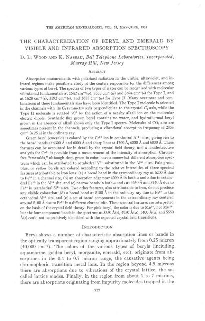
<strong>THE</strong> AMERICAN MINERALOGIST, VOL.53, MAY_JUNE, 1968<br />
<strong>THE</strong> <strong>CHARACTERIZATION</strong> <strong>OF</strong> <strong>BERYL</strong> <strong>AI</strong>{D <strong>EMERALD</strong> <strong>BY</strong><br />
<strong>VISIBLE</strong> AND INFRARED ABSORPTION SPECTROSCOPY<br />
D. L. Woop eNn K. Nesseu, BeIl Telephone Laboratories, Incorporated,<br />
Murray Hill, I{ew Jersey<br />
Assrnecr<br />
Absorption measurements with polarized radiation in the visible, ultraviolet, and infrared<br />
regions make possible a study of the centers responsible for the difierences among<br />
various types of beryl. The spectra of two types of water can be recognized with molecular<br />
vibrational fundamentals at 1542 cm-l(o), 3555 cm-1(o) and 3694 cm-r(e) for Type I, and<br />
at 1628 cm-l(e),3592 cm-r(e), and 3655 cm-r(o) for Type II. Many overtones and combinations<br />
of these fundamentals also have been identified. The Tlpe I molecule is oriented<br />
in the channels with its C2 symmetry axis perpendicular to the crystal C6-axis, while the<br />
Type II molecule is rotated 90o by the action of a nearby alkali ion on the molecular<br />
electric dipole. Synthetic flux grown beryl contains no water, and hydrothermal beryI<br />
grown in the absence of alkali shows only the Type I spectra. Molecules of COz also are<br />
sometimes present in the channels, producing a vibrational absorption frequency of 2353<br />
cm*r (4.25 p) in the ordinary ray.<br />
Green beryl (emerald) is colored by the Crs+ ion in octahedral Al3+ sites, giving rise to<br />
the broad bands at 4300 A and 6000 A and sharp lines at 4760 L,6800 A and 6830 A. These<br />
features can be accounted for in detail by the crystal field theory, and a nondestructive<br />
analysis for Cr3+ is possible from a measurement of the intensity of absorption. Chromefree<br />
"emeralds," although deep green in color, have a somewhat different absorption spectrum<br />
which can be attributed to octahedral V3+ substituted in the Als+ sites. PaIe green,<br />
blue, or yellow beryls are colored according to the relative intensities of three spectral<br />
features attributable to iron ions: (a) a broad band in the extraordinary tay at 6200 A due<br />
to Fe2+ in a channel site, (b) an absorption edge near 4000 A in both o and e due to octahedral<br />
Fe3+ in the Al3+ site, and (c) narrow bands in both o and e at 4650 A and 3740 A due to<br />
Fe3+ in tetrahedral Sia+ sites. Two other features, -also attributable to iron, do not produce<br />
any visible coloration: (d) a broad band at 8100 A in the ordinary ray due to Fe2+ in the<br />
octahedral Al3+ site, and (e) a set of broad components in the extraordinary ray centered<br />
around 8100 A due to Fez+ in a different channel site. These spectral features are interpreted<br />
on the basis of the crystal field theory. For pink beryl, the color is due to Mn2+, not Mn3+,<br />
but the four component bands in the spectrum at 3550 A(€), 4950 A(o), 5400 A(@) and 5550<br />
A(e) could not be positively identified with the expected crystal field transitions'<br />
INrnooucrroN<br />
Beryl shows a number of characteristic absorption lines or bands in<br />
the optically transparent region ranging approximately from 0.25 micron<br />
(40,000 cm-l).The colors of the various types of beryls (including<br />
aquamarine, golden beryl, morganite, emerald, etc). originate from absorptions<br />
in the 0.4 to 0.7 micron range, the causative agents being<br />
chromophoric transition metal ions. In the region beyond 4.5 microns<br />
there are absorptions due to vibrations of the crystal lattice, the socalled<br />
lattice modes. Finally, in the region from about 1 to 7 microns,<br />
there are absorptions originating from impurity molecules trapped in the
1z<br />
o F<br />
o<br />
cHROMOPHORES Fe,Cr<br />
JV<br />
D. L, WOOD AND K. NASSAU<br />
MoLECULAR vrBRATroNs HzO, COz<br />
LAfTICE VIERATIONS<br />
BeiA!2 SL6 016<br />
l'rc. 1. Composite spectrum of beryl showing: the region of lattice vibrations (4.5p<br />
toward longer wavelength), the region of molecular vibrations (water and COz from 0.84p<br />
to 6.5p), and the region of chromophoric absorptions (0.3 to 7.1p), u'ith ordinary ray polarization.<br />
beryl channels. In Figure 1 is shown a composite spectrum illustrating<br />
the various regions with specific assignments.<br />
Unpolarized room temperature absorption spectra of beryl have been<br />
reported before but we have extended these measurements to include<br />
data made with oriented sections, polarized radiation, and low temperatures,<br />
so that we are able to extend the interpretation of these spectra<br />
in terms of the energy levels of the chromophoric ions. We also review<br />
here the importance of the spectra of molecular species in the understanding<br />
of the structural details of beryl.<br />
ExppntueNt,tl,<br />
Specimens of beryl were obtained from a variety of sources, including<br />
commercial dealers, museums, private collections, and from various individuals.<br />
A list of the 79 crystals studied is presented in Table I which<br />
also gives the origin and spectral characters of the group of crystals.<br />
Plane sections were cut and polished with the optic axis parallel to the<br />
polished surfaces so that the ordinary (
INFRARED SPECTROSCOPY <strong>OF</strong> <strong>BERYL</strong><br />
TneLE 1. Bnnvr- Salpr-ns SruDrED<br />
1. Synthetic crystals (Emeralds) (with Cr)<br />
a) Without water (show only lattice modes and Cr lines)<br />
309* Chatham* (flux) M0* BTL" flux grown<br />
413 Chatham (flux) 450 Gilson" flux grown<br />
414 Chatham (flux) 476 Chatham (flux)<br />
436 Nackenb (flux) 484+ BTL flux grown<br />
437 Nacken (flux) 508 Gilson flux grown<br />
438 Nacken (flux) 509 Gilson flux grown<br />
439* Nacken (flux) 510 Gilson flux grorvn<br />
b) With water (show lattice modes, water type I, and Cr lines)<br />
435* Linde' hydrothermal<br />
457 Linde hydrothermal<br />
485 Lechleitner*hydrothermal<br />
2. Natural crystals<br />
a) Colorless (goshenite) (show lattice modes, water types I and II, some show COz,<br />
some show Fe2+)<br />
375 Mt. Antero, Colo. 481 Erongo Mts., S' W Africa<br />
430 Unknown 483 Newry, Maine<br />
431 Unknown 503 Unknown<br />
432*+ Unknown 1309 Acworth, N. H.<br />
433x* Unknown 10,202 Musinka, Siberia<br />
M3** Brazil lO,297 Stoneham, Me.<br />
444* Brazil 10,345 Pala, San Diego Co., Cal.<br />
M5+* Brazil 18,818a Minas Geraes, Brazil<br />
446+* Brazil 24,ll9b Erongo Mts., S. W. Africa<br />
451x Unknown 24,ll9c Erongo Mts., S. W. Africa<br />
b) Pale blue or green beryls (aquamarine) (show lattice modes, water types I and II,<br />
Fe2+, Fe3+, some show COs)<br />
434* Brazil 505 Unknown<br />
448** Madagascar 506 Unknown<br />
480 N. Carolina 10,228 Audon-Tschilon, Siberia<br />
486 Brazil 19,813 Klein Spitzkopje, S. W. Africa<br />
504 Unknown 512 Ural Mts., Russia<br />
513* Elba, Italyd<br />
c) Dark green beryls (emerald) (show lattice modes, water types I and II, CC+, some<br />
show Fe3+)<br />
415 Colombia 450d Colombia<br />
416a Colombia 450e Colombia<br />
416b Colombia 477 Braz\l<br />
4l6c Colombia 478a Bruzil<br />
416d, Colombia 478b Brazil<br />
449 Brazil 2379a Colombia<br />
450a Colombia 2379b Colombia<br />
450b Colombia<br />
d) Dark green beryls (chromium-free emerald) (show lattice modes, water types I and<br />
II,v)<br />
M9 Salininha, Brazil<br />
450c Salininha, Brazil<br />
779
i80 D. L. WOOD AND K. NASSAU<br />
Ttrrx, |-(continueil)<br />
459a Salininha, Brazil<br />
459b Salininha, Brazil<br />
e) Yellow to brown beryls (heiiodor)<br />
Fe3+, some show CO2)<br />
(show lattice modes, water types f and II, Fe2+<br />
477+* Brazil<br />
514a Nertschinsk,Russia<br />
29,611 Perm, Russia<br />
514b Nertschinsk,Russia<br />
f) Pink beryls (morganite) (show lattice modes, water types I and II, Mn)<br />
452 Unknown<br />
49O Bruzll<br />
487 Brazil<br />
491 Brazil<br />
488 Brazil<br />
507 Unknown<br />
489 Brazil<br />
515 Madagascar<br />
& The synthetic techniques are summarized by Flanigen et al. (1967).<br />
b Nacken used both hydrothermal (Van Praagh, 1947) and flux (Foerst, 1955) techniquesl<br />
our Nacken crystals are surely flux grown.<br />
" Linares et aJ. 1962\.<br />
d Vorobyeveite (Rosterite), containing 0 l7/e Cs,0.05/6 Na, and0.02/6Li.<br />
*<br />
COz line absent.<br />
** COz line Dresent.<br />
octahedra in the ratio of 6:3 : 2 to give the composition BeaAlrSioOrs. All<br />
the silicon tetrahedra occur in rings with hexagonal symmetry stacked<br />
above each other in a staggered arrangement producing channels enclosing<br />
the c-axes. This is shown in the unit cell projection of Figure 2 looking<br />
l.G ------ - - 9.r9i ---- --- -<br />
O oxveer.r<br />
Q rr-uvrr.ruv<br />
O srt-rcoN<br />
O <strong>BERYL</strong>LIUM<br />
,< c6 Axts<br />
Frc. 2. Projection of part of the structure of beryl on a plane perpendicular to the c axis.
INFRARED SPECTROSCOPY <strong>OF</strong> <strong>BERYL</strong><br />
down the hexagonal c-axis. The beryllium tetrahedra and aluminum<br />
octahedra connect the rings together. Figure 3 shows a partial projected<br />
cross-section of the oxygen walls of one of the channels.<br />
Based on the ionic radii of Table 2, it is clear that except perhaps for<br />
Li+, alkali ions would be expected to enter only the hexagonal channels.<br />
This has, in fact, been demonstrated in the case of Cs+ by Evans and<br />
Mrose (1966). The presence of channel alkali ions is made possible be-<br />
-l ^T,T"<br />
F-<br />
-t-<br />
C=g'igi<br />
__t_ \ zO<br />
z.ei<br />
LEVEL oF SL AToMs<br />
rN SL6OTB RINGS<br />
level or Be eruo<br />
A! AToMS<br />
HrO tvee t stre<br />
TYPE E srrE<br />
F'rC. 3. Partial cross-section of the channels in beryl showing two types of molecular water<br />
sites. The crystal c axis is vertical in the diagram'<br />
cause of missing negative charges in the lattice produced, for example,<br />
by the substitution of Fe2+ for Al3+ or by the omission of a Be2+ ion-<br />
SmaIIer impurity ions such as Fe3+ and Cr3+ are expected to substitute<br />
in the <strong>AI</strong> site on the basis of ionic radius. Various substitutions' some<br />
quite complex, have been suggested, but usually with no evidence other<br />
than that they are consistent with overall stoichiometry (Folinsbee,<br />
1941; Deer et aI.,1962).<br />
There are two major types of synthetic beryls, one produced by hydrothermal<br />
growth and the other from the flux. The hydrothermal technique,<br />
approximating closely the growth process occurring in nature, has been<br />
used by Nacken (Van Praagh,1947), Wyart and Sd6vincir (1957), Van<br />
781
D. L. WOOD AND I(. NASSAU<br />
Valkenburg and Weir (1957), and by Flanigen et al., (1965,1967); the<br />
last group of authors did not use any alkali in its syntheses.<br />
Several flux techniques have been used. Fluxes have usually been based<br />
on molybdenum oxide (Hautefeuille and Perry, 1888; Espig, 1960;<br />
Lefever et al.,1962, used also by Nacken (Forest, 1955)), or on vanadium<br />
oxide (Hautefeuille and Perry, 1888; Linares et al., 1962 Linares, 1967).<br />
Commercially available synthetic hydrothermal emeralds include the<br />
Linde product (Flanigen et al., 1965,1967) (solid growth on a thin seed)<br />
and the Lechleitner (thin overgrowth on a faceted natural beryl stone).<br />
Flux-grown synthetic emeralds prepared by undisclosed processes include<br />
the Chatham, Gilson and Zerfass products. Based on the absence of<br />
Mn'?+ 0.80<br />
Fe2+ O 74<br />
Fe3+ 0.64<br />
cc+ 0.63<br />
Tl.stn 2- DrlrnnsroNs or Rnr,rvelcn ro :rur Bpnvr, Srnucrrrnp<br />
(ionic radii in A)<br />
Lattice ions Channel sites<br />
Be2+ 0 .35<br />
A13+ 0. 51<br />
si4+<br />
0.+2<br />
Transition ions Alkali ions<br />
w+ 059<br />
v4+ 0.63<br />
v3+ o.74<br />
v2+ 0 .95<br />
Radius at silicate ring level 1.4 A<br />
Radius between rings 2 5 A<br />
Li+<br />
Na+<br />
K+<br />
Rb+<br />
Cs+<br />
0.68<br />
0.94<br />
IJJ<br />
148<br />
1.67<br />
Molecules (effective<br />
overall dimensions)<br />
HzO<br />
COz<br />
2 8x3.2x3.7 L<br />
2.8x2.8x5 0 A<br />
water (see below) and the occasional presence of lithium and molybdenum,<br />
the "Chatham created" emerald appears to be grown from a<br />
lithium molybdate-type flux. Synthetic products are extremely useful in<br />
spectroscopic studies since they provide reference material of known composition<br />
and growth conditions.<br />
Larrrcn, VrenlrroNs AND CARBoN DroxrlB<br />
From 4.5p on into the infrared are found a series of absorptions which<br />
are present in all beryl specimens. These are best seen in synthetic fluxgrown<br />
specimens, (Fig. 4a) which have no other absorptions in this region.<br />
These absorptions arise from overtones and combinations of a number<br />
of frequencies originating in the vibrations of atoms and groups of<br />
atoms of-the beryl structure. Well-recognized fundamental vibrations<br />
not shown in Figure 4 include the principal SiO+ bands at 8.2p, and 10.4p
INFRARED SPECTROSCOPY <strong>OF</strong> <strong>BERYL</strong><br />
(a)<br />
wavELENGTH (MlCmNS)<br />
783<br />
and a band at 12.5 p originating in the hexagonal silicate rings (Schaeffer<br />
et aI.,I934;Plyusnina ottd nukii, 1958; Saksena, 196l; Plyusnina, 1963)'<br />
The intensities of the combinations and overtones decrease with decreasing<br />
wavelength and they become unimportant below about 4'5 p in thin<br />
samples. Plyusnina (1964) has described changes in the silicate group<br />
vibrations caused by distortions due to the presence of alkali ions.<br />
We have observed a strongly dichroic line at 4'25 p which is present in
784 D. L. WOOD AND K. NASSAT]<br />
within the beryl structure and cannot be merely physically trapped in<br />
voids. Both CO and COz had been reported previously when beryl was<br />
heated (Biise, 1936) but the presence in the Iattice was not established.<br />
COz has also been reported recently for the closely related mineral cordierite<br />
by Farrell and Newnham (1967) and a similar infrared spectrum<br />
has been observed.<br />
The linear COz molecule is approximately 5 A lo.tg and 2.8 A u..orr,<br />
so there is adequate room for it in the channels between the silicate rings.<br />
Based on the dichroism, the molecule is oriented with its length perpen-<br />
dicular to the Co axis (Wood and Nasasu<br />
, 1967). Confirmation of the<br />
presence of COz was obtained by heating two specimens showing this line,<br />
free of any occlusions visible under the micrscope, to 1300.C. Both CO<br />
and COz were detected by mass-spectrometry in addition to water and<br />
hydrogen in the evolved gas. The CO and hydrogen may have been produced<br />
by the reduction of COz and H2O in contact with the molybdenum<br />
crucible used. we estimate that the amount of structural co2 present in<br />
these channels is at least 0.1 percent by weight. In addition to the COr,<br />
CO, H2, and the H:O discussed below, other molecules reported in the<br />
literature to be obtained by heating beryl are He, A, Nr, H2S and CHa,<br />
(Bdse, 1936; Aldritch and Nier, 1948). In the absence of clear-cut evidence<br />
such as dichroic absorption bands, one cannot positively Iocate<br />
such molecules within the structure as distinct from physical entrapment<br />
by inclusion in voids and cracks.<br />
Waran Sppc:rna<br />
The occurrence of water in beryl in the form of free molecules. has<br />
Iong been recognized. A detailed study by Wickersheim and Buchanan<br />
(1959) demonstrated that varying amounts of more than one type of<br />
spectrum were involved. one of these spectra (crystal 13) was interpreted<br />
as possibly demonstrating OH- (Wickersheim and Buchanan, 1959;<br />
Wickersheim and Buchanan, 1965), but was subsequently recognized as<br />
not being beryl (Wickersheim and Buchanan, 1968).<br />
We have examined 17 synthetic and 62 naturally occurring beryl<br />
crystals summarized in Table 1 and from the spectra can comment on the<br />
occurrence of various types of water spectra.<br />
Water spectrum, Type 1. All the naturally occurring beryls in our collection<br />
gave an infrared spectrum which was termed water Type I. The<br />
intensity of this spectrum varied from one crystal to another, but extremes<br />
were rare. This spectrum is also observed in the Linde hydrothermally<br />
grown emerald which is known to be free of alkali. We have identified<br />
the 16 lines in this spectrum as belonging to water molecures
INFRARED SPECTROSCOPY <strong>OF</strong> <strong>BERYL</strong><br />
aligned with the molecular symmetry axis perpendicular to the hexagonal<br />
Co axis and the fl-fl direction parallel to the Co axis (Wood and Nassau'<br />
1967) as shown in Figure 3. The fundamental vibrations are the deformation<br />
v2 at 1542 cm-l with perpendicular polari zation in the crystal (ordinary<br />
ray), the symmetric stretching v1 at 3555 cm-l also with perpendicular<br />
polarization, and the asymmetric stretching vs at 3694 cm-l with<br />
parallel polarization (extraordinary ray),<br />
The assignments of the fundamental and the various combination<br />
bands of Type I water are included with the spectrum in Figure 4b recorded<br />
from a natural beryl, ft445 from Brazil. An cr-polarized combining<br />
frequency (Wickersheim and Buchanan, 1965) of 170 cm-r is observed<br />
on each side of the e-polarized lines and the low frequency component<br />
disappears at 4.2oK (Wood and Nassau, 1967). This is interpreted as a<br />
more or less "free" rotation or libration of the water molecules. There is<br />
little shift in this combining frequency on lowering the temperature, so<br />
that the suggestion that many rotational levels are involved (Boutin<br />
et al.,1965) cannot apply. The Type I water appears to be present at<br />
Ieast to some extent in all the natural beryl spectra we have seen in the<br />
Iiterature.<br />
Water spectrwm, Type 11. We have observed a second type of spectrum<br />
(Wood and Nassau, 1967) termed Water Type II, which is present in<br />
many of the naturally occurring beryls, but in greatly varying intensity.<br />
For the lowest concentration specimen (#445) it was essentially absent,<br />
and this was also true of all the synthetic beryls tested. The strongest<br />
Water Type II spectrum observed was in crystal ff489, the spectrum of<br />
which is shown in Figure 4c.<br />
Alt the lines in the spectrum of Type II water can be identified as be-<br />
Ionging to water molecules aligned with the molecular symmetry axis<br />
parallel to the hexagonal G axis as shown in Figure 3. Here the fundamental<br />
vibrations are the deformation vz at 1628 cm-l with e-polarization<br />
in the crystal, the symmetrical stretching v1, at 3592 cm-1 also with<br />
e-polarization, and the asymmetric stretchingvt at 3655 with a polarization.<br />
The assignment of the fundamentals and the various combinations<br />
are included in Figure 4c, where the designations for the Type I water<br />
bands are omitted for clarity.<br />
We have also shown (Wood and Nassau, 1967) that the intensity of<br />
the type II water spectrum increases as the amount of alkali present in<br />
the beryl increases, and this has been interpreted in terms relating to the<br />
results of Bakakin and Belov (1952) and of Feklichev (1963). The Type<br />
II spectrum arises from water molecules which are adjacent to alkall<br />
metal ions in the channel, and they are rotated from the perpendicular<br />
/6J
786 D. L. WOOD AND K. NASSAU<br />
to the parallel position by the electric field of the charged alkali ion. As<br />
expected, the tighter binding due to the charged alkali ion produces a<br />
higher rotational combining frequency in the Type II spectrum; the<br />
deformation frequency z2 is also raised slightly, undoubtedly reflecting a<br />
closer approach of the water protons to the walls of the channel in this<br />
spectrum (Wood and Nassau, 1967).<br />
Location of the water molecules. The infrared spectrophotometric results<br />
are consistent with the Iocation of the water molecules in the channels and<br />
between the silicate rings as Feklichev (1963) concluded. It is difficult to<br />
accept the proposal of Bakakin and Belov (1962) that the water molecules<br />
are within the rings because of their size. The water molecule is irregular<br />
in shape but is not less than 2.8 A in one plane and,3.2 A to S.7 A in the<br />
others. Since the void within the ring has a diameter of about 2.8 A, the<br />
fit would be so tight as to drastically modify the water molecular frequencies.<br />
Such a modification is not observed, and we believe that the<br />
water is located between the rings. This is supported by recent X-ray<br />
studies bv Gibbs (unpublished results) and by Vorma et al., (1965).<br />
The Type II spectrum then arises from water molecules between the<br />
rings but with an alkali ion nearby to rotate the molecular dipole and<br />
produce dichroism opposite to that of the Type I spectrum.<br />
Vorma et al., (1955) and Evans and Mrose (1966) have suggested that<br />
the alkali ions are also located between the rings in the same position as<br />
the water molecules and at the level of Be and Al ions. This would require<br />
that the orienting effect of the alkali ion on Type II water molecules<br />
should extend over 4.6 A, and, if an alkali ion occurred with an HzO<br />
molecule on each side, both would give the Type II spectrum. The intensity<br />
ratio of Type I and Type II HrO absorption bands would then<br />
depend on the statistical distribution of alkali ions and water molecules<br />
in the channels. We have not attempted to evaluate the alkali position<br />
or distribution on the basis of the spectra since we feel that neither the<br />
analyses nor the assumptions about the alkali distribution are sufficiently<br />
reliable to make a quantitative interpretation meaningful. Our qualitative<br />
results do not conflict with those cited above, and we tentatively<br />
accept the conclusion that the alkali and the water are both in the positions<br />
between the rings. As Evans and Mrose (1966) have shown, Cs+<br />
ions fit closely in the voids; one would expect Li+ ions to take uncentered<br />
positions along the walls of the void because of their small size. An uncentered<br />
alkali ion would be expected to give a slightly different Type II<br />
H2O spectrum than a centered one but no evidence for this has been<br />
found.
INFRARED SPECTROSCOPY <strong>OF</strong> BDRYL<br />
Bind.ing of the water molecules. It would be natural to speculate that,<br />
since the Type I water is bound in the voids between the rings with the<br />
H-H line parallel to the channel axis, the binding force must be due to<br />
hydrogen bonding with the oxygen ions of the silicate rings. This is inconsistent,<br />
however, with the infrared spectrum since hydrogen bonding<br />
is known to drastically shift the oH stretching frequencies (Rundle and<br />
parasol, 1952;Lord and. Merrifield, 1953) and no such shift is observed.<br />
It must be concluded that the silicate oxygen atoms are mostly covalently<br />
bonded, and that the orienting forces on the Type I molecule are due to<br />
more remote electrostatic charge distributions.<br />
Another factor favoring unbonded water molecules in the Type I site<br />
is the following. If the structure of the channel is examined closely, one<br />
finds that a water molecule just fits with one H-atom next to the oxygen<br />
of one silicate ion in the ring above, and the other H-atom next to the<br />
oxygen of a silicate ion in the ring below the water molecule' These<br />
oxygen ions are rotated slightly with respect to each other, with a line<br />
thiough their centers making an angle of approximately 18o with the Ce<br />
axis. If the water molecule were hydrogen bonded to these likely looking<br />
sites, it then would have its H-H axis inclined to the crystal C6 axis by<br />
the same 180. The dichroic ratio of more than 25 to I observed in the<br />
absorption lines of the Type I site, however, requires that the H-H line<br />
be at an angle smaller than 4" to the Co axis of the crystal, so that this<br />
model of the water site involving hydrogen bonding cannot be correct.<br />
A number of attempts were made to modify the beryl infrared spectra<br />
by various treatments. Heating of crystal 1446 produced a weight loss of<br />
1.6 percent near 900oC, bfiff443 did not give off gases even at 1200oC<br />
andlt still retained the water infrared bands af ter this treatment. A temperature<br />
of 1350oC was necessary to release water and coz from the<br />
latter crystal and left a residue which no longer showed any water spectra.<br />
This high temperature stability appears to originate from a blocking<br />
of the channel by the alkali ions. These ions are presumably quite tightly<br />
bound. in the immediate neighborhood of the lattice ions for which they<br />
provide charge comPensation.<br />
The water is liberated gradually during heating, and differential thermal<br />
analysis on sample #446 did not show any discontinuities between<br />
room temperature and 1000oC. Heating crystal fr443 to 1000'C in air, or<br />
to 1200oC in vacuum, produced no appreciable changes in the water and<br />
CO2 spectra. The presence of an electric field parallel to the C axis during<br />
heating did not reiult in appreciable current flow as it does in the case of<br />
quartz (Wenden, 1967; Wood, 1960) nor did it produce any changes in<br />
the spectrum. we were unable to introduce water into flux-grown emer-
788 D. L. WOOD AND K. NASSAU<br />
ald even after 5 days under hydrothermal conditions at 35g'c and g,000<br />
psi since neither the weight nor the spectrum was changed by this treatment'<br />
The high stability of beryl with respect to these treatments indicates<br />
that the spectra described here can be used with some assurance to<br />
evaluate the conditions prevalent during the genesis of crystalline beryl.<br />
TUB Cnnouruu Spncrnulr<br />
The chromium ions replace the octahedrally coordinated aluminum<br />
ions, and this substitution occurs with no charge d.iscrepancy and very<br />
little size misfit. According to the structure refinement of Belov and<br />
Matveeva (1950), the octahedron is only slightly distorted, and it was<br />
necessary in an earlier report (wood, 1960) to invoke the influence of<br />
next nearest neighbors in order to explain the large ground state splitting<br />
of the cr3+ ion. rt has been found more recently by Gibbs (unpublished<br />
results) however, that the octahedron is actually appreciably distorted,<br />
and the ground state splitting is probably due entirely to this distortion.<br />
The full details of the interpretation of the porarized absorption spectra<br />
and their variation with temperature and magnetic field have been re-<br />
ported by one of us (Wood, 1965), where references to earlier work may<br />
also be found.<br />
The polarization of the absorption spectrum is the cause of the color<br />
change of emerald in polarized light (pleochroism) from yellowish-green<br />
for the ordinary ray to bluish-green for the extraordinary ray. The ab_<br />
sorption coefficients for crystals of known chromium content have been<br />
measured, and it is therefore possible to estimate the concentration in an<br />
unknown sample by the nondestructive absorption measurement which<br />
has been described in detail for ruby even for unoriented samples (Dodd<br />
et al., 1964). rf ,4 is the absorbance defined by A:logrolsf I for a given<br />
polarization of incident light of intensity rs and transmitted intensity ,I,<br />
and I the thickness of the sample in cm., then a:A/t is related to the<br />
concentration c in weight percent chromium by a:0.434 prc. The ab-<br />
sorption coeffi.cients<br />
p are given for the ordinary ray and extraordinary<br />
ray for emerald in Table 3.
INFRARED SPECTROSCOPY <strong>OF</strong> <strong>BERYL</strong><br />
(a) eenvl No,+rob (cr^3+)<br />
(b)eenvL No.+sea (v3+)<br />
o.3 0.4 0.5 0.6 0.7 0.8<br />
wAVELENGTH (urcnoNs)<br />
Frc. 5. Spectra of two dark green beryI crystals in the visible region of the spectrum:<br />
(a) emerald 1416b color due to cr3+ in the Al3+ site; (b) chrome-free emerald 1459a colored<br />
nearly the same but with V3+ in the <strong>AI</strong>3+ site. Dashed lines, extraordinary ray; full lines,<br />
ordinary ray.<br />
tz<br />
o<br />
I<br />
t<br />
o<br />
dl<br />
CnnouB-FnEE <strong>EMERALD</strong>S<br />
789<br />
Recently a source of emeralds has been discovered (Selig, 1965) in the<br />
state of Bahia in Brazil where beryl of green color very similar to that of<br />
"true" emerald is found, but these crystals do not contain sufficient<br />
chromium to account for the color (Leiper, 1965; Pough, 1967). From the<br />
Tesr-B 3. ArsonpttoN CorllrcrrNts or Elmner,o<br />
Irom | /t logo I o/ I :O.434 pc with I in cm and c in weight percent chromium<br />
p,A Polarization<br />
4160<br />
4350<br />
5970<br />
6300<br />
C<br />
@<br />
@<br />
e<br />
31<br />
34<br />
5l<br />
40
790 D. L. WOOD AND K. NASS<strong>AI</strong>J<br />
absorption coefficients just discussed for chromium in beryl the minimum<br />
detectable amount by optical absorption (logtolo/ I:0.05) or by visual<br />
observation of the color in a 2-cm thick piece of material would be 0.0012<br />
percent or 12 ppm. Of course, the minimum concentration to visibly<br />
color a crystal less than 2 cm thick would be correspondingly greater<br />
than 12 ppm. The analysis reported for the material from Salininha,<br />
Bahia, shows one quarter of this amount or 3 ppm (Leiper, 1965). Our<br />
analyses for "chrome-free emeralds" also show that the Cr content is less<br />
than this minimum detectable amount, and the crystals would be color-<br />
Iess if some other chromophore was not present.<br />
The spectrum of the chrome-free emeralds in the visible region is also<br />
different from that for ordinary emeralds as a comparison of Figure 5b<br />
with Figure 5a shows. There are two broad bands, one in the red and one<br />
in the violet in both spectra, but the frequencies and relative intensities<br />
are quite different for the two kinds of crystal. Probably the most important<br />
difference is the absence of the sharp R lines of chromium which<br />
are prominent in the spectrum of ordinary emerald. In a material whose<br />
coloration involved both chromium and another chromophore, the presence<br />
of these sharp lines would characterize the chromium, while the<br />
relative intensities of the broad bands would be different from that of<br />
ordinary emerald.<br />
The analyses given in Table 4 show that one element common to all<br />
the chrome-free emerald samples is vanadium, and we believe that it is<br />
possible to account for the green color on the basis of trivalent vanadium<br />
substituted in the Al3+ octahedral sites. The crystal field energy levels<br />
for this ion are known for <strong>AI</strong>zOa (McCIure, 1962) and for glasses (Kakabadse<br />
and Vassiliou, 1965) as well as for solutions (Orgel, 1955) and<br />
transitions 3T1(3F)-+3T2(3F) and 3T1(3F)---+3A2(3F) occur in the visible<br />
region. In the case of ALOa it is known that the crystal field is higher for<br />
the Cr3+ ion than for the same ion in the octahedral site in beryl (Wood,<br />
1965), and it is safe to assume that the same will be the case for the V3+<br />
ion. Thus on this basis one would expect that the crystal field parameter<br />
Dq would be about 1650 cm-1 in beryl, and this would predict from theory<br />
(Liehr and Ballhausen, 1959) that the V3+ bands would lie at 16,500 cm-l<br />
and 24,000 cm-l, which compares favorably with the observed values of<br />
15,300 cm+l and 25,000 cm-l.<br />
The fine structure present in the higher frequency band may be due to<br />
the presence of several singlet levels which may mix through spin-orbit<br />
coupling with the triplet giving composite levels to which transitions of<br />
considerable intensity may take place (Liehr and Ballhausen, 1959). The<br />
energy levels of V2+, V4+ and V5+ are also known for octahedral coordination,<br />
and their spectra do not resemble those of Figure 5b.
a<br />
rl<br />
z<br />
rl<br />
ti<br />
&<br />
z<br />
r.l<br />
E<br />
F<br />
INFRARED SPECTROSCOPY <strong>OF</strong> <strong>BERYL</strong><br />
,-r .=<br />
Fid<br />
=rn d<br />
JirJ''i<br />
c.:<br />
oJl<br />
Fq< .a n<br />
a<br />
d<<br />
u>ct<<br />
aqi<<br />
Q<br />
O<br />
O<br />
,t E ,9 4..s ?<br />
"i -9"-<br />
bA<br />
s<br />
4 ,9 iA i<br />
5<br />
-E"c'r<br />
;5<br />
-. 'Soci<br />
,1 tr 4().qt<br />
',a --ot> ,1<br />
i1= gtrrbi -"<br />
d- ^52 g<br />
N-A<br />
a- d (J bb<br />
*< z^ .e<br />
ovj<br />
(,<br />
o<br />
F<br />
OR<br />
ts:i<br />
Ov<br />
79r
792 D. L, WOOD AND K. NASSAU<br />
The fact that vanadium may color beryl green may seem inconsistent<br />
with the fact that synthetic beryl grown from a lithium vanadate or<br />
VzOs, flux is colorless (Linares et al., 1962) but we believe that this is a<br />
matter of the valence of the vanadium available to the growing crystal in<br />
the flux. If only the colorless pentavalent ion is present in the flux, then it<br />
may be accepted into the trivalent sites available only with difficulty. It<br />
must require, then, special conditions for the formation of the trivalent<br />
vanadium in the flux growth of chrome-free emerald. The fact that all<br />
chrome-free emeralds so far examined are very high in alkali and magnesium<br />
(Table 4) is not required for the explanation of the color, but it<br />
may have importance in the growth conditions establishing the trivalent<br />
vanadium ion during the genesis of the crystal. Many chromium containing<br />
emeralds show suficient vanadium content that the origin of their<br />
colors should be attributed to both chromium and. vanadium.<br />
InoN SpBcrna<br />
Many beryl crystals contain appreciable iron concentrations, and this<br />
can impart a blue, green, or yellow color as in aquamarine or heliodor. It<br />
may also leave the crystal colorless as in some goshenites, or it may modify<br />
the color due to other chromophores. Any variety of beryl, including<br />
vorobyeveite chara.cterized by a high Cs content, may exhibit iron colors<br />
in addition to their other properties. We have characterized fi.ve different<br />
spectral features attributable to iron ions, and these can be shown to be<br />
indepe_ndent of each other. The five features are: (1) a strong band at<br />
8100 A for the ordinary ray, (2) an €-component at the same wave-<br />
Iength_, (3) an e-component at 6200 A, 1+; u broad edge absorption near<br />
4000 A, and (5) line absorptions at 3740 A and 4650 A.<br />
6,000 to 8,000 A region. Most crystals whatever their color have a strong<br />
absorption near 8100 A in the ordinary ray as shown in Figure 6 and this<br />
feature has been studied by Grum-Grzhimailo et at. (1956, 1962). These<br />
workers have attributed the band to the Fe2+ ion, and in octahedral<br />
coordination this transition would correspond to 5T2(5D)->uE(uD) in the<br />
low spin configuration (Tanabe and Sugano, 1954). This would be the<br />
only spin-allowed transition expected in the spectral region covered, and<br />
the energy separation involved implies a crystal field parameter value of<br />
Dq:tZlS cm-1 (Liehr, unpublished results). This is a reasonable value<br />
for a divalent 3d transition metal ion with octahedral oxygen coordination<br />
(Jlrgensen, 1962). It is very unlikely that the transition could arise<br />
from tetrahedral Fe2+ replacing Be2+ or Sia+ from the point of view of both<br />
the magnitude oI Dq and the ionic radius. Tetrahedral Fe2+ would be expected<br />
to have the transition 5T2->58 near 6000 cm-r instead of 12.350
INFRARED SPECTROSCOPY <strong>OF</strong> <strong>BERYL</strong><br />
cm-l (Tanabe and Sugano, 1954) and Fe2+ has an ionic radius of 0'74 A<br />
compared with 0.35 for Be2+ and.O.42 A for Sia+. Thus the nearly octahedral<br />
site of Al3+ with ionic rad.ius 0.51 A is the most tikely substitutional<br />
site for Fe2+. It therefore seems reasonable to attribute this band to 5Tz<br />
--+5E of octahedral Fe2+ substituted for Al3+ in the structure.<br />
In some crystals there is also a component of absorption in the extraordinary<br />
ray in the same wavelength range, but the ratio of intensity of<br />
absorption for e and cu polarizations varies from one crystal to another.<br />
Comparison of Figures 6 and 7 shows this to be the case (and Figs. 1 and<br />
2 of Grum-Grzhimailo et aI., (1956) confirm it). The e-polarized absorp-<br />
WAVELENGTH (MICRONS)<br />
Fro. 6. Absorption spectrum of golden beryl {448 Dashed curve, extraordinary ray; full<br />
curve, ordinary ray.<br />
tion therefore arises from a different center than that of the o-spectrum.<br />
The frequency is appropriate to the 5Tz--+5E of an Fe2+ ion, and because<br />
the line is so broad we suggest that the ion responsible is located in the<br />
axial channel ways, probably at the level of the rings (Fig. 3) where the<br />
ionic distances would be smallest. This would lead to changes in the<br />
spectrum if water molecules or alkali ions were nearby, but we have found<br />
no definite evidence of such an effect.<br />
The third feature of the iron absorption is the broad band near 6200 A<br />
which is found as a shoulder in the e-spectrum in blue beryls. Such beryls<br />
look blue for the extraordinary ray, but yellow or colorless in the ordinary<br />
ray because the 6200 A band removes the red transmitted light.<br />
Figure 7 shows how this comes about, and comparison with Figure 8<br />
793
794 D, L. WOOD AND K. NASSAU<br />
where the band is absent shows that it is an independent feature. Again<br />
because the band is so broad we suggest that it arises from Fe2+ in the<br />
axial channels, but difierent from the preceding type, perhaps in a hydrated<br />
form. rt is certainly not Fe3+ or Fez+ in octahedral or tetrahedral<br />
sites.<br />
3,000 to 5,000 h region. One of the short wavelength features of the<br />
spectra of iron containing beryls is the broad absorption edge from 3200<br />
A to +SOO A i.r Figu.e 6 which is missing from Figure 8, and, *hi.h .urrr.,<br />
-"1*<br />
I<br />
(,<br />
-lrr<br />
tl<br />
d<br />
t<br />
,(= 2 5 Cm-r<br />
WAVELENGTH (MrcRoNs)<br />
1l 13<br />
Frc. 7. Absorption spectrum of blue beryl 1506. Dashed curve, extraordinary ray; fulr<br />
curve, ordinary ray.<br />
a yellow color when it alone is present. When both the 6200 A band of<br />
blue beryls and the short wavelength absorption edge of yelow beryls<br />
are present, the crystal has a greenish color, and all shades between blue<br />
and yellow are possible. When both features are absent as in the spectrum<br />
of Figure 8, the crystal appears colorless even though the 8100 A<br />
band is very strong. This is because there is essentially no absorption in<br />
the region to which the eye is sensitive (-4200 A to -6800 A). The short<br />
wavelength edge of Figure 6 can be assigned to Fe s+, not only because<br />
ferric salts are usually yellow or brown, but also because of heat treatment<br />
experiments whose results are shown in Figure 9. Here a yellow<br />
beryl fr447 was heated for 16 hours at 520oC in an atmosphere of flowing<br />
hydrogen gas. Before this strongly reducing treatment the short wave-
INFR-ARED SPECTROSCOPY <strong>OF</strong> <strong>BERYL</strong><br />
length cut-off was above 4000 A, and the 3100 A peaks due to Fe2+ were<br />
rather weak. After the treatment the short wavelength edge was shifted<br />
by a very considerable amount to 3500 A or below, while the Fe2+ absorptions<br />
at 8100 A beca-e more intense in both the e and (, spectra'<br />
Heat treatment in air at the same temperature does not change the edge.<br />
Thus both the Fe3+ short wavelength edge and the 8100 A Fe2+ assignments<br />
were confirmed by the decrease of the Fe3+ absorption and the increase<br />
in Fe2+ absorption in the reduced crystal.<br />
The nature of the short wavelength absorption edge of Fe3+ in an<br />
octahedral site in oxide crystals has been d.iscussed by clogston (1960),<br />
wickersheim and Lefever (1962), and by wood and Remeika (1967),<br />
and it is generally understocd to arise from an allowed charge transfer<br />
transition involving the motion of an electron from the 02- ligands to the<br />
central metal ion. Because it is a very strong absorption, its presence<br />
may be characteristic of only a small fraction of the ferric iron present.<br />
On the basis of charge and ionic radius the most likely site is that of the<br />
octahedral Al3+, but this evidence is not conclusive.<br />
The last feature of the iron absorption spectrum consists of two lines<br />
at3740 A and 4650 A in the ultraviolet and violet regions. These lines<br />
presumably arise from crystal field 3d-3d transitions in the ferric ion.<br />
'91*<br />
9<br />
o<br />
-lll<br />
6<br />
wAVELENGTH (vtcnous)<br />
Frc. 8. Absorption spectrum of colorless beryl 1503. Dashed curve, extraordinary ray; full<br />
curve, oronary ray.<br />
795
796 D. L. WOOD AND K. NASSAA<br />
t I<br />
z<br />
o<br />
r<br />
L<br />
t oq<br />
co<br />
- UNTREATED<br />
-HEAT TREATED<br />
\.-- ur'rrneare o<br />
ti HEAr TREATED<br />
i-<br />
i \ /)-- _'<br />
<strong>BERYL</strong> NO 447<br />
03 05 o.7 o.9 rr<br />
WAVELENGTH (MICRONS)<br />
Frc. 9. Changes in the absorption spectrum due to heating golden beryl ffM7 at S20oC<br />
in 1 atmosphere of pure H:. Dashed curves, extraordinary ray; full curves, ordinary ray.<br />
Because of their high frequency it is likely that they are characteristic of<br />
tetrahedrally coordinated Fe3+ and correspond to the 6,4.2--+4T1 (4650 A)<br />
and 6Az--+aT, (3740 A) transitions of the 3d5 configuration (Liehr and<br />
Ballahusen, 1959). on the basis of size alone the Sia+ site is more likely,<br />
but no conclusive evidence against the Be2+ site exists. These Fea+ lines<br />
do not contribute color to the crystal; they can be seen in the c,r and e<br />
spectra of Figure 7.<br />
Because the characteristic absorption coefficients for the various kinds<br />
of iron are not known individually, we have not been able to correlate<br />
the iron concentration determined by direct analysis with the five spectral<br />
features just discussed. Every natural crystal has very likely some of<br />
each type of center and probably there is a great disparity of the absorption<br />
per ion making the correlation diffi.cult. The five types of iron are<br />
summarized in Table 5.<br />
PrNr Bpnyrs<br />
Finally, we would like to add a few comments about pink beryl (Morganite).<br />
Analysis shows that all pink crystals in our collection (Table r)
INFRARED SPECTROSCOPV <strong>OF</strong> BERVL<br />
Optical characteristic<br />
Taslr 5. Frve Tvpns ol IRoN rN Brnvr<br />
8100 A o broad band, single component<br />
8100 A e broad band, more than one component<br />
6200 A e broad band, single component<br />
4000 A o and e edge absorption<br />
3740 ir o,4650 A , and €, narrow bands<br />
Possible assignment<br />
797<br />
Observed<br />
coloration<br />
Fd+ in oct. <strong>AI</strong> site<br />
none<br />
Fe2+ in channel site A none<br />
Fe2+ in channel site B Blue<br />
FeB+ in oct. Al site Yellow<br />
Fe3+ in tet. Si site none<br />
have one foreign element in common, namely manganese' The possibility<br />
thereiore exists that manganese may be the chromophoric ion'<br />
There are at least two Iikely valence states, Mn2+ and 1y[1a+, and two<br />
types of coordination: octahedral when substituted for Al3+, and tetrahedral<br />
when substituted for Be2+ or Sia+. The observed spectrum with<br />
for tetrahed,ral coordination (5Tr---+rE), and a single broad band near<br />
t2<br />
9 Fq<br />
E<br />
o<br />
@<br />
wavELENGTH (MTCRONS)<br />
Frc. 10. Absorption spectrum of pink beryI 1487. Dashed curve, extraordinary ray; full<br />
curve, ordrnary ray.
798 D, L. WOOD AND K. NASSAU<br />
7000 A for octahedral coordination (sE--+sfz). Neither of these fits the<br />
observations so that Mn3+ can be eliminated.<br />
For Mn2+, on the other hand, one expects three bands near the observed<br />
positions for the octahedrally coordinated ion, and a similar set<br />
for the tetrahedral ion but at somewhat higher frequency (uArrnTr,<br />
6Ar--+aTz, 6Ar-+aAr, aE). The difficulty with four observed components is<br />
removed if the reduced symmetry of the site is considered, but the relative<br />
positions of the bands do not agree well with the predictions of<br />
crystal field theory. The spacing from the 3550 A component to the other<br />
three is too large for the separation between the 4950 A and 5550 A coponents.<br />
It is possible that there is a superposition of more than one type<br />
of spectrum in our crystals, including the possibility of ions in the axial<br />
voids, and we do not feel confident that a reliable crystal field analysis<br />
can be made at this time. we do feel confident, however. that the color is<br />
caused by the manganese ion.<br />
AcrNowtnncurNr<br />
rt is a pleasure to acknowledge the help of the following in obtaining crystals: L G.<br />
van vitert, Bell Technical Lahoratoriesl F. H. pough, v. Manson, of the New york Museum<br />
of Natural History;L. Movd of the National Museum of canada, ottawa; J. s. white<br />
of the smithsonian Institution, washington, D. c., G R. crowningshierd, R. T. Liddicoat,<br />
Jr., and B' Krashes of the Gemolo3ical Institute of Amerr'ca; Linde company, Division of<br />
union carbjde; created Gemstones, Incorporated; ancl M R. Benedict company. valuable<br />
technical assistance was provided by Miss D. M. Dodd, Miss B. E. prescott, w. E.<br />
Burke, D. L. Nash, E. M. Kelly, and A. J. Caporaso<br />
Rrlrnrncrs<br />
Amnrrcu, L. T aNo A. o. Nrnn (1948) The occurrence of He3 in natural sources of helium.<br />
Phys. Rea ,74, 1590 1594.<br />
Bex,trrN, v. v. eno N. V. Brr.ov (1962) crystal chemistry of beryr.Geokhimi.ya,420 433.<br />
Brr.ov, N. V eNo R. G. Merv'rva (1950) Determination of the parameters of beryl by the<br />
method of partial projection Doht. Ahod..Ifaafr. SS.IR, 7J,2gg-3\2.<br />
Bcisn, R. (1963) optische und spektrographische untersuchugen an Beryllen, insbesondere<br />
bei hdheren Temperaturen. N eues J ahrb. M,ineral, Abt A, B eilage. 20, 467 -57 0.<br />
BourtN, H., G. J. Semono aNo H. R. DauNon (1965) Low frequency motions of H:O<br />
molecules in crystals. f . Chem. phys.,42, 1469 1470.<br />
Bnecc, W. L. aNo J. Wrsr (1926) The structure of beryl, BeaAhSioOri. proe. Roy. Soc.<br />
(London), Alll, 691-714<br />
cr,ocsroN, A M (1960) rnteraction of magnetic crystals with radiation in the range 10a<br />
105<br />
cm-t. J Appl. Phys.,3t,198S-20SS.<br />
Drrn, W. A., R. A Howrr ,lNo J Zussuan (1962), Rock-Jorming M.ineroLs, Vol. 1, John<br />
Wiley & Sons, New York, 256.<br />
Doon, D' M., D. L. woon, aNo R. L. BanNs (1964) spectrophotometric determination of<br />
chromium concentration in ruby. J . A p pl. p hy s. 35, 1 183-1 186.<br />
Esrrc, H. (1960) The synthesis of emeralds Chem. Technih,12,327-331.<br />
Evems, H. T' Jn. .LNl M. E. Mnosn (1966) crystal chemical studies of cesium bervr<br />
(abstt.) Geol,.<br />
Soc. Amer. Meet., p.63.
INFRARED SPECTROSCOPY <strong>OF</strong> <strong>BERYL</strong><br />
I.annrll, E. F. ern R. E. Nnwrw.tu (1967) Electronic and vibrational absorption spectra<br />
in cordierite. Amer. Mineral. 52' 380-388.<br />
Fnrrrcurv, V. G. (1963) Chemical composition of minerals of the beryl group. Geokhimi'a<br />
1963,391-401.<br />
Fr.enrcett, E. M., D. W Bnrcr, N R. Munn,lcn nNl A. M. Tevr-on (1965) New hydrothermal<br />
emerald. Gems Getnology, 11,259-264.<br />
D. W. Brucx, N. R. MuMsacn eno A. M. T,lvr'on (1967) Characteristics of synthetic<br />
emeralds . Amer. M ineral., 52, 7 M-7 72.<br />
Fonnsr, W. (1955) Smaragd-Synth ese. (Jllmans Eneyclopiidie d.er technischen Chemi'eYol' 6,<br />
Urban and Schwarzenberg, Munich-Berlin, p' 246.<br />
Folrrsnrn, R. E. (1954) Optic properties of cordierite in relation to alkalies in the cordierite-beryl<br />
structure. Amer. MineraI. 26' 485-500'<br />
Gnuu-Gtznrulrr.o, S. V., N. A. Bnrr,rr,lxrov, R' K. Svrtroove, O. N' Sukhanova and<br />
M. M. Kapitonova (1962) Absorption spectra of iron-colored beryls at temperatures<br />
from 290 to 1 7oK. Optics Spectr., 13' 133-134.<br />
--- AND L. A. Pevxnvl (1956) Absorption spectra of colored beryls and topazes'<br />
Trudy In:t. Kristallogr. Akad. Nauh..S,SSR, 12, 85-192.<br />
Hlurnnnurlr-u, P. .lxo A. Prnnnv (1888) Sur la reproduction de Ia phenacite et de<br />
I'Emeraude. C. R. Acad.. Sci. Paris.lC6' 18fi)-1802.<br />
JoncrNsax, C. K. (1962) Absorption spectra ond chemical bonding in eomplexes Addison-<br />
Wesley, Reading, Mass., p. 285.<br />
Kexnsensr, G. J. aNo E. Vassrr,tou (1965) The isolation of vanadium oxides in glasses<br />
P hys. Ckem. Glasses, 6, 33-37.<br />
LEIE\.ER, R. A., A. B. Cnasn ann L E. SoloN (1962) Synthetic emerald' Amer' Mineral"<br />
41,1450 1453<br />
LrEpER, H. N. (1965) Salininha emeralds are definitely shown to have chromium content'<br />
Laliilary J, 19, 990-991.<br />
Lrmn, A. D. axo C J. Bar.r.arrusrx (1959) Complete theorv of Ni (II) and V(III) in cubic<br />
crystalline fields. Anq. P hys. 6, 134-155.<br />
LrNanes, R. C. (1967) Growth of beryl from molten salt solutions, Amer' Minerol' 52'<br />
1554 1559.<br />
--, A. A. Bar-r,ueN eNn L. G Ve.N UmBnr (1962) Growth of beryl single crystals for<br />
microwave applications. I. A p pl. P hys., 33, 3209-3210.<br />
Lono, R. C. eNn R. E. Mrnnrnrnr-o (1953) Strong hydrogen bonds in crystals J Chem'<br />
Phys ,21, 166-t67.<br />
McCr-unr, D. S. (1962) Optical spectra of transition-metal ions in corundum' I' Chem'<br />
Phys.,36,2757 2779.<br />
oncnr, L. E. (1955) Spectra of transition-metal complexes. Electronic structures of<br />
transition-metal compiexes. Band widths in the spectra of manganous and other transiti<br />
on-metal compl exes. J . C h em. P hy s., lOO4-14 ; l8l9-23 ; 1824-29.<br />
Pr.r-usNrNa, I i. (1963) Infrared absorption spectra of beryllium minerals Geokhimia, 13,<br />
158-163.<br />
--- (1964) Infrared absorption spectra of beryls' Geokhimia, 14,31-41.<br />
eNn G. B. Barn (1958) The infrared reflection spectra of the cyclosilicates in the<br />
wavelength interval from 7 -15 p Sott. P hys. Crystall,ogr , 3, 7 6l-7 64.<br />
Poucn, F. H. (1967) Proceeclings of the 11th international Gemmological Conference in<br />
Barcelona. Lapidory J .,20, 1268-1277 .<br />
R.uxnrr, R. E. eNn M. Penesor- (1952) OH stretching frequencies in very short and pos-<br />
sibly symmetrical hydrogen bonds. -I. Chem. Phys.,2A, 1487.<br />
799
800 D. L. WOOD AND K. NASSAU<br />
SrrsrNa, B' D. (1961) Infrared absorption studies of some silicate structures. Trans. Faradoy<br />
Soc.,57,242-258.<br />
Srr,rc, B L. (1965) Caraniba emerald mine. Gems Mineral,s, 22_24.<br />
Scnallrrn, c., F. Marossr .q.xo wrnrz (1934) Das ultrarote Reflexionsspeletrum von<br />
Silikaten, Z. Phys., 89, 210-233.<br />
TrNeer, Y. eNo S. Sucaxo (1954) On the absorption spectra of complexions. f. phys. Soc.<br />
Japan., 9, 7 66-779.<br />
VeN Pnucn, G. (1947) Synthetic quartz crystals GeoI. Mag.,g4,gg_gg.<br />
ver't ver,rcNsuno, A. aNo c. E. wnrn (1957) Beryl studies 3Beo.Alzos.6Sioz. (abstr.).<br />
Bull. Geol. S oc. A mer, 68, 1808.<br />
vonu,l, A., T. G. sarreul eNn r. Hnerare (1965) Alkali position in the beryl structure.<br />
C. R. Soc. Geol. Fin.37,719-124.<br />
wnNnnN, H. E. (1957) ronic diffusion and the properties of qtattz in the direct current<br />
resistivity. Amer. Mineral, 42, 859-888.<br />
Wrcrrnsuoru, K. A. ann R. A. BucneNeN (1959) The near infrared spectrum of beryl.<br />
Amer. Mineral,. M, MO-444.<br />
-- AND -- (1965) Some remarks concerning the spectra of water and hydroxyl<br />
groups in beryl .r. Chern. P hys ., 42, 1468-1469.<br />
--- AND -- (1968) The near infrared spectrum of beryl, a correctj.on. Amer.<br />
Mineral.53.347.<br />
Woon, D. L. (1960) Infrared absorption of defects in quartz. J. phys.<br />
326-336.<br />
--- (1965) Absorption fluorescence andZeeman effect in emerald. -I.<br />
3404-3410.<br />
--- rNo K. Nrssau (1967) rnfrared spectra of foreign molecules in b eryl. J. chem. phys.,<br />
Chem. Solid.s, 13,<br />
Chem. Phys.,42,<br />
47,2220-2228.<br />
--- AND J P. Rno-rxa (1967) Etrect of impurities on the optical properties of yttrium<br />
iron garnet. -I. I ppl. Phys.,38,1038-1045.<br />
wvanr, J. eNo S. Scivnrcxn (1957) Synthese hydrothermale du beryl. BuJl.. soc. Franc.<br />
M iner al. Cristalo gr., 80, 395.<br />
Manuscri'pt recei'oeal, seprenber 11, 1967 ; accepreit for publicotion, December 15, 1962.


