Atlas occlusion diagnosis by bruxchecker
Atlas occlusion diagnosis by bruxchecker
Atlas occlusion diagnosis by bruxchecker

Create successful ePaper yourself
Turn your PDF publications into a flip-book with our unique Google optimized e-Paper software.
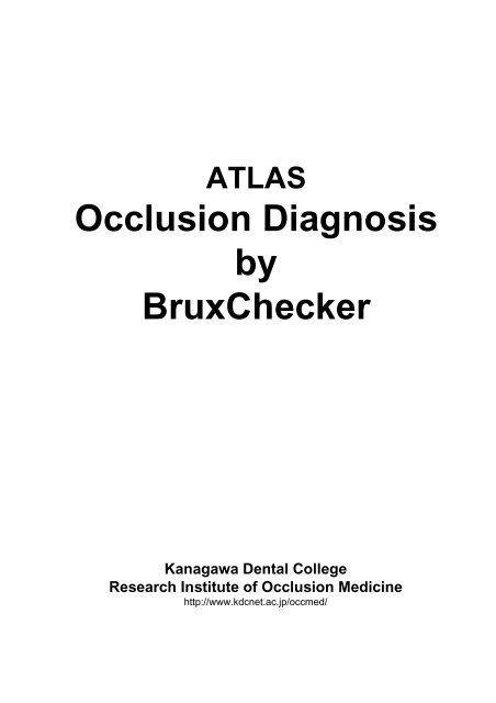
ATLAS<br />
Occlusion Diagnosis<br />
<strong>by</strong><br />
BruxChecker<br />
Kanagawa Dental College<br />
Research Institute of Occlusion Medicine<br />
http://www.kdcnet.ac.jp/occmed/
INTRODUCTION<br />
Applying all these concepts to the clinical is a principle, NOT an average value.<br />
The attainment of a healthy functional <strong>occlusion</strong>, should be based on the individualization of<br />
each patient’s condition and its complete understanding. That is, the form of skeletal frame,<br />
the form and the function of the temporomandibular joint, the inclination of the condyler path,<br />
the occlusal plane, the dental arch and the dental morphology. The <strong>occlusion</strong> treatment<br />
should be planned on that. “Needed treatment for the patient at that time” is the functional<br />
principle of the <strong>occlusion</strong>. If you do not know the functional meaning and apply it as well as<br />
the knowledge of the inclination of the tooth axis, the alignment of the teeth and the cant of<br />
the occlusal plane, even so, it is worthless. Therefore, once again, applying it to the clinical<br />
practice is a principle and not an average value.<br />
In Dentistry, the theory for <strong>occlusion</strong> refers to the occlusal patterns such as canine guidance<br />
and group function in general. However, those occlusal patterns can only work for<br />
mandibular border movements and when the mandible moves furthermore the limit; the<br />
scene in which these occlusal patterns are effective is not useful anymore. Since the<br />
mastication movement is not a border movement, this concept of the occlusal patterns can<br />
not be fitted for it. A real situation where the occlusal patterns are necessary is the sleep<br />
bruxism function. For instance, the term canine guidance is used for the grinding movement<br />
of the mandible, but during chewing movement, the upper and lower canines do not touch or<br />
g r i n d s o m u c h .<br />
Therefore, the term canine control should be used whenever we are thinking about the role<br />
of the canine during the mastication movement.<br />
Always when <strong>occlusion</strong> is constructed from a dental-medical perspective to the problem, one<br />
of the important considerations has to be the grinding movement of the mandible during the<br />
sleep bruxism. Because the force tangencies of teeth during the grinding movement of the<br />
mandible might strongly influence the masticatory muscle activity, it is closely related to the<br />
collapse of <strong>occlusion</strong>s and a lot of dental diseases. However, the clinical examination of the<br />
lateral grinding movement usually obtained is different from the actual grinding movement<br />
during sleep bruxism. So, the occlusal pattern at the sleep bruxism can not be reproduced in<br />
the clinical situation in most of the cases.<br />
Therefore, the evaluation and <strong>diagnosis</strong> in consideration of concrete approach for the sleep<br />
bruxism will constitute an important area which provides basis for the dental-medical<br />
t r e a t m e n t i n t h e n e a r f u t u r e .<br />
Currently, the treatment of hard tissue diseases, temporomandibular joint diseases and<br />
related diseases from which sleep bruxism is assumed to be the cause; is mainly passive<br />
treatment of decreasing the bruxism <strong>by</strong> using a night guard and the autosuggestion method,<br />
e t c .<br />
The sleep bruxism as an stress releaser has been reported as one of the important<br />
functions of the masticatory organ which maintains the homeostasis of the human body. If<br />
the sleep bruxism plays the key role for maintaining human life, a physiological dental<br />
<strong>occlusion</strong> which adapts to the reaction of the human body should be obtained; different from<br />
the concept of the mechanical dental <strong>occlusion</strong> from the past. For that reason, the<br />
examination, <strong>diagnosis</strong>, and treatment planning methods based on the occlusal pattern at<br />
t h e s l e e p b r u x i s m s h o u l d b e a c c o m p l i s h e d .<br />
June. 2005<br />
-1 -<br />
Sadao Sato<br />
Kanagawa Dental College<br />
Institute of <strong>occlusion</strong> medicine
I. Concept of Organic Occlusion<br />
The term Organic Occlusion recognized as a basis of <strong>occlusion</strong> is a concept<br />
based on mutual protection that works when the occlusal system is<br />
overloaded. The molars will protect the anterior teeth when clenching occurs<br />
and the anterior teeth will protect the molars when grinding occurs. This<br />
scene in which an excessive overload is present is chiefly known as bruxism<br />
f u n c t i o n .<br />
The bruxism is basically an appearing activity of the masticatory muscles as<br />
a response for a strong clenching and grinding movements of the mandible.<br />
When this occurs, abnormal contacts of the upper and lower teeth become a<br />
problem because of the muscle activity. The muscle activity according to the<br />
bruxism movement increases abnormally (hyperactivity) whenever there is a<br />
contact in the molar area. That represents a high risk to turn in a nonphysiological<br />
bruxism. Therefore, molar dis<strong>occlusion</strong> is indispensable during<br />
the bruxism to avoid any abnormal muscular activity.<br />
Thinking about the <strong>occlusion</strong> from such a point of view, the mal<strong>occlusion</strong> is<br />
an <strong>occlusion</strong> in which there are cuspid dental contacts of upper and lower<br />
teeth with a lack of occlusal support which generates a significant amount of<br />
the muscle activity during the strong clenching and grinding movements of<br />
b r u x i s m .<br />
-2 -
III. Relationship of CMS and Organic Occlusion<br />
An important factor which influences the mandibular position is the occlusal<br />
support. The masticatory organ is an unit that can be loaded <strong>by</strong> excessive<br />
forces of the bruxism and clenching, etc. Therefore, the <strong>occlusion</strong> should<br />
support these forces in such a function so that an excessive load could not<br />
compromise the temporomandibular joint (craniomandibular system).<br />
For instance, when a strong clenching is done, the temporomandibular joint<br />
should be supported <strong>by</strong> the dental <strong>occlusion</strong>. It should be a quiet down or<br />
repose state compared with the mastication and pronunciation functions’<br />
movements , etc. However, the occlusal support is often lost as<br />
consequence of a malintercuspation factor in the case of class II<br />
mal<strong>occlusion</strong>s and the premature lost of the molars syndrome. In those cases<br />
the generating powerful forces push the mandible in a retruded position<br />
caused <strong>by</strong> the increase of the muscular activity during the bruxism and<br />
clenching. On the other hand, a muscular group of the craniomandibular<br />
system is trying to maintain the mandible forward. That means that during the<br />
bruxism and the clenching, an important confrontation takes place between a<br />
muscular group which makes the mandible moves backward and the<br />
craniomandibular system muscular group which tries to locate mandible<br />
forward in the state where the occlusal support was lost. Therefore, the<br />
symptoms caused <strong>by</strong> an abnormal tension (tenderness) of these muscular<br />
groups appear in such a patients when waking up.<br />
-4 -
V. Physiological meaning of Bruxism<br />
STRESS<br />
Limbic<br />
system<br />
Visceral<br />
masticatory<br />
muscle activity<br />
Clenching<br />
Bruxism<br />
Hypothalamus<br />
Stress Syndrome<br />
Autonomic<br />
nervous system<br />
Pituitary<br />
adrenocortical<br />
system<br />
Occlusal pattern<br />
Balanced <strong>occlusion</strong><br />
Group function<br />
Canine guidance<br />
Dental disease<br />
Caries<br />
Periodontal disease<br />
Hypersensitivity<br />
Abfraction<br />
TMD<br />
Muscle symptoms<br />
Recently, research about the influence of the stress over the whole body<br />
constitutes an important concern whenever the area which should be called<br />
stress medicine is formed in modern medicine.<br />
Moreover, the stress syndrome is related to all fields of the medicine. On the<br />
other hand, the stress induces masticatory muscle activity and prevents the<br />
stress disease. From this point of view, the amount of the muscle activity is<br />
controlled <strong>by</strong> the contact pattern of upper and lower teeth. That is, strength<br />
of the masticatory muscle activity depends on the occlusal pattern though the<br />
sleep bruxism is basically a phenomenon subconsciously occurred.<br />
A significantly strong muscle activity is induced, and destructive forces will<br />
affect the teeth, periodontal tissue, and the temporomandibular joints, as a<br />
result in the <strong>occlusion</strong> of this type of molar contacts during the bruxism<br />
m o v e m e n t s .<br />
Therefore, a correct <strong>occlusion</strong> to maintain allostasis (stability or homeostasis<br />
through change) <strong>by</strong> a physiological bruxism, and to spend healthy life<br />
b e c o m e v e r y i m p o r t a n t .<br />
-6 -
VI. Basic concept of Functional Occlusion<br />
1. Passive Centric and Active Centric<br />
Passive centric line<br />
Active centric line<br />
Centric Stop<br />
The position of the mandible is mainly established <strong>by</strong> the teeth<br />
intercuspation of the maxilla and mandible. The contact points resulting<br />
from the engagement (<strong>occlusion</strong>) of the upper and lower teeth are<br />
c a l l e d c e n t r i c s t o p s .<br />
Passive Centric<br />
The centric stop of the maxilla that exists on the upper dentition in<br />
connection with the cranium. It is thought as a fixed static state<br />
compared with the dynamic movement of the mandible. Moreover, it is<br />
thought as a passive contact point which catches the functional cusp of<br />
m a n d i b u l a r t e e t h .<br />
Therefore, it is called passive centric.<br />
Active Centric<br />
The centric stop of the mandible moves dynamically compared with the<br />
passive centric of the maxilla. Therefore, a centric stop of the mandible<br />
i s c a l l e d a c t i v e c e n t r i c .<br />
Upper and lower centric stops are in a complete evenly and simultaneously<br />
relation. In class I <strong>occlusion</strong>, the passive centric stops with the upper first and<br />
second premolars are located on the mesial marginal ridges. The guidance at<br />
the lateral movement of the mandible passes through the mesial marginal<br />
ridge from this passive centric or very close to the labial cusp.<br />
-7 -
VI. Basic concept of Functional Occlusion<br />
2. Various lines on Occlusal Surface<br />
-8 -<br />
Functional<br />
Aesthetic line<br />
Passive centric line<br />
Stamp cusp line<br />
Sharing cusp line<br />
Contact line of<br />
stamp cusps<br />
Active centric line<br />
Passive centric line, Active centric line, etc.<br />
There are several functional lines on the dentition besides the passive<br />
centric line of the maxilla and the active centric line of the mandible.
VI. Basic concept of Functional Occlusion<br />
3. Intercuspation of the first molar -- 1<br />
The first molar erupts earliest in the permanent dentition, and plays an<br />
important role in maintaining the occlusal support and the mandibular position.<br />
The mesio-lingual cusp of the upper first molar and the disto-buccal cusp of<br />
the lower first molar are the largest cusps in the whole dentition.Occlusal<br />
support and the stability of the mandibular position are maintained <strong>by</strong> their<br />
e n g a g e m e n t ( o c c l u s i o n ) .<br />
-9 -
VI. Basic concept of Functional Occlusion<br />
3. Intercuspation of the first molar -- 2<br />
In examining the occlusal relationships in the permanent dentition, much<br />
attention is centered on the first molars.<br />
The engagement of an upper and lower first molar is the most important<br />
<strong>occlusion</strong> in the permanent dental articulation. The mesio-lingual cusp of the<br />
upper first molar occludes in the central fossa of the lower first molar, and the<br />
disto-buccal cusp of the lower first molar occludes in mesial with the<br />
transverse ridge of the upper first molar. Then, the contacts of cusps and<br />
ridges are obtained <strong>by</strong> these ABC contacts, and the Class I <strong>occlusion</strong> is<br />
maintained. Harmony with CMS is kept <strong>by</strong> such steady engagement.<br />
- 10 -<br />
C<br />
B<br />
A
VI. Basic concept of Functional Occlusion<br />
4. Passive Centric and Occlusal Guidance<br />
The active centric stops of the mandible occlude with the passive centric<br />
stops of the maxilla moving on the lingual surface of the incisors and canine<br />
and on the mesial marginal ridges of molars and premolars of the working<br />
side along the lateral excursions of the mandible. It moves consecutively on<br />
non-working side toward the lingual cusps.<br />
It moves from the passive centric line toward out side during the mandibular<br />
lateral excursions. Therefore, this area is called guiding area.<br />
The limit of the guiding area is the mesial marginal ridge of the upper first<br />
molars, and there is not guide posterior to this.<br />
- 11 -
VI. Basic concept of Functional Occlusion<br />
5. Retrusive Barrier (Transverse ridge)<br />
The disto-buccal cusp of the lower first molar is maintaining the class I<br />
<strong>occlusion</strong> <strong>by</strong> engaging in mesial with the transverse ridge of the upper first<br />
molar, and prevents the mandible from retreating.<br />
The transverse ridge of the upper first molar is an important structure to<br />
maintain the mandibular position in such a meaning.<br />
This ridge is especially called retrusive barrier.<br />
- 12 -
VI. Basic concept of Functional Occlusion<br />
6. Guidance and Mediotrusion<br />
In the analysis of the occlusal contacts, it is important to observe the relation<br />
between the guidance and mediotrusion.<br />
Especially, it is necessary to see carefully the relation between these in the<br />
Bruxchecker to examine the occlusal contact pattern during the bruxism.<br />
- 13 -
VII. Bruxism movement<br />
1. Occlusion during Sleep Bruxism -- 2<br />
- 15 -<br />
ICP<br />
ICP<br />
Grinding movement at the bruxism is basically a latero-retrusive movement,<br />
and the mandible does not move in a forward direction. There is a type<br />
including a centric stop at the intercuspal position and a type without a centric<br />
stop in the grinding area. Perhaps, this difference is thought to be dependent<br />
on the relation between the condylar inclination and the canine guidance. It is<br />
thought that grinding movement from an intercuspal position becomes difficult<br />
when the inclination of the canine guidance is too strong (steep) compared<br />
with the condylar inclination; it pass beyond to the canine cusp, and the<br />
b r u x i s m i s d o n e .
VII. Bruxism movement<br />
1. Occlusion during Sleep Bruxism -- 3<br />
Example of grinding during sleep bruxism <strong>by</strong> Bruxchecker.<br />
In this case, right and left sides, they both show contacts of the group<br />
function type. Moreover, the right and left second molar lingual cusps has<br />
c o m e s t r o n g l y i n c o n t a c t .<br />
- 16 -
VII. Bruxism movement<br />
2. Bruxism movement of the Mandible -- 1<br />
Bruxism is the grinding movement of upper and lower teeth caused <strong>by</strong> a<br />
strong activity of the closing muscles. Such a mandibular movement is one<br />
of the extremely important aspects to be considered in the treatment planning<br />
since it will controls the quality and stability of the outcomes in the clinical<br />
treatment of <strong>occlusion</strong>. In considering the influence of the bruxism on the<br />
generation of craniomandibular disorders, it is important to consider its<br />
examination and <strong>diagnosis</strong> in the clinical practice of dentistry.<br />
But, up to now, detailed information regarding the condylar movement<br />
according to the grinding pattern at bruxism has been hardly researched. In<br />
establishing the criteria for the optimal condylar movement evaluation at the<br />
bruxism which can be applied to the clinical, the analysis of the external<br />
condylar movements <strong>by</strong> Axiograph is very important, too.<br />
- 18 -
VII. Bruxism movement<br />
2. Bruxism movement of the Mandible -- 2<br />
Bruxism is the grinding movement of upper and lower teeth <strong>by</strong> a strong<br />
closing muscle activity. The mandibular movement is a whirling motion<br />
(rotational) of the non-working side condyle to make the working side condyle<br />
a rotation center. Such a movement demands a comparatively flat slope as a<br />
guiding path in the working side canine. When average tooth arrangement and<br />
the distance between the condylar heads to be 120 mm are assumed, the<br />
inclination on the canine slope becomes about 37 degrees. In this angle, the<br />
grinding movement which does not overwork from a geometrical relation<br />
between the condyle and the canine becomes possible. However, in a natural<br />
<strong>occlusion</strong>, there is an inclination which is about eight degrees steeper than a<br />
geometrical whirling motion, because the current average inclinations of the<br />
Japanese guiding path canine are about 45 degrees. Perhaps, it is thought<br />
that the inclination of canine means the limit of the canine guidance. It is<br />
necessary to note it because various problems on the occlusal function will be<br />
caused <strong>by</strong> a very strong inclination of canine.<br />
- 19 -
VII. Bruxism movement<br />
2. Bruxism movement of the Mandible -- 3<br />
As previously stated, the bruxism is grinding movement of upper and lower<br />
teeth <strong>by</strong> a strong closing muscle activity. The movement of the mandible is a<br />
whirling motion (rotational) of the non-working side condyle to make the<br />
working side condyle a rotation center. In such a movement, the laterotrusive<br />
movement of a lower canine is absorbed or dissipated. However, there are a<br />
lot of examples showing a latero-retrusive movement in an actual sleep<br />
bruxism pattern. This is directly influenced <strong>by</strong> a retrusive movement of the<br />
working side condyle and the Bennett movement of the mandible.<br />
- 20 -
VII. Bruxism movement<br />
3. Bruxism movement of the Mandible<br />
and Canine guidance<br />
44º<br />
44º<br />
Each average inclination of the guiding path of the canine and eminence for<br />
the Japanese is 44 degrees, and these are parallel relations. Smooth grinding<br />
of the mandible without any overwork because of that parallelism, becomes<br />
possible eventhough the bruxism is grinding movement of upper and lower<br />
teeth <strong>by</strong> a strong closing muscle activity.<br />
- 21 -
VII. Bruxism movement<br />
5. Bruxism movement of the Mandible<br />
and Working side Condyle<br />
Two types of laterotrusive grinding movements can occur during the bruxism.<br />
One with the presence of a working side condyle head retreated and another<br />
one without it. When the retreat of the working side condyle is accompanied,<br />
grinding movement on the occlusal surfaces becomes retrusive and lateral<br />
movement (latero-retrusive), and wide facets are formed. Moreover, the<br />
possibility of contacts on the non-working side increases, also.<br />
- 23 -
VIII. Bruxism and Occlusal pattern<br />
1. Compensation curves and Dis<strong>occlusion</strong><br />
Note:The curve of the canine is not a curve of Wilson .<br />
Several compensation curves exist in the dental <strong>occlusion</strong>. These are<br />
closely related to the cusp contact according to the grinding movement of<br />
the mandible. Curve of Spee is called sagittal compensating curve. A strong<br />
curve increases the possibility of molar contacts. Curve of Wilson is called<br />
lateral compensating curve. A strong curve of Wilson increases the<br />
possibility of non-working side contacts.<br />
- 24 -
VIII. Bruxism and Occlusal pattern<br />
2. Compensation curve : the Curve of Wilson -- 1<br />
Occlusal<br />
guidance<br />
Occlusal<br />
guidance<br />
Curve of Wilson<br />
Curve of Wilson<br />
The magnitude of curve of Wilson for each tooth is different according to the<br />
eruption pattern and the skeletal frame form. A negative curve of Wilson is<br />
o f t e n s h o w n i n t h e p r e m o l a r s .<br />
The degree of curvature of the curve of Wilson is an important factor which<br />
controls the inclination of occlusal guidance.<br />
- 25 -
VIII. Bruxism and Occlusal pattern<br />
2. Compensation curve : the Curve of Wilson -- 2<br />
Note:The curve of the canine is not a curve of Wilson .<br />
When viewed from the cross-section (frontal), the curve of Wilson of the<br />
maxillary teeth row has increased gradually from forward to backward. The<br />
curve of Wilson of the upper second premolar is positive though the curve of<br />
Wilson of the upper first premolar is a little negative. In addition, the curve of<br />
Wilson has intensified gradually as for rear teeth. The difference in<br />
the curve of Wilson for each tooth is deeply related with the degree of<br />
inclination of the dental occlusal guiding path. That is, the inclination of the<br />
occlusal guidance becomes flat according to an increment on Wilson’s curve.<br />
In general, the curve of Wilson accentuates in the skeletal class II cases, the<br />
inclination of the occlusal guidance strengthens weakly, and the occlusal<br />
pattern approaches the group function pattern. The curve of Wilson<br />
decreases in the skeletal class III cases, the inclination of the occlusal<br />
guidance weakens strongly, and the occlusal pattern approaches the canine<br />
d o m i n a n t p a t t e r n .<br />
- 26 -
VIII. Bruxism and Occlusal pattern<br />
2. Compensation curve : the Curve of Wilson -- 3<br />
The inclination of occlusal<br />
guidance increases when<br />
there is no curve of Wilson<br />
and the occlusal plane is<br />
f l a t .<br />
The inclination of occlusal<br />
guidance decreases when<br />
the curve of Wilson is<br />
s t r o n g .<br />
Molars comes to dis<strong>occlusion</strong><br />
easily at grinding movement<br />
when the curve of Wilson is<br />
weak, and the inclination of<br />
occlusal guidance is strong.<br />
The degree of curvature of the curve of Wilson is highly related with the<br />
inclination of the occlusal guidance. When there is an accentuated curve of<br />
Wilson, the inclination of the occlusal guidance becomes steep, and lingual<br />
cusps at non-working side can disocclude easily during lateral movements. In<br />
contrast, when the curve of Wilson is accentuated, the inclination of the<br />
occlusal guidance becomes flat, and for lingual cusps at non-working side is<br />
easy to come in contact during lateral movements.<br />
- 27 -
VIII. Bruxism and Occlusal pattern<br />
2. Compensation curve : the Curve of Wilson -- 4<br />
From a growing observation, the curve of Wilson during the first molar<br />
eruption is comparatively strong. The occlusal pattern while laterotrusive<br />
movement is balanced <strong>occlusion</strong>. Dis<strong>occlusion</strong> of the non-working side<br />
molar is achieved as shown in the figure <strong>by</strong> the premolar eruption.<br />
- 28 -
X. Occlusal Guidance<br />
Guidance from the front teeth to the molar is known as the guiding path<br />
inclination and distance to the rear molar and premolar from canine one <strong>by</strong><br />
one flatter and shorter. It is a condition for establish an easy dis<strong>occlusion</strong>.<br />
The condylar inclination and the lingual surface of canine incline almost in<br />
a p a r a l l e l r e l a t i o n .<br />
The condylar inclination and the lingual surface of canine incline almost in<br />
a p a r a l l e l r e l a t i o n .<br />
Therefore, molar dis<strong>occlusion</strong> at grinding movement is not the one obtained<br />
only <strong>by</strong> the strongly lingual surface inclination of canine. It should be<br />
recognized what should be achieved <strong>by</strong> canine guidance which harmonizes<br />
with the condyle path and guiding path inclination of anterior teeth, canine,<br />
p r e m o l a r s , a n d m o l a r s s e q u e n t i a l l y .<br />
- 31 -
XI. Occlusion analysis form<br />
- 32 -
References<br />
Sato S. and Slavicek R.: Bruxism as a stress management function of the masticatory organ. Bull.<br />
Kanagawa Dent. Coll. 29: 101-110, 2001.<br />
Sato S., Yuyama N., Tamura K., Tamaki K., Hori N., Kaneko M., Sasaguri K., Lee M. C-il.,<br />
Onozuka M. and Slavicek R.: The masticatory organ, brain function, stress-relase, and a<br />
proposal to add a new category to the taxonomy of the healing arts: Occlusion medicine.<br />
Bull Kanagawa Dent Coll 30: 2002.<br />
Sato S.: Role of masticatory organ and concept of functional <strong>occlusion</strong>. Hotetsurinsho (in<br />
Japanese)<br />
29;265-279,1996.<br />
Sato S. and Tamaki K.: Meaning of bruxism seen from functional <strong>occlusion</strong> restructuring. Nihon<br />
shika hyoron (in Japanese), 201-218,1997.<br />
Slavicek R.: The function of stress management. In: The Masticatory Organ – Function and<br />
Dysfunction, Slavicek, R. (Ed), Klosterneuburg, Gamma Medizinisch-wissenschaftliche<br />
Fortdungs-AG, pp. 281-291. 2002..<br />
Kulmer S, Ruzicka B, Niederwanger A, Moschen I. : Incline and length of guiding elements in<br />
untreated naturally grown dentition. J Oral Rehabilitation 26;650-660,1999.<br />
Leja W, Hilbe M, Stainer M, Kulmer S. : Nicht-kariöse zervikale läsionen in relation zum<br />
okklusionstypus und zur neigung der individullen führungselemente. Dtsch Aahnärztl Z,<br />
45;411-414,1999.<br />
Williamson E H, Lundquist D O.: Anterior guidance: its effect of electromyographic activity of the<br />
temporal and masseter muscles. J Prosthet Dent, 49: 816, 1983.<br />
Grubwieser G, Flatz A, Grunert I, Kofler M, Ulmer H, Gausch K, Kulmer S.: Quantitative analysis<br />
of masseter and temporalis EMGs : a comparison of anterior guided versus balanced<br />
occlusal concepts in patients wearing complete dentures. J Oral Rehabilitation, 26: 731-736,<br />
1999.<br />
Toubol, J-P., Michel, J-F.: le mouvement initial de Bennett. Experimentation clinique,<br />
Consequences therapeutiques. Les Cahiers Proth, 42 : 69-87, 1983.<br />
McHorris W H.: Focus on anterior guidance. J Gnathology, 8; 3-13, 1989.<br />
Sato S.: Relationship between <strong>occlusion</strong> and whole body seen from role of masticatory organ. (in<br />
Japanese) Nihon zenshin kougou academy J. 6(2):101-109,2000.<br />
Tamaki K: Occlusion and function of the Craniomandibular System. Bull of Kanagawa Dental<br />
College, 29(2), 111-119, 2001.<br />
Tamaki K, Ales G Celar, Toshio Teranaka, Sadao Sato, Slavicek R: Interdisciplinary Approach to<br />
the Patient with Mandibular Lateral Displacement and Complex Craniomandibular<br />
Symptoms. Information Orthop Kieferortho, (in press) 2005.<br />
Onodera K, Kawagoe T, Sasaguri K, Protacio-Quismundo C, Sato S.: Evaluation of the condylar<br />
movements in healthy and symptomatic temporomandibular joint patients during mastication<br />
and simulated bruxism utilizing condylograph. Stomatologie 101.8:187-190,2004.<br />
Slavicek R, Sato S: Bruxism—a function of the masticatory organ to cope with stress. Wien Med<br />
Wochenschr. 154: 584-9, 2004 (German)<br />
Onodera K., Kawagoe T., Sasaguri K. and Sato S.: Development of Bruxchecker - simple device<br />
for <strong>occlusion</strong> evaluation at sleep bruxism. (in Japanese) Kanagawashigaku 39:133-138,2004.<br />
Sato S. and Sasaguri K.: Physiology function and <strong>occlusion</strong> medical aspect of bruxism. (in<br />
Japanese)<br />
Nihon shika sangyo academy J. 18:3-10,2004.<br />
- 33 -