Ontogeny and distribution of Hygophum benoiti (Pisces ...
Ontogeny and distribution of Hygophum benoiti (Pisces ...
Ontogeny and distribution of Hygophum benoiti (Pisces ...

You also want an ePaper? Increase the reach of your titles
YUMPU automatically turns print PDFs into web optimized ePapers that Google loves.
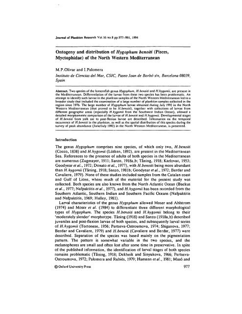
Journal <strong>of</strong> Plankton Research Vol.16 no.8 pp.977-991, 1994<br />
<strong>Ontogeny</strong> <strong>and</strong> <strong>distribution</strong> <strong>of</strong> <strong>Hygophum</strong> <strong>benoiti</strong> (<strong>Pisces</strong>,<br />
Myctophidae) <strong>of</strong> the North Western Mediterranean<br />
M.P.Olivar <strong>and</strong> I.Palomera<br />
Instituto de Ciencias del Mar, CS1C, Paseo Joan de Borbo sin, Barcelona 08039,<br />
Spain<br />
Abstract. Two species <strong>of</strong> the lanternfish genus <strong>Hygophum</strong>, H.<strong>benoiti</strong> <strong>and</strong> H.hygomii, are present in<br />
the Mediterranean. Differentiation <strong>of</strong> the larvae from these two species has been problematic. An<br />
attempt to identify such larvae in the plankton samples <strong>of</strong> the North Western Mediterranean led to a<br />
broader study that included the examination <strong>of</strong> a large number <strong>of</strong> plankton samples collected in the<br />
region since 1976. The large number <strong>of</strong> <strong>Hygophum</strong> larvae obtained during July 1992 in the North<br />
Western Mediterranean (that proved to be H.benoiii), together with collections <strong>of</strong> larvae from<br />
different geographic areas (especially H.hygomii from the Southwest Indian Ocean), allowed a<br />
detailed morphometric comparison <strong>of</strong> the larvae <strong>of</strong> H.<strong>benoiti</strong> <strong>and</strong> H.hygomii. Developmental stages<br />
<strong>of</strong> H.<strong>benoiti</strong> from yolk sac to post-flexion larvae are described. Information on the temporal<br />
occurrence <strong>of</strong> H.<strong>benoiti</strong> in the plankton, as well as the spatial <strong>distribution</strong> <strong>of</strong> this species during the<br />
survey <strong>of</strong> peak abundance (June/July 1992) in the North Western Mediterranean, is presented.<br />
Introduction<br />
The genus <strong>Hygophum</strong> comprises nine species, <strong>of</strong> which only two, H.<strong>benoiti</strong><br />
(Cocco, 1838) <strong>and</strong> H.hygomii (Liitken, 1892), are present in the Mediterranean<br />
Sea. References to the presence <strong>of</strong> adults <strong>of</strong> both species in the Mediterranean<br />
are numerous (Zugmayer, 1911; Sanzo, 1918a,b; Taning, 1918; Karlovac, 1953;<br />
Goodyear et al., 1972; Donato etal., 1977), with H.<strong>benoiti</strong> being more abundant<br />
than H.hygomii (Taning, 1918; Sanzo, 1981b; Goodyear et al., 1972; Berdar <strong>and</strong><br />
Cavaliere, 1979). None <strong>of</strong> these studies included samples from the Catalan coast<br />
<strong>and</strong> Gulf <strong>of</strong> Lions, where much <strong>of</strong> the material for the present study was<br />
collected. Both species are also known from the North Atlantic Ocean (Backus<br />
et al., 1977; Nafpaktitis et al., 1977), <strong>and</strong> H.hygomii has been recorded from the<br />
Southern Atlantic, Southern Indian <strong>and</strong> Southern Pacific Oceans (Nafpaktitis<br />
<strong>and</strong> Nafpaktitis, 1969; Hulley, 1981).<br />
Larval characteristics <strong>of</strong> the genus <strong>Hygophum</strong> allowed Moser <strong>and</strong> Ahlstrom<br />
(1974) <strong>and</strong> Moser et al. (1984) to differentiate three different morphological<br />
types <strong>of</strong> <strong>Hygophum</strong>. The species H.<strong>benoiti</strong> <strong>and</strong> H.hygomii belong to their<br />
'moderately slender' morphotype. Taning (1918) <strong>and</strong> Sanzo (1918a,b) described<br />
juveniles <strong>and</strong> post-flexion larvae <strong>of</strong> both species, <strong>and</strong> subsequently larval series<br />
<strong>of</strong> H.hygomii (Tortonese, 1956; Pertseva-Ostroumova, 1974; Shiganova, 1977;<br />
Berdar <strong>and</strong> Cavaliere, 1979) <strong>and</strong> H.<strong>benoiti</strong> (Cavaliere <strong>and</strong> Berdar, 1977) were<br />
described. Separation <strong>of</strong> the species was based mainly on the pigmentation<br />
pattern. The pattern is somewhat variable in the two species, <strong>and</strong> the<br />
melanophores are small <strong>and</strong> <strong>of</strong>ten lost after some time in preservative. In spite<br />
<strong>of</strong> the published information, the identification <strong>of</strong> larval stages <strong>of</strong> both species<br />
remains problematic (Taning, 1918; Dekhnik <strong>and</strong> Sinyukova, 1966; Pertseva-<br />
Ostroumova, 1972; Palomera <strong>and</strong> Rubins, 1979; Hamann etal., 1981; Mas6 <strong>and</strong><br />
© Oxford University Press 977
M.P.OIivar <strong>and</strong> I.Palomera<br />
Palomera, 1984; Sabates <strong>and</strong> Maso, 1990; Olivar <strong>and</strong> Fortuno, 1991; K.E.<br />
Hartel, personal communication).<br />
A large number <strong>of</strong> <strong>Hygophum</strong> larvae collected in the North Western<br />
Mediterranean during June-July 1992 supplied enough material to undertake a<br />
through study <strong>of</strong> the morphometry, as well as the pigmentation, <strong>of</strong> these larvae.<br />
Comparison <strong>of</strong> these larvae with other <strong>Hygophum</strong> larvae collected in different<br />
areas <strong>of</strong> the world led to the discovery <strong>of</strong> new characters by which larvae <strong>of</strong> the<br />
two species can be distinguished. In this paper, we describe our Mediterranean<br />
specimens, identified as H.<strong>benoiti</strong>, <strong>and</strong> provide diagnostic characters that<br />
differentiate them from H.hygomii larvae.<br />
Also, we discuss the temporal occurrence <strong>of</strong> H.<strong>benoiti</strong> in the North Western<br />
Mediterranean based on the presence <strong>of</strong> larvae from several cruises conducted<br />
from 1977 to 1992, <strong>and</strong> the spatial <strong>distribution</strong> during June/July 1992.<br />
Method<br />
The high number <strong>of</strong> H.<strong>benoiti</strong> larvae collected in the North Western Mediterranean<br />
during June-July 1992 (n = 1747) provided adequate material to<br />
describe the early stages <strong>of</strong> the development <strong>of</strong> this species. The survey covered,<br />
for the first time, the area between 39°30'N 00°30'E (south <strong>of</strong> the Ebro Delta)<br />
<strong>and</strong> 43°30'N 08°E (Gulf <strong>of</strong> Lions). Stations were sampled from inshore waters<br />
with depths <strong>of</strong> ~40 m to <strong>of</strong>fshore waters with depths <strong>of</strong> ~2000 m. This allowed<br />
the spatial <strong>distribution</strong> <strong>of</strong> larvae <strong>of</strong> H.<strong>benoiti</strong> along the Catalan coast <strong>and</strong> the<br />
southern coast <strong>of</strong> France to be presented (Figure 1). The survey consisted <strong>of</strong> a<br />
series <strong>of</strong> transects perpendicular to the coastline in which the stations were<br />
placed every 18 km, from very shallow waters (20 m depth) to beyond the slope<br />
(>2000 m depth). Samples were obtained with a Bongo net fitted with 0.3 mm<br />
mesh, hauled obliquely from 200 m to the surface, where the bottom depth<br />
41<br />
39<br />
EBROR.<br />
10 # E<br />
Fig. 1. Location <strong>of</strong> plankton stations sampled during June/July 1992 in the North Western<br />
Mediterranean.<br />
978
<strong>Ontogeny</strong> <strong>and</strong> <strong>distribution</strong> <strong>of</strong> H.benoUi in NW Mediterranean<br />
permitted. The volume <strong>of</strong> water filtered was estimated by means <strong>of</strong> a flowmeter<br />
mounted centrally in the mouth <strong>of</strong> one <strong>of</strong> the nets. Larvae were preserved in 5%<br />
buffered saline formalin <strong>and</strong> measurements were made at least 6 months after<br />
collection <strong>and</strong> fixation at sea. Measurements were performed to an accuracy <strong>of</strong><br />
0.1 mm. Larval nomenclature follows Kendall et al. (1984).<br />
The larvae described here are deposited in the Ichthyoplankton Collection <strong>of</strong><br />
the Instituto de Ciencias del Mar de Barcelona (ICICMB). Among the 1747<br />
larvae collected in the cruise, several measurements were taken from a selection<br />
<strong>of</strong> 60 larvae, covering all the larval size range [from 2.9 to 11.2 mm body length<br />
(BL)]. The following measurements were recorded: BL, the distance along the<br />
midline <strong>of</strong> the body from the tip <strong>of</strong> the snout to the tip <strong>of</strong> the notochord in preflexion<br />
larvae, <strong>and</strong> to the posterior margin <strong>of</strong> the hypural elements in postflexion<br />
stages; pre-anal length (PA), the distance along the midline <strong>of</strong> the body<br />
from the tip <strong>of</strong> the snout to the vent; head length (HL), the distance from the tip<br />
<strong>of</strong> the snout to the posterior margin <strong>of</strong> the cleithrum; head depth (HD),<br />
maximum depth <strong>of</strong> the head; body depth at pectoral (BDP), depth <strong>of</strong> the body at<br />
the base <strong>of</strong> the pectoral fin; dorsal fin distance (DD), the distance along the<br />
midline <strong>of</strong> the body from the tip <strong>of</strong> the snout to the origin <strong>of</strong> the dorsal fin; eye<br />
diameter (ED); eye depth (EDepth), eye depth including choroid tissue. The<br />
allometric relationships between the various body measurements <strong>and</strong> BL were<br />
calculated by use <strong>of</strong> the equation v = ax b , where x is BL, y is the other<br />
measurement being related, b is the allometric factor <strong>and</strong> a is the expected value<br />
<strong>of</strong> y at x = 1 (Gould, 1966). Confidence intervals (CI) were calculated at the<br />
95% level <strong>of</strong> significance.<br />
In order to study spatial <strong>distribution</strong>, the number <strong>of</strong> individuals collected in<br />
the June/July 1992 samples were st<strong>and</strong>ardized to numbers per 10 m 2 <strong>of</strong> surface<br />
sea. Temporal occurrence <strong>of</strong> H.<strong>benoiti</strong> larvae was based on samples obtained in<br />
several surveys carried out on the Catalan coast from 1976 to 1992 (Table I).<br />
Owing to the different methodology <strong>and</strong> bathymetric area covered, spatial<br />
<strong>distribution</strong> <strong>and</strong> abundances during these cruises are not straightforwardly<br />
comparable.<br />
For comparison, a large number (n = 1668) <strong>of</strong> H.hygomii larvae from the<br />
Southwest Indian Ocean were available (Beckley <strong>and</strong> van Ballegooyen, 1992).<br />
The methods <strong>of</strong> collection <strong>and</strong> preservation were similar to those described<br />
above for H.<strong>benoiti</strong>: oblique hauls with a Bongo net <strong>and</strong> fixation in buffered<br />
formalin at 5%. The methods for measurements <strong>and</strong> morphometric analysis <strong>of</strong><br />
H.hygomii larvae were the same as those described above for H.<strong>benoiti</strong>. Other<br />
<strong>Hygophum</strong> larvae from different museums or collections <strong>of</strong> the world have also<br />
been examined: H.hygomii from the Eastern Mediterranean (Centre de<br />
Recherches Marines, Jounieh, Lebanon), from the Central Western Atlantic<br />
(Museum <strong>of</strong> Comparative Zoology, Cambridge, MA), from the tropical<br />
Northeast Atlantic [Zoologisches Institut <strong>and</strong> Museum <strong>of</strong> Hamburg, Germany,<br />
<strong>and</strong> our material in the Instituto de Ciencias del Mar (ICM)] <strong>and</strong> from the<br />
southern Benguela region (our material in the ICM), <strong>and</strong> for H.<strong>benoiti</strong> from the<br />
Central Western Atlantic (Museum <strong>of</strong> Comparative Zoology, Cambridge) <strong>and</strong><br />
the tropical Northeast Atlantic (Zoologisches Institut <strong>and</strong> Museum <strong>of</strong> Hamburg,<br />
979
M.P.OIivar <strong>and</strong> I.Palomera<br />
Table I. Presence <strong>of</strong> H.<strong>benoiti</strong> larvae in the different cruises carried out in the North Western<br />
Mediterranean<br />
Month/year Latitude Presence<br />
August 1984 40°30'-42°30'N +<br />
September 1983 40°30'-42°30'N +<br />
September/October 1984 40"30'-42°30'N +<br />
October 1976 38°30'-42°30'N +<br />
October 1982 41°30'-42°30'N +<br />
October 1983 40°00'-41°30'N +<br />
October 1983 40°30'-42°30'N +<br />
December 1981 41°30'-42°30'N<br />
January 1982 41°30'-42°30'N<br />
March 1982 41°30'-42°30'N<br />
April 1983 40°30'-42°30'N<br />
May 1983 40 o 00'-41 o 30'N +<br />
May 1983 40°30'-42°30'N +<br />
May 1990 39°0O'-41°30'N +<br />
May 1992 42°05'-42°00'N +<br />
June 1983 40°30'-42°30'N +<br />
June 1992 42°05'-42 1> 20'N . +<br />
July 1983 40°30'-42 o 30'N +<br />
June/July 1992 39°30'-43°30'N +<br />
<strong>and</strong> our material in the ICM). Comparative material <strong>of</strong> H.proximum was also<br />
obtained from Indo-Pacific collections from the waters around Australia<br />
(Australian Museum) <strong>and</strong> Pacific collections from the waters around Japan<br />
(Faculty <strong>of</strong> Fisheries, Kagoshima University).<br />
Results<br />
Based on pigmentation pattern, which was mainly useful in recently collected<br />
material, we identified the larvae collected in June-July 1992 as H.<strong>benoiti</strong><br />
(Figure 2). Morphometric data on H.hygomii larvae from the Southwest Indian<br />
Ocean used for comparative purposes (Figure 3) also showed differences<br />
between H.<strong>benoiti</strong> <strong>and</strong> H.hygomii, which permitted us to re-identify all larvae <strong>of</strong><br />
<strong>Hygophum</strong> collected in previous cruises carried out in the North Western<br />
Mediterranean <strong>and</strong> determine the spawning period <strong>of</strong> H.<strong>benoiti</strong>.<br />
Morphology <strong>and</strong> morphometry<br />
Larvae <strong>of</strong> H.<strong>benoiti</strong> were moderately slender, especially in pre-flexion stages.<br />
As a proportion <strong>of</strong> body length, body depth at the pectoral base <strong>and</strong> head depth<br />
showed significant allometric increases, with development (Figure 4). Relative<br />
body depth <strong>and</strong> head depth were slightly smaller in H.<strong>benoiti</strong> than in H.hygomii<br />
larvae (Figure 5).<br />
In H.<strong>benoiti</strong>, the gut was slightly curved with a prominent fold-terminated<br />
section. Pre-anal length versus body length was relatively constant with<br />
development (Figure 4). The anterior section <strong>of</strong> the gut did not have transverse<br />
rugae <strong>and</strong> was conspicuously thinner than the posterior section. The anterior<br />
section was relatively longer in pre-flexion <strong>and</strong> flexion larvae (representing ~l/3<br />
980
<strong>Ontogeny</strong> <strong>and</strong> <strong>distribution</strong> <strong>of</strong> H.<strong>benoiti</strong> in N W Mediterranean<br />
Fig. 2. Larval development <strong>of</strong> H.<strong>benoiti</strong>. (A) 2.9 mm; (B) 5.5 mm; (C) 7.8 mm, (D) 9.2 mm.<br />
<strong>of</strong> gut length) than in later stages (Figure 2). Pre-anal length versus body length<br />
was one <strong>of</strong> the important characters that differentiates H.<strong>benoiti</strong> from<br />
H.hygomii (Figure 5). Pre-anal length was significantly shorter (P < 0.0001) in<br />
the latter (always 60% <strong>of</strong> BL). Furthermore,<br />
in H.hygomii the gut was more curved <strong>and</strong> the foregut was shorter (25-27% <strong>of</strong><br />
cleithnim to anus distance) than in H.<strong>benoiti</strong> (32-34% <strong>of</strong> cleithrum to anus<br />
distance).<br />
Head length in H.<strong>benoiti</strong> was almost isometrical with development: from 25%<br />
981
M.P.OIivar <strong>and</strong> I.Palomera<br />
B<br />
Fig. 3. Larval development <strong>of</strong> H.hygomii. (A) 5.3 mm; (B) 7.0 mm; (C) 9.9 mm; (D) 13.0 mm.<br />
<strong>of</strong> BL at 3 mm to 29% at 11 mm. Small teeth were discernible in the upper jaw<br />
in early larvae as small as 3 mm.<br />
Pre-dorsal length in H.<strong>benoiti</strong> decreased with development. A significantly<br />
negative allometnc relationship was observed between pre-dorsal length <strong>and</strong><br />
body length (Figure 4). For the size range considered, pre-dorsal distance varied<br />
from 56 to 49% <strong>of</strong> BL. In H.hygomii,.this distance was smaller (changing from<br />
50 to 44% <strong>of</strong> BL for the same range <strong>of</strong> sizes) (Figure 5).<br />
Eyes were elliptical during all larval stages studied in both species (Figures 2<br />
<strong>and</strong> 3). A conical mass <strong>of</strong> brown choroid tissue, similar in the two species, was<br />
always visible on the ventral edge <strong>of</strong> the eye.<br />
Notochordal flexion started at ~5 mm <strong>and</strong> was completed at 5.5 mm in<br />
H.<strong>benoiti</strong>. In H.hygomii, notochordal flexion started at ~6 mm BL <strong>and</strong> was<br />
completed at 7 mm BL.<br />
982
HP (mm)<br />
BDP (mm)<br />
4.0<br />
5.0<br />
HD • 0.0594 SL 1 ' 6662<br />
Cfb • 0.1113<br />
• 0.9740<br />
1.6600<br />
BDP • 0.0602 SL<br />
Ob • a 1204<br />
r • 0.0671<br />
4.0<br />
3.0"<br />
3.0<br />
2.0"<br />
2.0<br />
1.0<br />
1.0<br />
n • 46<br />
n • 55<br />
3.0 4.0 5.0 6.0 7.0 8.0 0.0 10.0 11.0<br />
SL (mm)<br />
3.0 4.0 5.0 6.0 7.0 8.0 9.0 10.0 11.0<br />
SL (mm)<br />
O<br />
DD (mm)<br />
PA (mm)<br />
7.0<br />
641<br />
• a8682 SL°'<br />
00<br />
••' +<br />
6.0<br />
0.9314<br />
r •<br />
1.1108<br />
PA • 0.6342 SL<br />
Ob • 0.0381<br />
r - 0.9916<br />
^<br />
¥~~**~~<br />
6.0<br />
ir<br />
I<br />
.+••<br />
•*"""" +<br />
' .. .^ •<br />
4.0<br />
•31<br />
n<br />
n • 60<br />
9.6"<br />
8.6"<br />
7.5"<br />
6.5"<br />
5.5-<br />
4.5-<br />
3.5"<br />
2.5"<br />
S.<br />
-•— H<br />
10.0 11.0<br />
2.0 —i—<br />
H<br />
H<br />
6.0 7.0 8.0 9.0<br />
SL (mm)<br />
4.0 5.0 6.0 7.0 8.0 9.0 10.0 11.0<br />
SL (mm)<br />
Fig. 4. Morphology <strong>of</strong> H.benoiii larvae. Relationship between body length (SL) <strong>and</strong> body depth at pectoral (BDP), head depth (HD), pre-anal length (PA)<br />
<strong>and</strong> dorsal distance (DD). Continuous line: fitted curve. Dashed lines: confidence intervals (at the 95% level <strong>of</strong> significance) for estimating y.
HP (mm)<br />
BDP (mm)<br />
1.4060<br />
3.0+ HD-0.0822 SL<br />
Ob - 0.0842<br />
r • 0.9784<br />
1.6707<br />
3.0 + BDP-0.0699 SL<br />
Ob • a0838<br />
r -0.0800<br />
2.0 •<br />
2.0-<br />
1o<br />
1.0-•<br />
1.0-•<br />
n-52<br />
n-ee<br />
0.0<br />
3.0 4.0 5.0 6.0 7.0 8.0 0.0 10.0 11.0 12.0 13.0<br />
SL (mm)<br />
0.0<br />
3.0 4.0 5.0 6.0 7.0 8.0 0.0 10.0 11.0 12.0 13.0<br />
SL(mm)<br />
DD (mm)<br />
O7062<br />
6.0 •}• DO-0.8908 SL<br />
Ob • 0.0920<br />
r • 0.S469<br />
PA (mm)<br />
8.0<br />
1.0192<br />
7 0 •• PA-0.5577 SL<br />
Qb - 0.0209<br />
6.0+ r-0.8952<br />
5.0-<br />
n • 31<br />
n-92<br />
11.0 12.0 13.0<br />
3.0<br />
7.0 8.0 ' 0.0 10.0<br />
SL(mm)<br />
4 1-<br />
2.5 3.5 4.5 6.5 &6 7.5 8.5 0.5 10.5 11.5 12.6 13.5<br />
SL(mm)<br />
Fig. S. Morphology <strong>of</strong> H.hygomii larvae. Relationship between body length (SL) <strong>and</strong> body depth at pectoral (BDP), head depth (HD), pre-anal length<br />
(PA) <strong>and</strong> dorsal distance (DD). Continuous line: fitted curve. Dashed lines: confidence intervals (at the 95% level <strong>of</strong> significance) for estimating y.
Fin development<br />
<strong>Ontogeny</strong> <strong>and</strong> <strong>distribution</strong> <strong>of</strong> H.<strong>benoiti</strong> in NW Mediterranean<br />
In post-flexion H.<strong>benoiti</strong> larvae, the finfold was still present at the dorsal margin<br />
<strong>of</strong> the body <strong>and</strong> was somewhat inflated anteriorly. The caudal fin developed at<br />
small size <strong>and</strong> all rays were visible at ~6.5 mm BL. The anal fin base started<br />
forming at ~5.5 mm BL; at 6 mm BL ~15 pterygiophores were present, at<br />
~8 mm BL all pterygiophores were forming, <strong>and</strong> at ~9 mm BL all rays (19)<br />
were visible. The dorsal fin started forming at larger larval size than the anal fin.<br />
At ~6 mm BL a rudimentary dorsal fin base developed in the finfold, at 7.7 mm<br />
BL 9 pterygiophores were visible, at 11.2 mm BL (our largest larvae) 8 or 9 rays<br />
were visible. Pelvic fin buds were visible at ~9 mm BL. The adipose fin was<br />
visible in larvae from ~11 mm BL.<br />
Development <strong>of</strong> the caudal fin was similar in H.hygomii (all rays visible at<br />
~7 mm). Development <strong>of</strong> the anal fin started at 5.5-6 mm BL <strong>and</strong> at 9-10 mm<br />
BL all rays were formed. Like H.<strong>benoiti</strong>, the dorsal fin base started to develop at<br />
slightly larger sizes, at 7 mm, <strong>and</strong> all rays were present at ~13 mm BL.<br />
Photophores<br />
The Br2 photophores developed at slightly smaller sizes,in H.<strong>benoiti</strong> larvae, at<br />
~7 mm BL, <strong>and</strong> were the only photophores visible in our largest larvae<br />
(~11 mm), which is typical for most myctophid larvae. In H.hygomii <strong>of</strong> the<br />
Southwest Indian Ocean, the Br2 photophores appeared at ~7.5 mm BL, <strong>and</strong><br />
the same was observed in H.hygomii larvae from the Eastern Mediterranean (<strong>of</strong>f<br />
Lebanon).<br />
Myomeres<br />
The number <strong>of</strong> myomeres was similar in both species, with ranges from 34 to 37<br />
(from 15 to 18 pre-anal) in H.<strong>benoiti</strong>, <strong>and</strong> 35-37 myomeres in H.hygomii.<br />
Pigmentation<br />
The pigmentation pattern that is diagnostic for the genus <strong>Hygophum</strong> (Moser et<br />
al., 1984) was evident in all larvae examined: melanophores at the cleithral<br />
symphysis <strong>and</strong> isthmus region.<br />
Lateral gut spots were present in all H.<strong>benoiti</strong> larvae. Large melanophores<br />
occurred over the terminal portion <strong>of</strong> the gut. Generally, two lateral gut spots<br />
were present over the area <strong>of</strong> the transverse rugae, <strong>and</strong> one anterior spot was<br />
present at the foregut. No pigment was visible on the lower jaw in pre-flexion<br />
stages, but was present in some flexion <strong>and</strong> post-flexion larvae. At any stage <strong>of</strong><br />
development, melanophores could be found scattered over the pectoral fin rays,<br />
as shown in H.hygomii larvae (Sanzo, 1918b; Tortonese, 1956; Pertseva-<br />
Ostroumova, 1974; Berdar <strong>and</strong> Cavaliere, 1979; <strong>and</strong> our observations).<br />
In H.<strong>benoiti</strong> larvae >9 mm, internal pigmentation was present over the<br />
cleithral region. In the smallest larvae, ~3 mm, there was pigmentation on the<br />
finfold. One melanophore was present in the dorsal finfold at about the level <strong>of</strong><br />
the anus, another melanophore was located on the post-anal membrane, ~5<br />
985
M.P.OIivar <strong>and</strong> I.Palomera<br />
myomeres posterior to the anus, <strong>and</strong> two groups <strong>of</strong> melanophores were present<br />
at the dorsal <strong>and</strong> ventral borders <strong>of</strong> the caudal finfold. This pigmentation was<br />
not always visible due to the difficulty <strong>of</strong> collecting small larvae with intact<br />
fmfolds. In pre-flexion larvae, but not in flexion or post-flexion stages, 1-4 small<br />
spots were sometimes present on the post-anal midventral line <strong>of</strong> the tail. This<br />
pigmentation differed from the post-anal melanophore present in pre-flexion<br />
<strong>and</strong> some flexion H.hygomii larvae. This large stellate melanophore <strong>of</strong>ten<br />
extended onto the myosepta, <strong>and</strong> was always located between 6 <strong>and</strong> 8 myomeres<br />
posterior to the anus [also noted by Berdar <strong>and</strong> Cavaliere (1979)].<br />
The dorsal <strong>and</strong> ventral caudal finfold pigment which was sometimes noted in<br />
pre-flexion H.<strong>benoiti</strong> was never observed in H.hygomii. In some post-flexion<br />
stages <strong>of</strong> H.<strong>benoiti</strong>, a spot was visible at the tip <strong>of</strong> notochord, <strong>and</strong> in some larvae<br />
a medial spot was located at the caudal bases. In a few larvae, both<br />
melanophores were present. Finally, a group <strong>of</strong> many spots was scattered more<br />
or less distally in the ventral rays <strong>of</strong> the caudal fin. The pigmentation in these<br />
three areas was never present in larvae <strong>of</strong> H.hygomii.<br />
The diagnostic characteristics <strong>of</strong> H.<strong>benoiti</strong> larvae that differentiated them<br />
from H.hygomii were: moderately slender body, with the gut extending to<br />
>60% <strong>of</strong> BL, a thin foregut that comprised —1/3 <strong>of</strong> the gut length in pre-flexion<br />
<strong>and</strong> flexion larvae, <strong>and</strong> a dorsal fin that originated behind the midpoint <strong>of</strong> the<br />
body. Most larvae had caudal pigmentation, but lacked both the stellate<br />
melanophore on the ventral midline <strong>of</strong> the tail <strong>and</strong> the pigmentation on the<br />
pectoral rays. In H.hygomii, pre-anal length <strong>and</strong> foregut length were shorter,<br />
<strong>and</strong> the dorsal fin originated anteriorly to the midpoint <strong>of</strong> the body. Caudal<br />
pigmentation was never present, but melanophores were generally scattered<br />
over the pectoral fin rays <strong>and</strong> a large stellate melanophore was always present on<br />
the ventral midline <strong>of</strong> the tail in pre-flexion larvae.<br />
A controversial point is when the Br2 photophores appear. According to<br />
Taning (1918), Cavaliere <strong>and</strong> Berdar (1977) <strong>and</strong> Berdar <strong>and</strong> Cavaliere (1979),<br />
these photophores are present in smaller H.<strong>benoiti</strong> (~8.5 mm BL) than in<br />
H.hygomii (~ 11 mm BL). However, Sanzo (1918a,b) states just the opposite:<br />
Br2 appear later in larger H.<strong>benoiti</strong> [at 14 mm total length (TL)] than H.hygomii<br />
(at 10.3 mm TL). The results presented here show no difference in the stage at<br />
formation <strong>of</strong> Br2 photophores in either species. Tortonese (1956), who reviewed<br />
Taning's (1918) <strong>and</strong> Sanzo's (1918a,b) descriptions, pointed out that the<br />
progressive development <strong>of</strong> the different photophores is not identical in all<br />
individuals.<br />
A summary <strong>of</strong> the main features that differentiate the larvae <strong>of</strong> all the<br />
<strong>Hygophum</strong> species <strong>of</strong> the 'moderately slender type' <strong>of</strong> the world is included in<br />
Table II.<br />
Larval <strong>distribution</strong> <strong>of</strong> H. <strong>benoiti</strong> in the North Western Mediterranean<br />
Larvae <strong>of</strong> H.<strong>benoiti</strong> appeared in the plankton from May (northern spring) to<br />
October (northern autumn). During the survey <strong>of</strong> June/July 1992, the highest<br />
abundances <strong>of</strong> larvae <strong>of</strong> this species were obtained (Figure 6). No larvae <strong>of</strong> this<br />
986
<strong>Ontogeny</strong> <strong>and</strong> <strong>distribution</strong> <strong>of</strong> H.<strong>benoiti</strong> in NW Mediterranean<br />
Table II. Main characters that differentiate the larvae <strong>of</strong> <strong>Hygophum</strong> species <strong>of</strong> the 'moderately<br />
slender type' in pre-flexion-flexion stages<br />
H.<strong>benoiti</strong><br />
H.bruuni<br />
H.hanseni<br />
H.hygomii<br />
H.proximum<br />
%PA/BL<br />
62-66<br />
52-61<br />
53-57<br />
57-58<br />
61-63<br />
Pigmentation<br />
Lower jaw Tail Caudal fin Pectoral fin<br />
- In larvae
M.P.OIivar <strong>and</strong> I.Palomera<br />
43<br />
41<br />
39<br />
43<br />
41<br />
39<br />
43<br />
41<br />
39<br />
1000'<br />
II I 1 I<br />
•
<strong>Ontogeny</strong> <strong>and</strong> <strong>distribution</strong> <strong>of</strong> H.<strong>benoiti</strong> in NW Mediterranean<br />
During June/July 1992, two patterns <strong>of</strong> <strong>distribution</strong> <strong>of</strong> H.<strong>benoiti</strong> larvae were<br />
obvious. Larvae were most abundant in deep waters <strong>of</strong>f the Catalan coast, but<br />
were largely absent from the Gulf <strong>of</strong> Lions in the northern part <strong>of</strong> the survey<br />
region. This difference could not be related to differences in temperature <strong>and</strong><br />
salinity between the Catalan <strong>and</strong> French coasts, since these environmental<br />
factors showed little variation between the slope areas <strong>of</strong> the two sectors.<br />
The main concentrations <strong>of</strong> H.<strong>benoiti</strong> larvae at deeper stations <strong>of</strong>f the Catalan<br />
coast match the oceanic habits <strong>of</strong> the adults (Taning, 1918; Goodyear et al.,<br />
1972; Hulley, 1981). As pointed out by Sabates <strong>and</strong> Maso (1990), the circulating<br />
pattern in the area has a major effect on the <strong>distribution</strong> <strong>of</strong> mesopelagic larvae.<br />
Current flow in the region is southwesterly along the edge <strong>of</strong> the continental<br />
shelf. Associated with this current is a typical shelf-slope front separating less<br />
saline inshore waters from the open sea (Font et al., 1988). Just north <strong>of</strong> the<br />
Ebro Delta, the current penetrates to the wide continental shelf (Font et al.,<br />
1990), <strong>and</strong> the concentration <strong>of</strong> larvae closer to the coast in this area is probably<br />
related to this circulation pattern. Thus, the nearshore <strong>distribution</strong> <strong>of</strong> H.<strong>benoiti</strong><br />
observed in this study may be closely related to both hydrographic <strong>and</strong><br />
topographic features in the area.<br />
Acknowledgements<br />
The authors wish to thank Dr H.G.Moser for the revisions <strong>and</strong> useful comments<br />
on the manuscript, <strong>and</strong> advice in its preparation. Larvae used for comparison<br />
were generously made available by the following persons, to whom we are<br />
especially grateful: A.Aboussouan (Faculte des Sciences de Luminy, Marseille),<br />
L.Beckley (Oceanographic Research Institute, Durban), K.E.Hartel (Museum<br />
<strong>of</strong> Comparative Zoology, Cambridge), W.F.H<strong>of</strong>fman (National Museum <strong>of</strong><br />
Natural History, Smithsonian Institution), H.Ch.John <strong>and</strong> C.Zelck (Zoologisches<br />
Institut <strong>and</strong> Museum, Hamburg), J.Leis <strong>and</strong> T.Trnski (Australian<br />
Museum, Sydney), T.Ozawa (Kagashima University), P.Rubies <strong>and</strong> A.SabatSs<br />
(Instituto de Ciencias del Mar, Barcelona), R.Zeidane (Centre de Recherches<br />
Marines, Lebanon). We are also grateful to G.Fuster <strong>and</strong> A.Carpena for sorting<br />
the samples. We also appreciate the comments <strong>and</strong> suggestions made by<br />
P.Rubies while preparing the manuscript. J.Corbera prepared the figures <strong>of</strong> the<br />
larvae.<br />
References<br />
Backus.R.J., Craddock.J.E., Haedrich.R.L. <strong>and</strong> Robison.B.H. (1977) Atlantic mesopelagic<br />
zoogeography. In Gibbs.R.H. Jr (ed.), Fishes <strong>of</strong> the Western North Atlantic, Sears Found. Mar.<br />
Res. Yale Univ. Mem., 1, 266-287.<br />
Balbontin.F. <strong>and</strong> Orellana.M.C. (1983) Descripci6n de las larvas del pez linterna <strong>Hygophum</strong> bruuni<br />
del 4rea de Valparaiso, Chile (<strong>Pisces</strong>: Myctophidae). Rev. Biol. Mar., 19, 205-216.<br />
Beckley.LE. <strong>and</strong> van Ballegooyen.R.C. (1992) Oceanographic conditions during three ichthyoplankton<br />
surveys <strong>of</strong> the Agulhas current in 1990/91. In Payne,A.I.L., Brink,K.H., Mann.K.H.<br />
<strong>and</strong> Hilborn.R. (eds), Benguela Trophic Functioning. S. Afr. J. Mar. Sci., 12, 83-93.<br />
Berdar.A. <strong>and</strong> Cavaliere.A. (1979) Stadi larvali e postlarvali di mict<strong>of</strong>idi: <strong>Hygophum</strong> hygomi<br />
(Lutken). Mem. Biol. Mar. Ocean., 9, 167-173.<br />
989
M.P.Olivar <strong>and</strong> I.Palomera<br />
Cavaliere.A. <strong>and</strong> Berdar.A. (1977) Stadi larvali e postlarvali di mia<strong>of</strong>idi: <strong>Hygophum</strong> <strong>benoiti</strong><br />
(Cocco). Atti Soc. Peloriiana, 23, 141-149.<br />
Dekhnik.T.V. <strong>and</strong> Sinyukova.V.I. (1966) Distribution <strong>of</strong> the pelagic fish eggs <strong>and</strong> larvae in the<br />
Mediterranean Sea. Part II. On the reproduction <strong>and</strong> ecology <strong>of</strong> larvae in Mediterranean<br />
Myctophidae. In Studies on the Plankton on Southern Seas. Nauka, Moskow, pp. 1-210 (in<br />
Russian).<br />
Donato.A., Contini.A., Giannetto.S. <strong>and</strong> Berdar.A. (1977) Esame biometrico di una popolazione<br />
di <strong>Hygophum</strong> <strong>benoiti</strong> Cocco e ritrovamento di alcuni semplari di <strong>Hygophum</strong> hygomii Lutken<br />
(<strong>Pisces</strong>: Myctophidae). Riv. Biol. Norm. Pawl., 3, 133-146.<br />
Font.J., Salat,J. <strong>and</strong> Tintore\J. (1988) Permanent features in the circulation <strong>of</strong> the Catalan sea. In<br />
Minas.H.J. <strong>and</strong> Nival,P. (eds), Oceanographie Pel&gique Miditerraniene. Oceanol. Acta, Spec.<br />
Iss. 9, 51-57.<br />
Font.J., Julia,A., Rovira.J., SalatJ. <strong>and</strong> S5nchez-Pardo,J. (1990) Circulaci6n marina en la<br />
plataforma continental del Ebro determinada apartir de la <strong>distribution</strong> de masas de agua y los<br />
microcontaminantes orgSnicos en el sedimento. Acta Geol. Hisp., 21/22, 483-489.<br />
Goodyear.R.H., Zahuranec.B.J., Pugh.W.L. <strong>and</strong> Gibbs,R.H.,Jr (1972) Ecology <strong>and</strong> vertical<br />
<strong>distribution</strong> <strong>of</strong> Mediterranean midwater fishes. In Goodyear.R.H., Gibbs,R.H.,Jr, Roper,<br />
C.F.E., Kleckner.R.C, Sweeney.M.J., Zahuranec.B.J. <strong>and</strong> Pugh.W.L. (eds), Mediterr. Biol.<br />
Stud. Final Rep., 3, 91-229.<br />
Gould,S.J. (1966) Allometry <strong>and</strong> size in ontogeny <strong>and</strong> phylogeny. Biol. Rev., 41, 587-640.<br />
Hamann.I., John.H.Ch. <strong>and</strong> Mittelstaedt.E. (1981) Hydrography <strong>and</strong> its effect on fish larvae in the<br />
Mauritanian upwelling area. Deep-Sea Res., 28, 561-575.<br />
Hulley.P.A. (1981) Results <strong>of</strong> the research cruise <strong>of</strong> FRV'Walter Herwig'to South America. Family<br />
Myctophidae (Osteichthyes, Myctophiformes). Arch. Fischereiwiss., 31, 1-300.<br />
Karlovac,J. (1953) Sternoptychidae, Stomiatidae, <strong>and</strong> Scopelidae in the Adriatic Sea. hv. Insl.<br />
Oceanogr. Split, 5, 1-44.<br />
Kendall, A.W.,Jr, Ahlstrom.E.H. <strong>and</strong> Moser.H.G. (1984) Early life history stages <strong>of</strong> fishes <strong>and</strong> their<br />
characters. In Moser.H.G., Richards.W.J., Cohen.D.M., Fahay.M.P., Kendall,A.W.,Jr <strong>and</strong><br />
Richardson.S.L. (eds), <strong>Ontogeny</strong> <strong>and</strong> Systematics <strong>of</strong> Fishes. American Society <strong>of</strong> Ichthyologists<br />
<strong>and</strong> Herpetologists, Spec. Publ. No. 1, pp. 11-22.<br />
Moser.H.G. <strong>and</strong> Ahlstrom.E.H. (1974) Role <strong>of</strong> larval stages in systematic investigations <strong>of</strong> marine<br />
teleosts: the Myctophidae, a case study. Fish. Bull. US, 72, 391-413.<br />
Moser.H.G., Ahlstrom.E.H. <strong>and</strong> Paxton.J.R. (1984) Myctophidae: Development. In Moser.H.G.,<br />
Richards.W.J., Cohen.D.M., Fahay.M.P., Kendall,A.W.,Jr <strong>and</strong> Richardson.S.L. (eds).<br />
<strong>Ontogeny</strong> <strong>and</strong> Systematics <strong>of</strong> Fishes. American Society <strong>of</strong> Ichthyologists <strong>and</strong> Herpetologists, Spec.<br />
Publ. No. 1, pp. 218-239.<br />
Maso.M. <strong>and</strong> Palomera.l. (1984) Distribution vertical de fases larvarias de peces meso y<br />
batipelagicos del MediterrSneo occidental. Invest. Pesq., 48, 455-468.<br />
Nafpaktitis.B.G. <strong>and</strong> Nafpaktitis.M. (1969) Lanternfishes (Family Myctophidae) collected during<br />
cruises 3 <strong>and</strong> 6 <strong>of</strong> the R/V Anton Bruun in the Indian Ocean. Bull. Los Angeles Co. Mus. Nat.<br />
Hist. Sci., 5, 1-79.<br />
Nafpaktitis.B.G., Backus.R.H., Craddock,J.E., Haedrich.R.L., Robison.B.H. <strong>and</strong> Karnella.C.<br />
(1977) Family Myctophidae. In Gibbs,R.H.,Jr (ed.), Fishes <strong>of</strong> the Western North Atlantic. Sears<br />
Found. Mar. Res. Yale Univ. Mem., 1, 13-265.<br />
Olivar.M.P. <strong>and</strong> Fortuno.J.M. (1991) Guide to ichthyoplankton <strong>of</strong> the Southeast Atlantic (Benguela<br />
Current region). Sci. Mar., 55, 1-383.<br />
Ozawa.T. (1986) Early life history <strong>of</strong> the family Myctophidae in the ocean <strong>of</strong>f southern Japan. In<br />
Ozawa.T. (ed.), Studies on the Oceanic Ichthyoplankton in the Western North Pacific. Kyushu<br />
University Press, Fukuoka, Japan, pp. 114-187.<br />
Palomera.I. <strong>and</strong> Rubids.P. (1979) Ichthyoplankton de la mer Catalane. Larves de poissons recoltees<br />
sur des stations fixes devant Barcelona au cours d'un cycle annuel (1975-1976). Rapp. P.-V. Riun<br />
Comm. Int. Explor. Sci. Mer Mediterr., 25126, 201-206.<br />
Pertseva-Ostroumova.T.A. (1972) The larvae <strong>of</strong> lanternfish (Myctophidae) collected by Australian<br />
Expeditions on H.M.A.S. Gascoyne <strong>and</strong> H.M.A.S. Diamantina. J. Ichthyol., 12, 634-643 (in<br />
Russian).<br />
Pertseva-Ostroumova.T.A. (1974) New data on lanternfish larvae (Myctophidae, <strong>Pisces</strong>) with oval<br />
eyes from the Indian <strong>and</strong> Pacific Oceans. Tr. Inst. Okeanol. Akad. Nauk SSSR, 96, 77-142 (in<br />
Russian).<br />
Sabates.A. <strong>and</strong> Maso.M. (1990) Effect <strong>of</strong> a shelf-slope front on the spatial <strong>distribution</strong> <strong>of</strong><br />
mesopelagic fish larvae in the western Mediterranean. Deep-Sea Res., 37, 1085-1098.<br />
990
<strong>Ontogeny</strong> <strong>and</strong> <strong>distribution</strong> <strong>of</strong> H.<strong>benoiti</strong> in NW Mediterranean<br />
Sanzo.L. (1918a) Contributo alia conoscenza dello svilluppo post-embrionale degli Scopelini Muller.<br />
Nota V. M.Benoiti (Cocco). Mem. R. Com. Talassogr. Ital., 66, 23-25.<br />
Sanzo.L. (1918b) Contributo alia conoscenza dello svilluppo post-embrionale degli Scopelini Muller.<br />
Nota VI. M.Benoiti Hygomi (Lutken) A.Brauer. Mem. R. Com. Talassogr. Ital., 66, 27-31.<br />
Scotto di Carlo,B., Costanzo,G., Fresi,E.,Guglielmo,L. <strong>and</strong> Ianora.A. (1982) Feeding ecology <strong>and</strong><br />
str<strong>and</strong>ing mechanisms in two lantemfishes, <strong>Hygophum</strong> <strong>benoiti</strong> <strong>and</strong> Myctophum punctatum. Mar.<br />
Ecol. Prog. Ser.,9, 13-24.<br />
Shiganova.T.A. (1977) Larvae <strong>and</strong> juveniles <strong>of</strong> the lantemfishes (Myctophidae, <strong>Pisces</strong>) <strong>of</strong> the<br />
Atlantic Ocean. Tr. Inst. Okeanol. Akad. Nauk SSSR, 109, 42-112 (in Russian).<br />
Taning.A.V. (1918) Mediterranean Scopelidae (Saurus Aulopus, Chlorphthalmus, <strong>and</strong> Myctophum).<br />
In Schmidt.J. (ed.). Rep. Dan. Oceanogr. Exped. Mediterr., Host, Copenhagen, 2, pp.<br />
1-154.<br />
Tortonese.E. (1956) Myctophidae. In Uova, larve e stadi giovanili di teleostei. Fauna e Flora di Golfo<br />
di Napoli. Monogr. 38, Edizione della Stazione Zoologica di Napoli, pp. 916-959.<br />
Zugmayer,E. (1911) Diagnoses des poissons nouveaux provenant des campagnes du yacht 'Princesse<br />
Alice' (1901 a 1910). Bull. Inst. Oceanogr. Monaco, 193, 1-14.<br />
Received on September 21, 1993; accepted on March 16, 1994<br />
991